5IKB
 
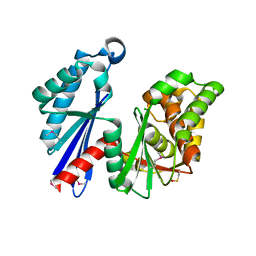
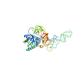
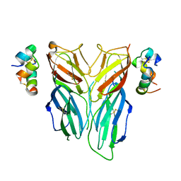
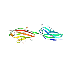
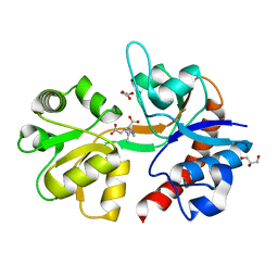 | | Crystal structure of the kainate receptor GluK4 ligand binding domain in complex with kainate | | 分子名称: | 3-(CARBOXYMETHYL)-4-ISOPROPENYLPROLINE, GLYCEROL, Glutamate receptor ionotropic, ... | | 著者 | Kristensen, O, Kristensen, L.B, Frydenvang, K, Kastrup, J.S. | | 登録日 | 2016-03-03 | | 公開日 | 2016-08-24 | | 最終更新日 | 2024-01-10 | | 実験手法 | X-RAY DIFFRACTION (2.05 Å) | | 主引用文献 | The Structure of a High-Affinity Kainate Receptor: GluK4 Ligand-Binding Domain Crystallized with Kainate.
Structure, 24, 2016
|
|
5DRV
 | |
2J4R
 | |
1T6C
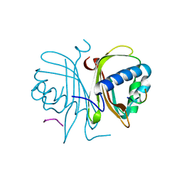
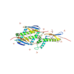
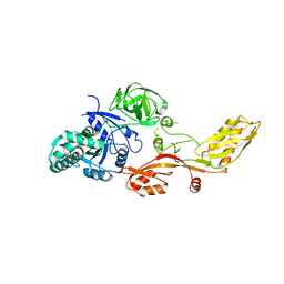
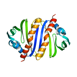
 | | Structural characterization of the Ppx/GppA protein family: crystal structure of the Aquifex aeolicus family member | | 分子名称: | (4S)-2-METHYL-2,4-PENTANEDIOL, CALCIUM ION, CHLORIDE ION, ... | | 著者 | Kristensen, O, Laurberg, M, Liljas, A, Kastrup, J.S, Gajhede, M. | | 登録日 | 2004-05-06 | | 公開日 | 2004-08-03 | | 最終更新日 | 2024-04-03 | | 実験手法 | X-RAY DIFFRACTION (1.53 Å) | | 主引用文献 | Structural characterization of the stringent response related exopolyphosphatase/guanosine pentaphosphate phosphohydrolase protein family
Biochemistry, 43, 2004
|
|
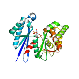
1T6D
 | | MIRAS phasing of the Aquifex aeolicus Ppx/GppA phosphatase: crystal structure of the type II variant | | 分子名称: | 2-AMINO-2-HYDROXYMETHYL-PROPANE-1,3-DIOL, CHLORIDE ION, exopolyphosphatase | | 著者 | Kristensen, O, Laurberg, M, Liljas, A, Kastrup, J.S, Gajhede, M. | | 登録日 | 2004-05-06 | | 公開日 | 2004-08-03 | | 最終更新日 | 2021-11-10 | | 実験手法 | X-RAY DIFFRACTION (2.15 Å) | | 主引用文献 | Structural characterization of the stringent response related exopolyphosphatase/guanosine pentaphosphate phosphohydrolase protein family
Biochemistry, 43, 2004
|
|
1P9Y
 | |
1OMS
 | |
2FPF
 | |
2FPD
 | |
1QJH
 | |
1CQN
 | |
1CQM
 | |
1OB2
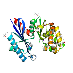
 | | E. coli elongation factor EF-Tu complexed with the antibiotic kirromycin, a GTP analog, and Phe-tRNA | | 分子名称: | ELONGATION FACTOR TU, KIRROMYCIN, MAGNESIUM ION, ... | | 著者 | Kristensen, O, Nissen, P, Nyborg, J. | | 登録日 | 2003-01-24 | | 公開日 | 2004-05-27 | | 最終更新日 | 2023-12-13 | | 実験手法 | X-RAY DIFFRACTION (3.35 Å) | | 主引用文献 | Kirromycin Defines a Specific Domain Arrangement of Elongation Factor EF-TU
To be Published
|
|
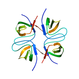
2FPE
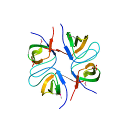
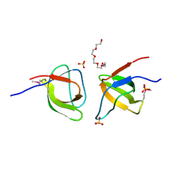 | | Conserved dimerization of the ib1 src-homology 3 domain | | 分子名称: | C-jun-amino-terminal kinase interacting protein 1, HEXAETHYLENE GLYCOL, SULFATE ION, ... | | 著者 | Guenat, S, Dar, I, Bonny, C, Kastrup, J.S, Gajhede, M, Kristensen, O. | | 登録日 | 2006-01-16 | | 公開日 | 2006-02-28 | | 最終更新日 | 2024-10-16 | | 実験手法 | X-RAY DIFFRACTION (1.75 Å) | | 主引用文献 | A unique set of SH3-SH3 interactions controls IB1 homodimerization
Embo J., 25, 2006
|
|
2DQU
 | | Crystal form II: high resolution crystal structure of the complex of the hydrolytic antibody Fab 6D9 and a transition-state analog | | 分子名称: | IMMUNOGLOBULIN 6D9, [1-(3-DIMETHYLAMINO-PROPYL)-3-ETHYL-UREIDO]-[4-(2,2,2-TRIFLUORO-ACETYLAMINO)-BENZYL]PHOSPHINIC ACID-2-(2,2-DIHYDRO-ACETYLAMINO)-3-HYDROXY-1-(4-NITROPHENYL)-PROPYL ESTER | | 著者 | Kristensen, O, Vassylyev, D.G, Tanaka, F, Ito, N, Morikawa, K, Fujii, I. | | 登録日 | 2006-05-30 | | 公開日 | 2006-06-20 | | 最終更新日 | 2024-10-16 | | 実験手法 | X-RAY DIFFRACTION (1.7 Å) | | 主引用文献 | Thermodynamic and structural basis for transition-state stabilization in antibody-catalyzed hydrolysis
J.Mol.Biol., 369, 2007
|
|
2DQT
 | | High resolution crystal structure of the complex of the hydrolytic antibody Fab 6D9 and a transition-state analog | | 分子名称: | IMMUNOGLOBULIN 6D9, [1-(3-DIMETHYLAMINO-PROPYL)-3-ETHYL-UREIDO]-[4-(2,2,2-TRIFLUORO-ACETYLAMINO)-BENZYL]PHOSPHINIC ACID-2-(2,2-DIHYDRO-ACETYLAMINO)-3-HYDROXY-1-(4-NITROPHENYL)-PROPYL ESTER | | 著者 | Kristensen, O, Vassylyev, D.G, Tanaka, F, Ito, N, Morikawa, K, Fujii, I. | | 登録日 | 2006-05-30 | | 公開日 | 2006-06-20 | | 最終更新日 | 2024-10-23 | | 実験手法 | X-RAY DIFFRACTION (1.8 Å) | | 主引用文献 | Thermodynamic and structural basis for transition-state stabilization in antibody-catalyzed hydrolysis
J.Mol.Biol., 369, 2007
|
|
4ZG1
 | |
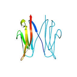
6FF3
 | | Crystal structure of Drosophila neural ectodermal development factor Imp-L1 with Human IGF-I | | 分子名称: | Insulin-like growth factor I, Neural/ectodermal development factor IMP-L2 | | 著者 | Brzozowski, A.M, Kulahin, N, Kristensen, O, Schluckebier, G, Meyts, P.D, Viola, C.M. | | 登録日 | 2018-01-03 | | 公開日 | 2018-09-26 | | 最終更新日 | 2024-11-13 | | 実験手法 | X-RAY DIFFRACTION (2.57 Å) | | 主引用文献 | Structures of insect Imp-L2 suggest an alternative strategy for regulating the bioavailability of insulin-like hormones.
Nat Commun, 9, 2018
|
|
6FEY
 | | Crystal structure of Drosophila neural ectodermal development factor Imp-L2 with Drosophila DILP5 insulin | | 分子名称: | Neural/ectodermal development factor IMP-L2, Probable insulin-like peptide 5 | | 著者 | Brzozowski, A.M, Kulahin, N, Kristensen, O, Schluckebier, G, Meyts, P.D. | | 登録日 | 2018-01-03 | | 公開日 | 2018-09-26 | | 最終更新日 | 2024-10-23 | | 実験手法 | X-RAY DIFFRACTION (3.48 Å) | | 主引用文献 | Structures of insect Imp-L2 suggest an alternative strategy for regulating the bioavailability of insulin-like hormones.
Nat Commun, 9, 2018
|
|
4IIA
 | |
1KTV
 | |
2UUS
 | | Crystal structure of the rat FGF1-sucrose octasulfate (SOS) complex. | | 分子名称: | 1,3,4,6-tetra-O-sulfo-beta-D-fructofuranose-(2-1)-2,3,4,6-tetra-O-sulfonato-alpha-D-glucopyranose, HEPARIN-BINDING GROWTH FACTOR 1 | | 著者 | Kulahin, N, Kiselyov, V, Kochoyan, A, Kristensen, O, Berezin, V, Bock, E, Gajhede, M. | | 登録日 | 2007-03-07 | | 公開日 | 2008-05-13 | | 最終更新日 | 2023-12-13 | | 実験手法 | X-RAY DIFFRACTION (2.2 Å) | | 主引用文献 | Dimerization Effect of Sucrose Octasulfate on Rat Fgf1.
Acta Crystallogr.,Sect.F, 64, 2008
|
|
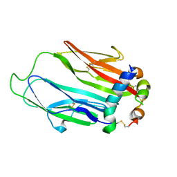
2V5T
 | | Crystal structure of NCAM2 Ig2-3 | | 分子名称: | 2-acetamido-2-deoxy-beta-D-glucopyranose, GLYCEROL, NEURAL CELL ADHESION MOLECULE 2, ... | | 著者 | Kulahin, N, Rasmussen, K.K, Kristensen, O, Berezin, V, Bock, E, Walmod, P.S, Gajhede, M. | | 登録日 | 2007-07-10 | | 公開日 | 2008-07-29 | | 最終更新日 | 2024-11-20 | | 実験手法 | X-RAY DIFFRACTION (2 Å) | | 主引用文献 | Structural Model and Trans-Interaction of the Entire Ectodomain of the Olfactory Cell Adhesion Molecule.
Structure, 19, 2011
|
|
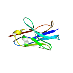
2VAJ
 | | Crystal structure of NCAM2 Ig1 (I4122 cell unit) | | 分子名称: | NEURAL CELL ADHESION MOLECULE 2 | | 著者 | Kulahin, N, Rasmussen, K.K, Kristensen, O, Kastrup, J.S, Navarro-Poulsen, J.-C, Berezin, V, Bock, E, Walmod, P.S, Gajhede, M. | | 登録日 | 2007-08-31 | | 公開日 | 2008-08-26 | | 最終更新日 | 2024-10-23 | | 実験手法 | X-RAY DIFFRACTION (2.701 Å) | | 主引用文献 | Crystal Structure of the Ig1 Domain of the Neural Cell Adhesion Molecule Ncam2 Displays Domain Swapping.
J.Mol.Biol., 382, 2008
|
|
3IJT
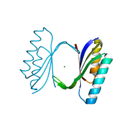
 | | Structural Characterization of SMU.440, a Hypothetical Protein from Streptococcus mutans | | 分子名称: | Putative uncharacterized protein | | 著者 | Nan, J, Brostromer, E, Kristensen, O, Su, X.-D. | | 登録日 | 2009-08-05 | | 公開日 | 2009-08-25 | | 最終更新日 | 2024-03-20 | | 実験手法 | X-RAY DIFFRACTION (2.377 Å) | | 主引用文献 | Bioinformatics and structural characterization of a hypothetical protein from Streptococcus mutans: implication of antibiotic resistance
Plos One, 4, 2009
|
|


