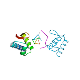
5B16
 
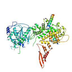 | | X-ray structure of DROSHA in complex with the C-terminal tail of DGCR8. | | 分子名称: | Microprocessor complex subunit DGCR8, Ribonuclease 3,DROSHA,Ribonuclease 3,DROSHA,Ribonuclease 3, ZINC ION | | 著者 | Kwon, S.C, Nguyen, T.A, Choi, Y.G, Jo, M.H, Hohng, S, Kim, V.N, Woo, J.S. | | 登録日 | 2015-11-23 | | 公開日 | 2016-02-03 | | 実験手法 | X-RAY DIFFRACTION (3.2 Å) | | 主引用文献 | Structure of Human DROSHA
Cell, 164, 2016
|
|
6JM9
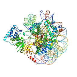 | |
6JMA
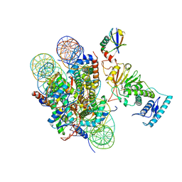 | |
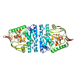
4ICS
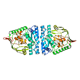 | | Crystal structure of PepS from Streptococcus pneumoniae in complex with a substrate | | 分子名称: | Aminopeptidase PepS, GLYCINE, TRYPTOPHAN, ... | | 著者 | Lee, S, Kim, K.K, Ta, M.H. | | 登録日 | 2012-12-11 | | 公開日 | 2013-10-23 | | 最終更新日 | 2024-02-28 | | 実験手法 | X-RAY DIFFRACTION (1.97 Å) | | 主引用文献 | Structure-based elucidation of the regulatory mechanism for aminopeptidase activity.
Acta Crystallogr.,Sect.D, 69, 2013
|
|
4ICQ
 | |
4ICR
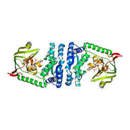 | | Structural basis for substrate recognition and reaction mechanism of bacterial aminopeptidase peps | | 分子名称: | Aminopeptidase PepS, CACODYLATE ION, ZINC ION | | 著者 | Lee, S, Kim, K.K, Ta, M.H. | | 登録日 | 2012-12-11 | | 公開日 | 2013-10-23 | | 最終更新日 | 2024-02-28 | | 実験手法 | X-RAY DIFFRACTION (2.17 Å) | | 主引用文献 | Structure-based elucidation of the regulatory mechanism for aminopeptidase activity.
Acta Crystallogr.,Sect.D, 69, 2013
|
|
4KMF
 | |


