1KLV
 
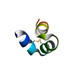
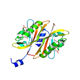
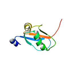 | | Solution Structure and Backbone Dynamics of GABARAP, GABAA Receptor associated protein | | 分子名称: | GABA(A) Receptor associated protein | | 著者 | Kouno, T, Miura, K, Tada, M, Kanematsu, T, Tate, S, Shirakawa, M, Hirata, M, Kawano, K. | | 登録日 | 2001-12-13 | | 公開日 | 2003-10-07 | | 最終更新日 | 2024-05-29 | | 実験手法 | SOLUTION NMR | | 主引用文献 | 1H, 13C and '5N resonance assignments of GABARAP, GABAA receptor associated protein.
J.Biomol.Nmr, 22, 2002
|
|
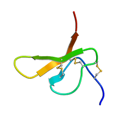
1KM7
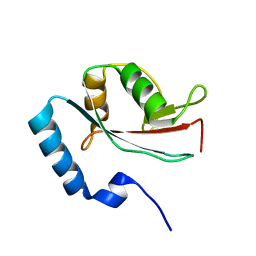
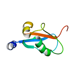 | | Solution Structure and Backbone Dynamics of GABARAP, GABAA Receptor Associated Protein | | 分子名称: | GABA(A) Receptor Associated Protein | | 著者 | Kouno, T, Miura, K, Tada, M, Kanematsu, T, Tate, S, Shirakawa, M, Hirata, M, Kawano, K. | | 登録日 | 2001-12-14 | | 公開日 | 2003-10-07 | | 最終更新日 | 2024-05-29 | | 実験手法 | SOLUTION NMR | | 主引用文献 | 1H, 13C and '5N resonance assignments of GABARAP, GABAA receptor associated protein.
J.Biomol.Nmr, 22, 2002
|
|
8I74
 | |
8I75
 | |
8I76
 | |
1LYP
 | |
1V49
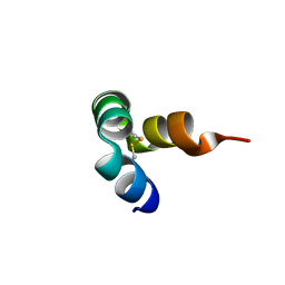
 | | Solution structure of microtubule-associated protein light chain-3 | | 分子名称: | Microtubule-associated proteins 1A/1B light chain 3B | | 著者 | Kouno, T, Mizuguchi, M, Tanida, I, Ueno, T, Kominami, E, Kawano, K. | | 登録日 | 2003-11-11 | | 公開日 | 2004-12-28 | | 最終更新日 | 2023-12-27 | | 実験手法 | SOLUTION NMR | | 主引用文献 | Solution structure of microtubule-associated protein light chain 3 and identification of its functional subdomains.
J.Biol.Chem., 280, 2005
|
|
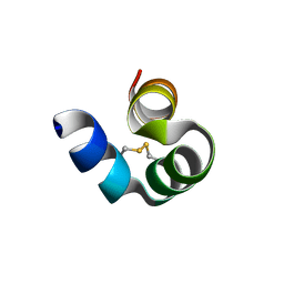
1HAK
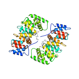 | | CRYSTAL STRUCTURE OF RECOMBINANT HUMAN PLACENTAL ANNEXIN V COMPLEXED WITH K-201 AS A CALCIUM CHANNEL ACTIVITY INHIBITOR | | 分子名称: | 4-[3-{1-(4-BENZYL)PIPERODINYL}PROPIONYL]-7-METHOXY-2,3,4,5-TERTRAHYDRO-1,4-BENZOTHIAZEPINE, ANNEXIN V | | 著者 | Ago, H, Inagaki, E, Miyano, M. | | 登録日 | 1997-12-10 | | 公開日 | 1999-02-16 | | 最終更新日 | 2024-05-22 | | 実験手法 | X-RAY DIFFRACTION (3 Å) | | 主引用文献 | Crystal structure of annexin V with its ligand K-201 as a calcium channel activity inhibitor.
J.Mol.Biol., 274, 1997
|
|
2Z6D
 | |
2Z6C
 | |
2DCW
 | |
2DCV
 | |
7CFZ
 | |


