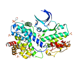
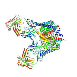
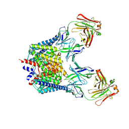
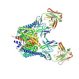
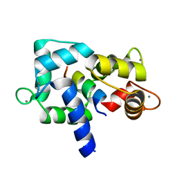
8H4R
 
 | | The Crystal Structure of CDK3 and CyclinE1 Complex with Dinaciclib from Biortus | | 分子名称: | 2-(N-MORPHOLINO)-ETHANESULFONIC ACID, 3-[({3-ethyl-5-[(2S)-2-(2-hydroxyethyl)piperidin-1-yl]pyrazolo[1,5-a]pyrimidin-7-yl}amino)methyl]-1-hydroxypyridinium, G1/S-specific cyclin-E1, ... | | 著者 | Gui, W, Wang, F, Cheng, W, Gao, J, Huang, Y, Ouyang, Z. | | 登録日 | 2022-10-11 | | 公開日 | 2023-10-11 | | 実験手法 | X-RAY DIFFRACTION (2.75 Å) | | 主引用文献 | The Crystal Structure of CDK3 and CyclinE1 Complex with Dinaciclib from Biortus
To Be Published
|
|
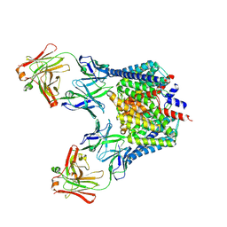
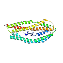
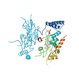
7XQK
 | | The Crystal Structure of CDK3 and CyclinE1 Complex from Biortus. | | 分子名称: | 2-(N-MORPHOLINO)-ETHANESULFONIC ACID, G1/S-specific cyclin-E1, GLYCEROL, ... | | 著者 | Gui, W, Wang, F, Cheng, W, Gao, J, Huang, Y. | | 登録日 | 2022-05-07 | | 公開日 | 2023-05-17 | | 最終更新日 | 2023-11-29 | | 実験手法 | X-RAY DIFFRACTION (2.25 Å) | | 主引用文献 | The Crystal Structure of CDK3 and CyclinE1 Complex from Biortus.
To Be Published
|
|
7F2F
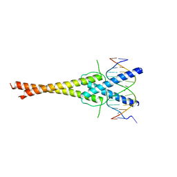 | | The complex of DNA with the C-terminal domain of TYE7 from Saccharomyces cerevisiae. | | 分子名称: | DNA (5'-D(*CP*AP*GP*AP*TP*CP*AP*TP*GP*TP*GP*TP*GP*CP*C)-3'), DNA (5'-D(*GP*GP*GP*CP*AP*CP*AP*CP*AP*TP*GP*AP*TP*CP*T)-3'), Serine-rich protein TYE7 | | 著者 | Gui, W. | | 登録日 | 2021-06-10 | | 公開日 | 2021-10-13 | | 最終更新日 | 2023-11-29 | | 実験手法 | X-RAY DIFFRACTION (2.55 Å) | | 主引用文献 | Crystal structure of the complex of DNA with the C-terminal domain of TYE7 from Saccharomyces cerevisiae.
Acta Crystallogr.,Sect.F, 77, 2021
|
|
2H2P
 | |
2H2S
 | |
2R9H
 | |
8J2K
 | | Crystal structure of a bright green fluorescent protein (StayGold) with double mutation (N137A, Q140S) in jellyfish Cytaeis uchidae from Biortus | | 分子名称: | 1,2-ETHANEDIOL, StayGold(N137A, Q140S) | | 著者 | Wu, J, Wang, F, Gui, W, Cheng, W, Yang, Y. | | 登録日 | 2023-04-14 | | 公開日 | 2023-12-13 | | 実験手法 | X-RAY DIFFRACTION (1.7 Å) | | 主引用文献 | Crystal structure of a bright green fluorescent protein (StayGold) in jellyfish Cytaeis uchidae from Biortus
To Be Published
|
|
8J2J
 | | Crystal structure of a bright green fluorescent protein (StayGold) with single mutation (Y187F) in jellyfish Cytaeis uchidae from Biortus | | 分子名称: | 1,2-ETHANEDIOL, SULFATE ION, StayGold(Y187F) | | 著者 | Wu, J, Wang, F, Gui, W, Cheng, W, Yang, Y. | | 登録日 | 2023-04-14 | | 公開日 | 2023-12-13 | | 実験手法 | X-RAY DIFFRACTION (1.9 Å) | | 主引用文献 | Crystal structure of a bright green fluorescent protein (StayGold) in jellyfish Cytaeis uchidae from Biortus
To Be Published
|
|
8J2I
 | | Crystal structure of a bright green fluorescent protein (StayGold) with single mutation (Q140S) in jellyfish Cytaeis uchidae from Biortus | | 分子名称: | 1,2-ETHANEDIOL, StayGold(Q140S) | | 著者 | Wu, J, Wang, F, Gui, W, Cheng, W, Yang, Y. | | 登録日 | 2023-04-14 | | 公開日 | 2023-12-13 | | 最終更新日 | 2024-10-09 | | 実験手法 | X-RAY DIFFRACTION (1.75 Å) | | 主引用文献 | Crystal structure of a bright green fluorescent protein (StayGold) in jellyfish Cytaeis uchidae from Biortus
To Be Published
|
|
8J3J
 | | Crystal structure of a bright green fluorescent protein (StayGold) with double mutations (Q140S, Y187F) in jellyfish Cytaeis uchidae from Biortus | | 分子名称: | 1,2-ETHANEDIOL, StayGold(Q140S, Y187F) | | 著者 | Wu, J, Wang, F, Gui, W, Cheng, W, Yang, Y. | | 登録日 | 2023-04-17 | | 公開日 | 2023-12-13 | | 実験手法 | X-RAY DIFFRACTION (1.7 Å) | | 主引用文献 | Crystal structure of a bright green fluorescent protein (StayGold) in jellyfish Cytaeis uchidae from Biortus
To Be Published
|
|
8J2L
 | | Crystal structure of a bright green fluorescent protein (StayGold) with double mutations (N137A, Y187F) in jellyfish Cytaeis uchidae from Biortus | | 分子名称: | 4-(2-HYDROXYETHYL)-1-PIPERAZINE ETHANESULFONIC ACID, GLYCEROL, SODIUM ION, ... | | 著者 | Wu, J, Wang, F, Gui, W, Cheng, W, Yang, Y. | | 登録日 | 2023-04-14 | | 公開日 | 2023-12-13 | | 実験手法 | X-RAY DIFFRACTION (1.7 Å) | | 主引用文献 | Crystal structure of a bright green fluorescent protein (StayGold) in jellyfish Cytaeis uchidae from Biortus
To Be Published
|
|
8J2H
 | | Crystal structure of a bright green fluorescent protein (StayGold) with single mutation (N137A) in jellyfish Cytaeis uchidae from Biortus | | 分子名称: | GLYCEROL, SODIUM ION, StayGold(N137A) | | 著者 | Wu, J, Wang, F, Gui, W, Cheng, W, Yang, Y. | | 登録日 | 2023-04-14 | | 公開日 | 2023-12-13 | | 実験手法 | X-RAY DIFFRACTION (1.7 Å) | | 主引用文献 | Crystal structure of a bright green fluorescent protein (StayGold) in jellyfish Cytaeis uchidae from Biortus
To Be Published
|
|
8GYF
 | | Crystal structure of a bright green fluorescent protein (StayGold) with single mutation (K192Y) in jellyfish Cytaeis uchidae from Biortus | | 分子名称: | 1,2-ETHANEDIOL, staygold(K192Y) | | 著者 | Wu, J, Wang, F, Gui, W, Cheng, W, Yang, Y. | | 登録日 | 2022-09-22 | | 公開日 | 2023-10-04 | | 最終更新日 | 2023-11-15 | | 実験手法 | X-RAY DIFFRACTION (2 Å) | | 主引用文献 | Crystal structure of a bright green fluorescent protein (StayGold) with single mutation (K192Y) in jellyfish Cytaeis uchidae from Biortus
To Be Published
|
|
7Y40
 | | Crystal structure of a bright green fluorescent protein (StayGold) in jellyfish Cytaeis uchidae from Biortus | | 分子名称: | 1,2-ETHANEDIOL, staygold | | 著者 | Wu, J, Wang, F, Gui, W, Cheng, W, Yang, Y. | | 登録日 | 2022-06-13 | | 公開日 | 2023-07-05 | | 最終更新日 | 2024-04-03 | | 実験手法 | X-RAY DIFFRACTION (1.7 Å) | | 主引用文献 | Crystal structure of a bright green fluorescent protein (StayGold) in jellyfish Cytaeis uchidae from Biortus
To Be Published
|
|
7YAO
 | |
7YRE
 | | Crystal structure of a bright green fluorescent protein (StayGold) with triple mutations (N137A, Q140S, Y187F) in jellyfish Cytaeis uchidae from Biortus | | 分子名称: | 1,2-ETHANEDIOL, staygold(N137A,Q140S,Y187F) | | 著者 | Wu, J, Wang, F, Gui, W, Cheng, W, Yang, Y. | | 登録日 | 2022-08-09 | | 公開日 | 2023-08-16 | | 最終更新日 | 2023-11-15 | | 実験手法 | X-RAY DIFFRACTION (2.3 Å) | | 主引用文献 | Crystal structure of a bright green fluorescent protein (StayGold) with triple mutations (N137A, Q140S, Y187F) in jellyfish Cytaeis uchidae from Biortus
To Be Published
|
|
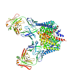
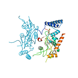
2FEE
 | | Structure of the Cl-/H+ exchanger CLC-ec1 from E.Coli in NaBr | | 分子名称: | Fab fragment, heavy chain, light chain, ... | | 著者 | Accardi, A, Walden, M.P, Nguitragool, W, Jayaram, H, Williams, C, Miller, C. | | 登録日 | 2005-12-15 | | 公開日 | 2006-01-03 | | 最終更新日 | 2023-08-30 | | 実験手法 | X-RAY DIFFRACTION (3.2 Å) | | 主引用文献 | Separate ion pathways in a Cl-/H+ exchanger
J.Gen.Physiol., 126, 2005
|
|
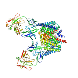
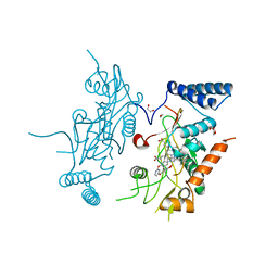
2FED
 | | Structure of the E203Q mutant of the Cl-/H+ exchanger CLC-ec1 from E.Coli | | 分子名称: | Fab fragment, heavy chain, light chain, ... | | 著者 | Accardi, A, Walden, M.P, Nguitragool, W, Jayaram, H, Williams, C, Miller, C. | | 登録日 | 2005-12-15 | | 公開日 | 2006-01-03 | | 最終更新日 | 2023-08-30 | | 実験手法 | X-RAY DIFFRACTION (3.317 Å) | | 主引用文献 | Separate ion pathways in a Cl-/H+ exchanger
J.Gen.Physiol., 126, 2005
|
|
2FEC
 | | Structure of the E203Q mutant of the Cl-/H+ exchanger CLC-ec1 from E.Coli | | 分子名称: | Fab fragment, heavy chain, light chain, ... | | 著者 | Accardi, A, Walden, M.P, Nguitragool, W, Jayaram, H, Williams, C, Miller, C. | | 登録日 | 2005-12-15 | | 公開日 | 2006-01-03 | | 最終更新日 | 2021-10-20 | | 実験手法 | X-RAY DIFFRACTION (3.967 Å) | | 主引用文献 | Separate ion pathways in a Cl-/H+ exchanger
J.Gen.Physiol., 126, 2005
|
|
3EWV
 | | Crystal Structure of calmodulin complexed with a peptide | | 分子名称: | CALCIUM ION, Calmodulin, Tumor necrosis factor receptor superfamily member 16 | | 著者 | Jiang, T, Cao, P, Gong, Y, Yu, H.J, Gui, W.J, Zhang, W.T. | | 登録日 | 2008-10-16 | | 公開日 | 2009-10-20 | | 最終更新日 | 2023-11-01 | | 実験手法 | X-RAY DIFFRACTION (2.6 Å) | | 主引用文献 | Structural insights into the mechanism of calmodulin binding to death receptors.
Acta Crystallogr.,Sect.D, 70, 2014
|
|
3EWT
 | | Crystal Structure of calmodulin complexed with a peptide | | 分子名称: | CALCIUM ION, Calmodulin, Tumor necrosis factor receptor superfamily member 6 | | 著者 | Jiang, T, Cao, P, Gong, Y, Yu, H.J, Gui, W.J, Zhang, W.T. | | 登録日 | 2008-10-16 | | 公開日 | 2009-10-20 | | 最終更新日 | 2023-11-01 | | 実験手法 | X-RAY DIFFRACTION (2.4 Å) | | 主引用文献 | Structural insights into the mechanism of calmodulin binding to death receptors.
Acta Crystallogr.,Sect.D, 70, 2014
|
|
4Z8N
 | |
2ORQ
 | | Murine Inducible Nitric Oxide Synthase Oxygenase Domain (DELTA 114) 4-(imidazol-1-yl)phenol and piperonylamine Complex | | 分子名称: | 1-(1,3-BENZODIOXOL-5-YL)METHANAMINE, 4-(1H-IMIDAZOL-1-YL)PHENOL, Nitric oxide synthase, ... | | 著者 | Adler, M, Whitlow, M. | | 登録日 | 2007-02-04 | | 公開日 | 2007-04-17 | | 最終更新日 | 2024-02-21 | | 実験手法 | X-RAY DIFFRACTION (2.1 Å) | | 主引用文献 | Design, Synthesis, and Activity of 2-Imidazol-1-ylpyrimidine Derived Inducible Nitric Oxide Synthase Dimerization Inhibitors
J.Med.Chem., 50, 2007
|
|
2ORR
 | | Murine Inducible Nitric Oxide Synthase Oxygenase Domain (Delta 114) 4-(Benzo[1,3]dioxol-5-yloxy)-2-(4-imidazol-1-yl-phenoxy)-pyrimidine Complex | | 分子名称: | 1,2-ETHANEDIOL, 4-(1,3-BENZODIOXOL-5-YLOXY)-2-[4-(1H-IMIDAZOL-1-YL)PHENOXY]PYRIMIDINE, Nitric oxide synthase, ... | | 著者 | Adler, M, Whitlow, M. | | 登録日 | 2007-02-04 | | 公開日 | 2007-04-17 | | 最終更新日 | 2024-02-21 | | 実験手法 | X-RAY DIFFRACTION (2 Å) | | 主引用文献 | Design, Synthesis, and Activity of 2-Imidazol-1-ylpyrimidine Derived Inducible Nitric Oxide Synthase Dimerization Inhibitors
J.Med.Chem., 50, 2007
|
|
2ORT
 | | Murine Inducible Nitric Oxide Synthase Oxygenase Domain (Delta 114) 1-Benzo[1,3]dioxol-5-ylmethyl-3S-(4-imidazol-1-yl-phenoxy)-piperidine Complex | | 分子名称: | (3S)-1-(1,3-BENZODIOXOL-5-YLMETHYL)-3-[4-(1H-IMIDAZOL-1-YL)PHENOXY]PIPERIDINE, Nitric oxide synthase, inducible, ... | | 著者 | Adler, M, Whitlow, M. | | 登録日 | 2007-02-04 | | 公開日 | 2007-04-17 | | 最終更新日 | 2023-08-30 | | 実験手法 | X-RAY DIFFRACTION (1.87 Å) | | 主引用文献 | Design, Synthesis, and Activity of 2-Imidazol-1-ylpyrimidine Derived Inducible Nitric Oxide Synthase Dimerization Inhibitors
J.Med.Chem., 50, 2007
|
|


