Loading
PDBj
メニューPDBj@FacebookPDBj@TwitterPDBj@YouTubewwPDB FoundationwwPDB
RCSB PDBPDBeBMRBAdv. SearchSearch help
Search by PDB author
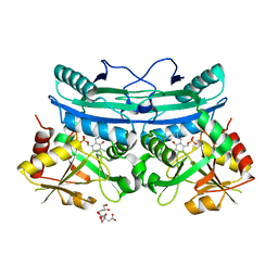
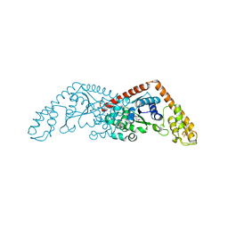
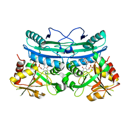
6Q1Q
DownloadVisualize
BU of 6q1q by Molmil
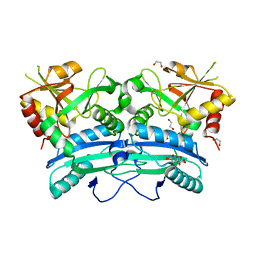
A hypothetical aminotransferase from Mycobacterium tuberculosis, apo form
分子名称: 1,2-ETHANEDIOL, CITRIC ACID, Probable amino acid aminotransferase
著者Duan, L, Sacchettini, J.
登録日2019-08-05
公開日2020-09-23
最終更新日2023-10-11
実験手法X-RAY DIFFRACTION (2.4 Å)
主引用文献A hypothetical aminotransferase from Mycobacterium tuberculosis
To Be Published
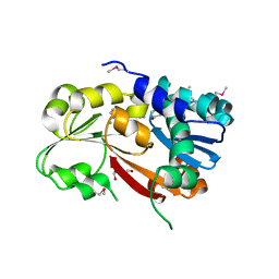
6Q1S
DownloadVisualize
BU of 6q1s by Molmil
A hypothetical aminotransferase from Mycobacterium tuberculosis, alpha-ketoglutarate and PMP bound form
分子名称: 2-OXOGLUTARIC ACID, 4'-DEOXY-4'-AMINOPYRIDOXAL-5'-PHOSPHATE, CITRIC ACID, ...
著者Duan, L, Sacchettini, J.
登録日2019-08-05
公開日2020-09-23
最終更新日2023-10-11
実験手法X-RAY DIFFRACTION (2.3 Å)
主引用文献A hypothetical aminotransferase from Mycobacterium tuberculosis
To Be Published
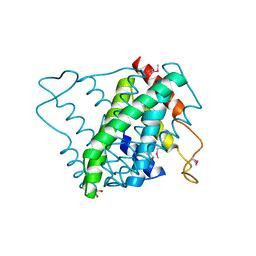
6Q1R
DownloadVisualize
BU of 6q1r by Molmil
A hypothetical aminotransferase from Mycobacterium tuberculosis, PLP-bound form
分子名称: CITRIC ACID, GLYCEROL, PYRIDOXAL-5'-PHOSPHATE, ...
著者Duan, L, Sacchettini, J.
登録日2019-08-05
公開日2020-09-23
最終更新日2023-10-11
実験手法X-RAY DIFFRACTION (2.702 Å)
主引用文献A hypothetical aminotransferase from Mycobacterium tuberculosis
To Be Published
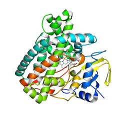
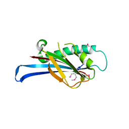
5L1P
DownloadVisualize
BU of 5l1p by Molmil
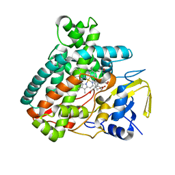
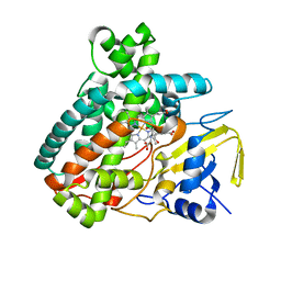
X-ray Structure of Cytochrome P450 PntM with Pentalenolactone
分子名称: PROTOPORPHYRIN IX CONTAINING FE, Pentalenolactone synthase, pentalenolactone
著者Duan, L, Jogl, G, Cane, D.E.
登録日2016-07-29
公開日2016-09-14
最終更新日2023-10-04
実験手法X-RAY DIFFRACTION (2.28 Å)
主引用文献The Cytochrome P450-Catalyzed Oxidative Rearrangement in the Final Step of Pentalenolactone Biosynthesis: Substrate Structure Determines Mechanism.
J.Am.Chem.Soc., 138, 2016
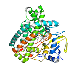
5L1O
DownloadVisualize
BU of 5l1o by Molmil
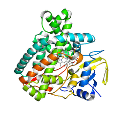
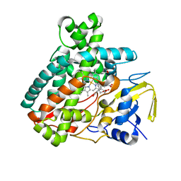
X-ray Structure of Cytochrome P450 PntM with Pentalenolactone F
分子名称: PROTOPORPHYRIN IX CONTAINING FE, Pentalenolactone synthase, pentalenolactone F
著者Duan, L, Jogl, G, Cane, D.E.
登録日2016-07-29
公開日2016-09-14
最終更新日2023-10-04
実験手法X-RAY DIFFRACTION (2.03 Å)
主引用文献The Cytochrome P450-Catalyzed Oxidative Rearrangement in the Final Step of Pentalenolactone Biosynthesis: Substrate Structure Determines Mechanism.
J.Am.Chem.Soc., 138, 2016
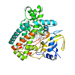
5L1Q
DownloadVisualize
BU of 5l1q by Molmil
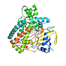
X-ray Structure of Cytochrome P450 PntM with Dihydropentalenolactone F
分子名称: Dihydropentalenolactone F, PROTOPORPHYRIN IX CONTAINING FE, Pentalenolactone synthase
著者Duan, L, Jogl, G, Cane, D.E.
登録日2016-07-29
公開日2016-09-14
最終更新日2023-10-04
実験手法X-RAY DIFFRACTION (2.03 Å)
主引用文献The Cytochrome P450-Catalyzed Oxidative Rearrangement in the Final Step of Pentalenolactone Biosynthesis: Substrate Structure Determines Mechanism.
J.Am.Chem.Soc., 138, 2016
5L1S
DownloadVisualize
BU of 5l1s by Molmil
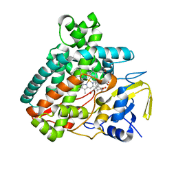
X-ray Structure of F232L mutant of Cytochrome P450 PntM with pentalenolactone F
分子名称: PROTOPORPHYRIN IX CONTAINING FE, Pentalenolactone synthase, pentalenolactone F
著者Duan, L, Jogl, G, Cane, D.E.
登録日2016-07-29
公開日2016-09-14
最終更新日2023-10-04
実験手法X-RAY DIFFRACTION (2.08 Å)
主引用文献The Cytochrome P450-Catalyzed Oxidative Rearrangement in the Final Step of Pentalenolactone Biosynthesis: Substrate Structure Determines Mechanism.
J.Am.Chem.Soc., 138, 2016
5L1W
DownloadVisualize
BU of 5l1w by Molmil
X-ray Structure of 2-Mercaptoethanol modified M81C mutant of Cytochrome P450 PntM with pentalenolactone F
分子名称: PROTOPORPHYRIN IX CONTAINING FE, Pentalenolactone synthase, pentalenolactone F
著者Duan, L, Jogl, G, Cane, D.E.
登録日2016-07-29
公開日2016-09-14
最終更新日2023-10-04
実験手法X-RAY DIFFRACTION (2.06 Å)
主引用文献The Cytochrome P450-Catalyzed Oxidative Rearrangement in the Final Step of Pentalenolactone Biosynthesis: Substrate Structure Determines Mechanism.
J.Am.Chem.Soc., 138, 2016
5L1V
DownloadVisualize
BU of 5l1v by Molmil
X-ray Structure of M81C mutant of Cytochrome P450 PntM with pentalenolactone F
分子名称: PROTOPORPHYRIN IX CONTAINING FE, Pentalenolactone synthase, pentalenolactone F
著者Duan, L, Jogl, G, Cane, D.E.
登録日2016-07-29
公開日2016-09-14
最終更新日2023-10-04
実験手法X-RAY DIFFRACTION (2.12 Å)
主引用文献The Cytochrome P450-Catalyzed Oxidative Rearrangement in the Final Step of Pentalenolactone Biosynthesis: Substrate Structure Determines Mechanism.
J.Am.Chem.Soc., 138, 2016
5L1U
DownloadVisualize
BU of 5l1u by Molmil
X-ray Structure of M81A mutant of Cytochrome P450 PntM with pentalenolactone F
分子名称: PROTOPORPHYRIN IX CONTAINING FE, Pentalenolactone synthase, pentalenolactone F
著者Duan, L, Jogl, G, Cane, D.E.
登録日2016-07-29
公開日2016-09-14
最終更新日2023-10-04
実験手法X-RAY DIFFRACTION (2.074 Å)
主引用文献The Cytochrome P450-Catalyzed Oxidative Rearrangement in the Final Step of Pentalenolactone Biosynthesis: Substrate Structure Determines Mechanism.
J.Am.Chem.Soc., 138, 2016
5L1R
DownloadVisualize
BU of 5l1r by Molmil
X-ray Structure of the Substrate-free Cytochrome P450 PntM
分子名称: BICINE, PROTOPORPHYRIN IX CONTAINING FE, Pentalenolactone synthase
著者Duan, L, Jogl, G, Cane, D.E.
登録日2016-07-29
公開日2016-09-14
最終更新日2023-10-04
実験手法X-RAY DIFFRACTION (2 Å)
主引用文献The Cytochrome P450-Catalyzed Oxidative Rearrangement in the Final Step of Pentalenolactone Biosynthesis: Substrate Structure Determines Mechanism.
J.Am.Chem.Soc., 138, 2016
5L1T
DownloadVisualize
BU of 5l1t by Molmil
X-ray Structure of M77S mutant of Cytochrome P450 PntM with pentalenolactone F
分子名称: PROTOPORPHYRIN IX CONTAINING FE, Pentalenolactone synthase, pentalenolactone F
著者Duan, L, Jogl, G, Cane, D.E.
登録日2016-07-29
公開日2016-09-14
最終更新日2023-10-04
実験手法X-RAY DIFFRACTION (2.082 Å)
主引用文献The Cytochrome P450-Catalyzed Oxidative Rearrangement in the Final Step of Pentalenolactone Biosynthesis: Substrate Structure Determines Mechanism.
J.Am.Chem.Soc., 138, 2016
5V93
DownloadVisualize
BU of 5v93 by Molmil
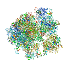
Cryo-EM structure of the 70S ribosome from Mycobacterium tuberculosis bound with Capreomycin
分子名称: 16S rRNA, 23S rRNA, 30S ribosomal protein S10, ...
著者Yang, K, Chang, J.-Y, Cui, Z, Li, X, Meng, R, Duan, L, Thongchol, J, Jakana, J, Huwe, C, Sacchettini, J, Zhang, J.
登録日2017-03-22
公開日2017-09-20
最終更新日2020-08-12
実験手法ELECTRON MICROSCOPY (4 Å)
主引用文献Structural insights into species-specific features of the ribosome from the human pathogen Mycobacterium tuberculosis.
Nucleic Acids Res., 45, 2017
5FID
DownloadVisualize
BU of 5fid by Molmil
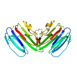
Crystal structure of the protein elicitor MoHrip2 from Magnaporthe oryzae
分子名称: Elicitor protein Hrip2
著者Liu, M, Duan, L, Qiu, D, Liu, X.
登録日2015-12-23
公開日2016-12-07
実験手法X-RAY DIFFRACTION (1.809 Å)
主引用文献Crystal Structure Analysis and the Identification of Distinctive Functional Regions of the Protein Elicitor Mohrip2
Front Plant Sci, 7, 2016
8K20
DownloadVisualize
BU of 8k20 by Molmil
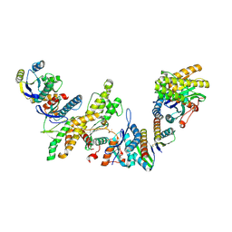
Cryo-EM structure of KEOPS complex from Arabidopsis thaliana
分子名称: At4g34412, At5g53043, FE (III) ION, ...
著者Zheng, X.X, Zhu, L, Duan, L, Zhang, W.H.
登録日2023-07-11
公開日2024-04-03
最終更新日2024-05-22
実験手法ELECTRON MICROSCOPY (3.7 Å)
主引用文献Molecular basis of A. thaliana KEOPS complex in biosynthesizing tRNA t6A.
Nucleic Acids Res., 52, 2024
3BOS
DownloadVisualize
BU of 3bos by Molmil
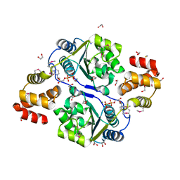
Crystal structure of a putative dna replication regulator HDA (SAMA_1916) from Shewanella amazonensis sb2b at 1.75 A resolution
分子名称: 1,2-ETHANEDIOL, CHLORIDE ION, CYTIDINE-5'-DIPHOSPHATE, ...
著者Joint Center for Structural Genomics (JCSG)
登録日2007-12-17
公開日2008-01-15
最終更新日2023-01-25
実験手法X-RAY DIFFRACTION (1.75 Å)
主引用文献A structural basis for the regulatory inactivation of DnaA.
J.Mol.Biol., 385, 2009
1VR8
DownloadVisualize
BU of 1vr8 by Molmil
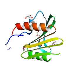
Crystal structure of GTP binding regulator (TM1622) from Thermotoga Maritima at 1.75 A resolution
分子名称: AZIDE ION, GLYCEROL, GTP binding regulator
著者Joint Center for Structural Genomics (JCSG)
登録日2005-02-16
公開日2005-03-15
最終更新日2023-01-25
実験手法X-RAY DIFFRACTION (1.75 Å)
主引用文献Crystal structure of an ORFan protein (TM1622) from Thermotoga maritima at 1.75 A resolution reveals a fold similar to the Ran-binding protein Mog1p.
Proteins, 65, 2006
2FNA
DownloadVisualize
BU of 2fna by Molmil
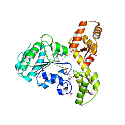
Crystal structure of an archaeal aaa+ atpase (sso1545) from sulfolobus solfataricus p2 at 2.00 A resolution
分子名称: 1,2-ETHANEDIOL, ADENOSINE-5'-DIPHOSPHATE, Conserved hypothetical protein, ...
著者Joint Center for Structural Genomics (JCSG)
登録日2006-01-10
公開日2006-02-07
最終更新日2024-10-09
実験手法X-RAY DIFFRACTION (2 Å)
主引用文献Crystal structure of a novel archaeal AAA+ ATPase SSO1545 from Sulfolobus solfataricus.
Proteins, 74, 2009
2G36
DownloadVisualize
BU of 2g36 by Molmil
Crystal structure of Tryptophanyl-tRNA synthetase (EC 6.1.1.2) (Tryptophan-tRNA ligase)(TrpRS) (tm0492) from THERMOTOGA MARITIMA at 2.50 A resolution
分子名称: IRON/SULFUR CLUSTER, TRYPTOPHAN, Tryptophanyl-tRNA synthetase
著者Joint Center for Structural Genomics (JCSG)
登録日2006-02-17
公開日2006-03-28
最終更新日2024-04-03
実験手法X-RAY DIFFRACTION (2.5 Å)
主引用文献Structure of a tryptophanyl-tRNA synthetase containing an iron-sulfur cluster.
Acta Crystallogr.,Sect.F, 66, 2010
3L5O
DownloadVisualize
BU of 3l5o by Molmil
Crystal structure of protein with unknown function from DUF364 family (ZP_00559375.1) from Desulfitobacterium hafniense DCB-2 at 2.01 A resolution
分子名称: 1,2-ETHANEDIOL, CHLORIDE ION, IMIDAZOLE, ...
著者Joint Center for Structural Genomics, Joint Center for Structural Genomics (JCSG)
登録日2009-12-22
公開日2010-02-02
最終更新日2023-02-01
実験手法X-RAY DIFFRACTION (2.01 Å)
主引用文献Structure of the first representative of Pfam family PF04016 (DUF364) reveals enolase and Rossmann-like folds that combine to form a unique active site with a possible role in heavy-metal chelation.
Acta Crystallogr.,Sect.F, 66, 2010
3NL9
DownloadVisualize
BU of 3nl9 by Molmil
Crystal structure of a putative NTP pyrophosphohydrolase (Exig_1061) from EXIGUOBACTERIUM SP. 255-15 at 1.78 A resolution
分子名称: 1,2-ETHANEDIOL, putative NTP pyrophosphohydrolase
著者Joint Center for Structural Genomics (JCSG)
登録日2010-06-21
公開日2010-07-21
最終更新日2024-10-09
実験手法X-RAY DIFFRACTION (1.78 Å)
主引用文献Structure of a putative NTP pyrophosphohydrolase: YP_001813558.1 from Exiguobacterium sibiricum 255-15.
Acta Crystallogr.,Sect.F, 66, 2010
2QTP
DownloadVisualize
BU of 2qtp by Molmil
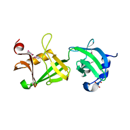
Crystal structure of a duf1185 family protein (spo0826) from silicibacter pomeroyi dss-3 at 2.10 A resolution
分子名称: Uncharacterized protein
著者Joint Center for Structural Genomics (JCSG)
登録日2007-08-02
公開日2007-08-21
最終更新日2023-01-25
実験手法X-RAY DIFFRACTION (2.1 Å)
主引用文献Structures of the first representatives of Pfam family PF06684 (DUF1185) reveal a novel variant of the Bacillus chorismate mutase fold and suggest a role in amino-acid metabolism.
Acta Crystallogr.,Sect.F, 66, 2010
2RA9
DownloadVisualize
BU of 2ra9 by Molmil
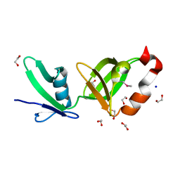
Crystal structure of a duf1285 family protein (sbal_2486) from shewanella baltica os155 at 1.40 A resolution
分子名称: 1,2-ETHANEDIOL, SODIUM ION, Uncharacterized protein DUF1285
著者Joint Center for Structural Genomics (JCSG)
登録日2007-09-14
公開日2007-10-16
最終更新日2023-01-25
実験手法X-RAY DIFFRACTION (1.4 Å)
主引用文献Structures of the first representatives of Pfam family PF06938 (DUF1285) reveal a new fold with repeated structural motifs and possible involvement in signal transduction.
Acta Crystallogr.,Sect.F, 66, 2010
2RE3
DownloadVisualize
BU of 2re3 by Molmil
CRYSTAL STRUCTURE OF a DUF1285 family protein (SPO_0140) FROM SILICIBACTER POMEROYI DSS-3 AT 2.50 A RESOLUTION
分子名称: GLYCEROL, Uncharacterized protein
著者Joint Center for Structural Genomics (JCSG)
登録日2007-09-25
公開日2007-10-09
最終更新日2024-10-09
実験手法X-RAY DIFFRACTION (2.5 Å)
主引用文献Structures of the first representatives of Pfam family PF06938 (DUF1285) reveal a new fold with repeated structural motifs and possible involvement in signal transduction.
Acta Crystallogr.,Sect.F, 66, 2010
1ZKG
DownloadVisualize
BU of 1zkg by Molmil
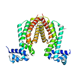
Crystal structure of Transcriptional regulator, TETR family (tm1030) from Thermotoga maritima at 2.30 A resolution
分子名称: UNKNOWN LIGAND, transcriptional regulator, TetR family
著者Joint Center for Structural Genomics (JCSG)
登録日2005-05-02
公開日2005-07-26
最終更新日2023-01-25
実験手法X-RAY DIFFRACTION (2.3 Å)
主引用文献Crystal structure of TM1030 from Thermotoga maritima at 2.3 A resolution reveals molecular details of its transcription repressor function.
Proteins, 68, 2007


1234>

225946

件を2024-10-09に公開中

PDB statisticsPDBj update infoContact PDBjnumon