7O1N
 
 | |
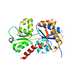
1AF7
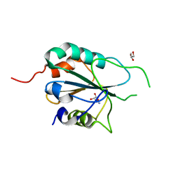
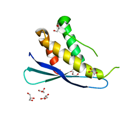
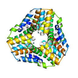
 | | CHER FROM SALMONELLA TYPHIMURIUM | | 分子名称: | CHEMOTAXIS RECEPTOR METHYLTRANSFERASE CHER, S-ADENOSYL-L-HOMOCYSTEINE | | 著者 | Djordjevic, S, Stock, A.M. | | 登録日 | 1997-03-22 | | 公開日 | 1998-01-28 | | 最終更新日 | 2024-02-07 | | 実験手法 | X-RAY DIFFRACTION (2 Å) | | 主引用文献 | Crystal structure of the chemotaxis receptor methyltransferase CheR suggests a conserved structural motif for binding S-adenosylmethionine.
Structure, 5, 1997
|
|
1A2O
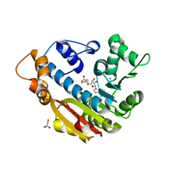
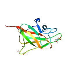
 | | STRUCTURAL BASIS FOR METHYLESTERASE CHEB REGULATION BY A PHOSPHORYLATION-ACTIVATED DOMAIN | | 分子名称: | CHEB METHYLESTERASE | | 著者 | Djordjevic, S, Goudreau, P.N, Xu, Q, Stock, A.M, West, A.H. | | 登録日 | 1998-01-06 | | 公開日 | 1998-04-29 | | 最終更新日 | 2024-05-22 | | 実験手法 | X-RAY DIFFRACTION (2.4 Å) | | 主引用文献 | Structural basis for methylesterase CheB regulation by a phosphorylation-activated domain.
Proc.Natl.Acad.Sci.USA, 95, 1998
|
|
1BC5
 | |
1BUC
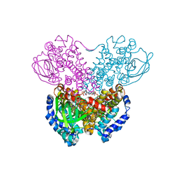 | | THREE-DIMENSIONAL STRUCTURE OF BUTYRYL-COA DEHYDROGENASE FROM MEGASPHAERA ELSDENII | | 分子名称: | ACETOACETYL-COENZYME A, BUTYRYL-COA DEHYDROGENASE, FLAVIN-ADENINE DINUCLEOTIDE | | 著者 | Djordjevic, S, Pace, C.P, Stankovich, M.T, Kim, J.J.P. | | 登録日 | 1994-09-06 | | 公開日 | 1995-04-20 | | 最終更新日 | 2024-02-07 | | 実験手法 | X-RAY DIFFRACTION (2.5 Å) | | 主引用文献 | Three-dimensional structure of butyryl-CoA dehydrogenase from Megasphaera elsdenii.
Biochemistry, 34, 1995
|
|
1J04
 | | Structural mechanism of enzyme mistargeting in hereditary kidney stone disease in vitro | | 分子名称: | (AMINOOXY)ACETIC ACID, GLYCEROL, alanine--glyoxylate aminotransferase | | 著者 | Zhang, X, Djordjevic, S, Bartlam, M, Ye, S, Rao, Z, Danpure, C.J. | | 登録日 | 2002-10-30 | | 公開日 | 2003-11-11 | | 最終更新日 | 2023-11-15 | | 実験手法 | X-RAY DIFFRACTION (2.6 Å) | | 主引用文献 | Structural implications of a G170R mutation of alanine:glyoxylate aminotransferase that is associated with peroxisome-to-mitochondrion mistargeting.
Acta Crystallogr.,Sect.F, 66, 2010
|
|
6ESK
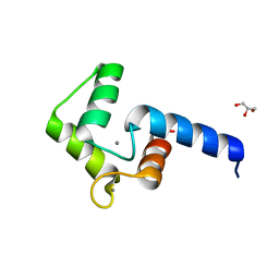
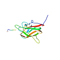
 | | Structure of the apo form of AioX from Rhizobium sp. str. NT-26 | | 分子名称: | GLYCEROL, Putative periplasmic phosphite-binding-like protein (Pbl) PtxB-like protein designated AioX | | 著者 | Djordjevic, S, Badilla, C, Cole, A, Santini, J. | | 登録日 | 2017-10-20 | | 公開日 | 2018-10-17 | | 最終更新日 | 2024-01-17 | | 実験手法 | X-RAY DIFFRACTION (1.75 Å) | | 主引用文献 | A new family of periplasmic-binding proteins that sense arsenic oxyanions.
Sci Rep, 8, 2018
|
|
6EU7
 | |
6ESV
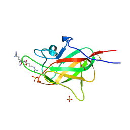
 | | Structure of the phosphate-bound form of AioX from Rhizobium sp. str. NT-26 | | 分子名称: | PHOSPHATE ION, Putative periplasmic phosphite-binding-like protein (Pbl) PtxB-like protein designated AioX | | 著者 | Djordjevic, S, Badilla, C, Cole, A, Santini, J. | | 登録日 | 2017-10-24 | | 公開日 | 2018-10-17 | | 最終更新日 | 2019-10-16 | | 実験手法 | X-RAY DIFFRACTION (1.78 Å) | | 主引用文献 | A new family of periplasmic-binding proteins that sense arsenic oxyanions.
Sci Rep, 8, 2018
|
|
5DN2
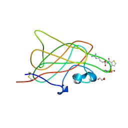
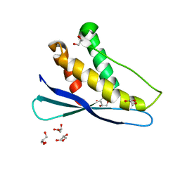
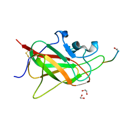
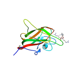 | | Human NRP2 b1 domain in complex with the peptide corresponding to the C-terminus of VEGF-A | | 分子名称: | 1,4-DIETHYLENE DIOXIDE, GLYCEROL, Neuropilin-2, ... | | 著者 | Tsai, Y.C.I, Frankel, P, Fotinou, C, Rana, R, Zachary, I, Djordjevic, S. | | 登録日 | 2015-09-09 | | 公開日 | 2016-07-20 | | 最終更新日 | 2024-01-10 | | 実験手法 | X-RAY DIFFRACTION (1.95 Å) | | 主引用文献 | Structural studies of neuropilin-2 reveal a zinc ion binding site remote from the vascular endothelial growth factor binding pocket.
Febs J., 283, 2016
|
|
3E0U
 | | Crystal structure of T. cruzi GPX1 | | 分子名称: | AMMONIUM ION, GLYCEROL, Glutathione peroxidase | | 著者 | Patel, S.H, Hussain, S, Harris, R, Driscoll, P, Djordjevic, S. | | 登録日 | 2008-08-01 | | 公開日 | 2009-08-04 | | 最終更新日 | 2023-08-30 | | 実験手法 | X-RAY DIFFRACTION (2.3 Å) | | 主引用文献 | Structural insights into the catalytic mechanism of Trypanosoma cruzi GPXI (glutathione peroxidase-like enzyme I).
Biochem.J., 425, 2010
|
|
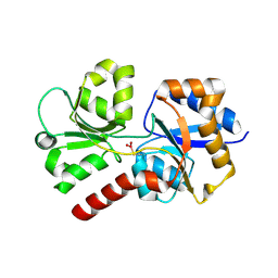
3O7W
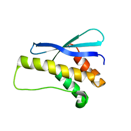
 | | The Crystal Structure of Human Leucine Carboxyl Methyltransferase 1 | | 分子名称: | GLYCEROL, Leucine carboxyl methyltransferase 1, S-ADENOSYLMETHIONINE, ... | | 著者 | Tsai, M.L, Cronin, N, Djordjevic, S. | | 登録日 | 2010-08-01 | | 公開日 | 2010-09-08 | | 最終更新日 | 2024-03-20 | | 実験手法 | X-RAY DIFFRACTION (2 Å) | | 主引用文献 | The structure of human leucine carboxyl methyltransferase 1 that regulates protein phosphatase PP2A
Acta Crystallogr.,Sect.D, 67, 2011
|
|
3I97
 | |
1Q31
 | | Crystal Structure of the Tobacco Etch Virus Protease C151A mutant | | 分子名称: | BETA-MERCAPTOETHANOL, Nuclear inclusion protein A | | 著者 | Nunn, C.M, Djordjevic, S, George, R.R, Urquhart, G.T, Chao, L.H, Tsuchiya, Y. | | 登録日 | 2003-07-28 | | 公開日 | 2004-11-02 | | 最終更新日 | 2023-10-25 | | 実験手法 | X-RAY DIFFRACTION (2.7 Å) | | 主引用文献 | Crystal structure of tobacco etch virus protease shows the protein C terminus bound within the active site.
J.Mol.Biol., 350, 2005
|
|
5DQ0
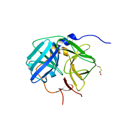
 | | Structure of human neuropilin-2 b1 domain with novel and unique zinc binding site | | 分子名称: | 1,2-ETHANEDIOL, 2-(N-MORPHOLINO)-ETHANESULFONIC ACID, CHLORIDE ION, ... | | 著者 | Tsai, Y.I, Rana, R.R, Zachary, I, Djordjevic, S. | | 登録日 | 2015-09-14 | | 公開日 | 2016-09-28 | | 最終更新日 | 2024-01-10 | | 実験手法 | X-RAY DIFFRACTION (1.8 Å) | | 主引用文献 | Structure of human neuropilin-2 b1 domain with novel and unique zinc binding site
To Be Published
|
|
4RN5
 | | B1 domain of human Neuropilin-1 with acetate ion in a ligand-binding site | | 分子名称: | ACETATE ION, GLYCEROL, Neuropilin-1, ... | | 著者 | Allerston, C.K, Yelland, T.S, Jarvis, A, Jenkins, K, Winfield, N, Cheng, L, Jia, H, Zachary, I, Selwood, D.L, Djordjevic, S. | | 登録日 | 2014-10-23 | | 公開日 | 2015-10-28 | | 実験手法 | X-RAY DIFFRACTION (1.73 Å) | | 主引用文献 | Conserved water molecules in a ligand-binding site of neuropilin-1
To be Published
|
|
2I08
 | |
2AR5
 | |
2REA
 | |
2RED
 | | Crystal structures of C2ALPHA-PI3 kinase PX-domain domain indicate conformational change associated with ligand binding. | | 分子名称: | GLYCEROL, Phosphatidylinositol-4-phosphate 3-kinase C2 domain-containing alpha polypeptide | | 著者 | Parkinson, G.N, Vines, D, Driscoll, P.C, Djordjevic, S. | | 登録日 | 2007-09-26 | | 公開日 | 2007-11-27 | | 最終更新日 | 2023-08-30 | | 実験手法 | X-RAY DIFFRACTION (2.1 Å) | | 主引用文献 | Crystal structures of PI3K-C2alpha PX domain indicate conformational change associated with ligand binding
Bmc Struct.Biol., 8, 2008
|
|
1EVV
 | |
6TDB
 | | Neuropilin2-b1 domain in a complex with the C-terminal VEGFB167 peptide | | 分子名称: | 1,2-ETHANEDIOL, ACETYL GROUP, C-terminal VEGFB167 peptide, ... | | 著者 | Eldrid, C, Yu, L, Yelland, T, Fotinou, C, Djordjevic, S. | | 登録日 | 2019-11-08 | | 公開日 | 2020-11-18 | | 最終更新日 | 2024-01-24 | | 実験手法 | X-RAY DIFFRACTION (2.45 Å) | | 主引用文献 | NRP2-b1 domain in a complex with the C-terminal VEGFB167 peptide
To Be Published
|
|
6TJT
 | | Neuropilin2-b1 domain in a complex with the C-terminal VEGFC peptide | | 分子名称: | 1,2-ETHANEDIOL, Neuropilin-2, SULFATE ION, ... | | 著者 | Eldrid, C, Yu, L, Yelland, T, Fotinou, C, Djordjevic, S. | | 登録日 | 2019-11-26 | | 公開日 | 2020-12-16 | | 最終更新日 | 2024-01-24 | | 実験手法 | X-RAY DIFFRACTION (1.31 Å) | | 主引用文献 | NRP2-b1 domain in a complex with the C-terminal VEGFC peptide
To Be Published
|
|
6TKK
 | | Neuropilin 1-b1 domain in a complex with the C-terminal VEGFB186 peptide | | 分子名称: | 1,2-ETHANEDIOL, ACE-ARG-PRO-GLN-PRO-ARG, Neuropilin-1 | | 著者 | Eldrid, C, Yu, L, Yelland, T, Fotinou, C, Djordjevic, S. | | 登録日 | 2019-11-28 | | 公開日 | 2020-12-16 | | 最終更新日 | 2024-01-24 | | 実験手法 | X-RAY DIFFRACTION (1.06 Å) | | 主引用文献 | Neuropilin 1-b1 domain in a complex with the C-terminal VEGFB186 peptide
To Be Published
|
|
1GU9
 | | Crystal Structure of Mycobacterium tuberculosis Alkylperoxidase AhpD | | 分子名称: | ALKYLHYDROPEROXIDASE D | | 著者 | Nunn, C.M, Djordjevic, S, Hillas, P.J, Nishida, C, Ortiz de Montellano, P.R. | | 登録日 | 2002-01-24 | | 公開日 | 2002-02-14 | | 最終更新日 | 2018-02-07 | | 実験手法 | X-RAY DIFFRACTION (1.9 Å) | | 主引用文献 | The Crystal Structure of Mycobacterium Tuberculosis Alkylhydroperoxidase Ahpd, a Potential Target for Antitubercular Drug Design
J.Biol.Chem., 277, 2002
|
|


