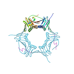
5T9D
 
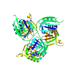 | |
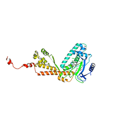
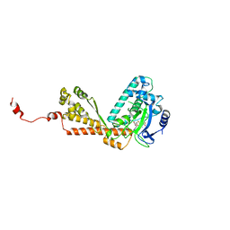
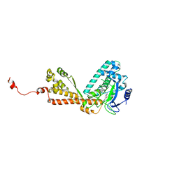
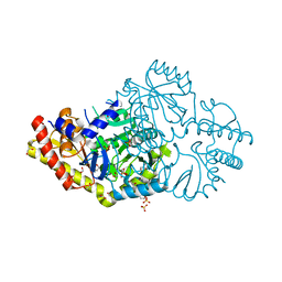
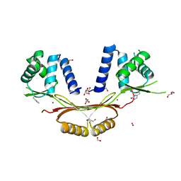
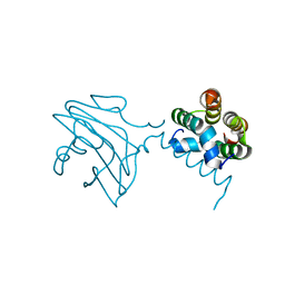
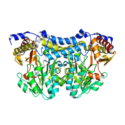
1MRQ
 | | Crystal structure of human 20alpha-HSD in ternary complex with NADP and 20alpha-hydroxy-progesterone | | 分子名称: | Aldo-keto reductase family 1 member C1, BETA-MERCAPTOETHANOL, NADP NICOTINAMIDE-ADENINE-DINUCLEOTIDE PHOSPHATE, ... | | 著者 | Couture, J.F, Legrand, P, Cantin, L, Luu-The, V, Labrie, F, Breton, R. | | 登録日 | 2002-09-18 | | 公開日 | 2003-09-30 | | 最終更新日 | 2024-02-14 | | 実験手法 | X-RAY DIFFRACTION (1.59 Å) | | 主引用文献 | Human 20alpha-hydroxysteroid dehydrogenase: crystallographic and site-directed mutagenesis studies lead to the identification of an alternative binding site for C21-steroids.
J.Mol.Biol., 331, 2003
|
|
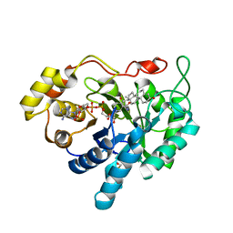
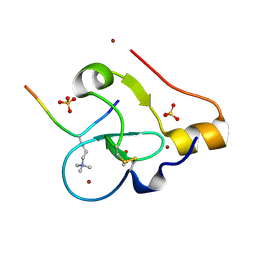
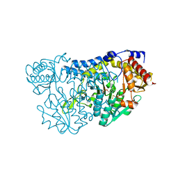
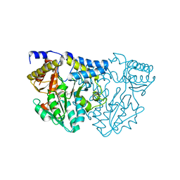
1Q5M
 | | Binary complex of rabbit 20alpha-hydroxysteroid dehydrogenase with NADPH | | 分子名称: | NADPH DIHYDRO-NICOTINAMIDE-ADENINE-DINUCLEOTIDE PHOSPHATE, Prostaglandin-E2 9-reductase, SULFATE ION | | 著者 | Couture, J.F, Legrand, P, Cantin, L, Labrie, F, Luu-The, V, Breton, R. | | 登録日 | 2003-08-08 | | 公開日 | 2004-05-18 | | 最終更新日 | 2024-02-14 | | 実験手法 | X-RAY DIFFRACTION (1.32 Å) | | 主引用文献 | Loop Relaxation, A Mechanism that Explains the Reduced Specificity of Rabbit 20alpha-Hydroxysteroid Dehydrogenase, A Member of the Aldo-Keto Reductase Superfamily.
J.Mol.Biol., 339, 2004
|
|
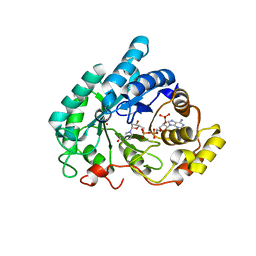
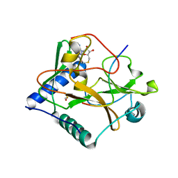
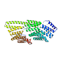
2H23
 | | Structure of Rubisco LSMT bound to Trimethyllysine and AdoHcy | | 分子名称: | N-TRIMETHYLLYSINE, Ribulose-1,5 bisphosphate carboxylase/oxygenase large subunit N-methyltransferase, S-ADENOSYL-L-HOMOCYSTEINE | | 著者 | Couture, J.F, Hauk, G, Trievel, R.C. | | 登録日 | 2006-05-17 | | 公開日 | 2006-05-30 | | 最終更新日 | 2024-02-14 | | 実験手法 | X-RAY DIFFRACTION (2.45 Å) | | 主引用文献 | Catalytic Roles for Carbon-Oxygen Hydrogen Bonding in SET Domain Lysine Methyltransferases.
J.Biol.Chem., 281, 2006
|
|
2H2E
 | | Structure of Rubisco LSMT bound to AzaAdoMet and Lysine | | 分子名称: | LYSINE, Ribulose-1,5 bisphosphate carboxylase/oxygenase large subunit N-methyltransferase, S-5'-AZAMETHIONINE-5'-DEOXYADENOSINE | | 著者 | Couture, J.F, Hauk, G, Trievel, R.C. | | 登録日 | 2006-05-18 | | 公開日 | 2006-05-30 | | 最終更新日 | 2024-02-14 | | 実験手法 | X-RAY DIFFRACTION (2.6 Å) | | 主引用文献 | Catalytic Roles for Carbon-Oxygen Hydrogen Bonding in SET Domain Lysine Methyltransferases.
J.Biol.Chem., 281, 2006
|
|
2H13
 | |
2H21
 | |
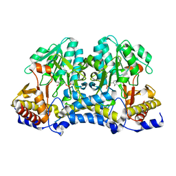
2H2J
 | | Structure of Rubisco LSMT bound to Sinefungin and Monomethyllysine | | 分子名称: | N-METHYL-LYSINE, Ribulose-1,5 bisphosphate carboxylase/oxygenase large subunit N-methyltransferase, SINEFUNGIN | | 著者 | Couture, J.F, Hauk, G, Trievel, R.C. | | 登録日 | 2006-05-18 | | 公開日 | 2006-05-30 | | 最終更新日 | 2024-02-14 | | 実験手法 | X-RAY DIFFRACTION (2.45 Å) | | 主引用文献 | Catalytic Roles for Carbon-Oxygen Hydrogen Bonding in SET Domain Lysine Methyltransferases.
J.Biol.Chem., 281, 2006
|
|
2H14
 | |
6O09
 | |
6DVS
 | | Crystal structure of Pseudomonas stutzeri D-phenylglycine aminotransferase | | 分子名称: | 1,2-ETHANEDIOL, 2-AMINO-2-HYDROXYMETHYL-PROPANE-1,3-DIOL, ACETATE ION, ... | | 著者 | Couture, J.F, Chica, R. | | 登録日 | 2018-06-25 | | 公開日 | 2018-09-12 | | 最終更新日 | 2024-03-13 | | 実験手法 | X-RAY DIFFRACTION (1.821 Å) | | 主引用文献 | Structural Determinants of the Stereoinverting Activity of Pseudomonas stutzeri d-Phenylglycine Aminotransferase.
Biochemistry, 57, 2018
|
|
9C0O
 | |
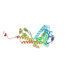
4O30
 | | Crystal structure of ATXR5 in complex with histone H3.1 and AdoHcy | | 分子名称: | BETA-MERCAPTOETHANOL, DIMETHYL SULFOXIDE, Histone-lysine N-methyltransferase ATXR6, ... | | 著者 | Bergamin, E, Mongeon, V, Couture, J.F. | | 登録日 | 2013-12-17 | | 公開日 | 2014-03-26 | | 最終更新日 | 2024-02-28 | | 実験手法 | X-RAY DIFFRACTION (2.1 Å) | | 主引用文献 | Selective methylation of histone H3 variant H3.1 regulates heterochromatin replication.
Science, 343, 2014
|
|
6D57
 | | Campylobacter jejuni ferric uptake regulator S1 metalated | | 分子名称: | FORMIC ACID, Ferric uptake regulation protein, GLYCEROL, ... | | 著者 | Sarvan, S, Brunzelle, J.S, Couture, J.F. | | 登録日 | 2018-04-19 | | 公開日 | 2018-05-23 | | 最終更新日 | 2020-01-08 | | 実験手法 | X-RAY DIFFRACTION (1.81 Å) | | 主引用文献 | Functional insights into the interplay between DNA interaction and metal coordination in ferric uptake regulators.
Sci Rep, 8, 2018
|
|
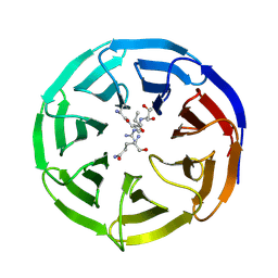
8SKJ
 | | Crystal structure of a Nanobody bound to the V5 peptide. | | 分子名称: | NbA1, V5 Epitope Tag Peptide | | 著者 | Zaghal, M, Matte, K, Venes, A, Patel, S, Laroche, G, Sarvan, S, Joshi, M, Couture, J.F, Giguere, P.M. | | 登録日 | 2023-04-19 | | 公開日 | 2023-11-22 | | 実験手法 | X-RAY DIFFRACTION (2.01 Å) | | 主引用文献 | Development of a V5-tag-directed nanobody and its implementation as an intracellular biosensor of GPCR signaling.
J.Biol.Chem., 299, 2023
|
|
5VAC
 | | Crystal Structure of ATXR5 SET domain in complex with K36me3 histone H3 peptide | | 分子名称: | DIMETHYL SULFOXIDE, Histone H3.2, Probable Histone-lysine N-methyltransferase ATXR5, ... | | 著者 | Bergamin, E, Sarvan, S, Malette, J, Eram, M, Yeung, S, Mongeon, V, Joshi, M, Brunzelle, J.S, Michaels, S.D, Blais, A, Vedadi, M, Couture, J.F. | | 登録日 | 2017-03-24 | | 公開日 | 2017-04-19 | | 最終更新日 | 2023-10-04 | | 実験手法 | X-RAY DIFFRACTION (1.949 Å) | | 主引用文献 | Molecular basis for the methylation specificity of ATXR5 for histone H3.
Nucleic Acids Res., 45, 2017
|
|
6VHF
 | |
6E2H
 | |
7T7T
 | |
7TLM
 | |
7TLR
 | |
7TLQ
 | |
7TLP
 | | Structure of Atopobium parvulum SufS K235R | | 分子名称: | Cysteine desulfurase, N-GLYCINE-[3-HYDROXY-2-METHYL-5-PHOSPHONOOXYMETHYL-PYRIDIN-4-YL-METHANE] | | 著者 | Karunakaran, G, Couture, J.F. | | 登録日 | 2022-01-18 | | 公開日 | 2022-02-16 | | 最終更新日 | 2023-10-18 | | 実験手法 | X-RAY DIFFRACTION (2.99 Å) | | 主引用文献 | Structural analysis of Atopobium parvulum SufS cysteine desulfurase linked to Crohn's disease.
Febs Lett., 596, 2022
|
|
6DK4
 | | Crystal structure of Campylobacter jejuni peroxide stress regulator | | 分子名称: | (4S)-2-METHYL-2,4-PENTANEDIOL, Ferric uptake regulation protein, MANGANESE (II) ION, ... | | 著者 | Sarvan, S, Brunzelle, J.S, Couture, J.F. | | 登録日 | 2018-05-28 | | 公開日 | 2018-06-13 | | 最終更新日 | 2024-03-06 | | 実験手法 | X-RAY DIFFRACTION (2.71 Å) | | 主引用文献 | Crystal structure of Campylobacter jejuni peroxide regulator.
FEBS Lett., 592, 2018
|
|
6E29
 | |


