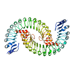
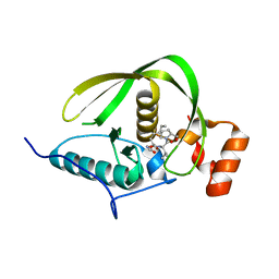
2RHJ
 
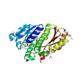 | | Synthetic Gene Encoded Bacillus Subtilis FtsZ with Two Sulfate Ions and Sodium Ion in the Nucleotide Pocket | | 分子名称: | ACETATE ION, Cell Division Protein ftsZ, SODIUM ION, ... | | 著者 | Lovell, S, Halloran, Z, Hjerrild, K, Sheridan, D, Burgin, A, Stewart, L, Accelerated Technologies Center for Gene to 3D Structure (ATCG3D) | | 登録日 | 2007-10-09 | | 公開日 | 2008-10-21 | | 最終更新日 | 2024-02-21 | | 実験手法 | X-RAY DIFFRACTION (1.761 Å) | | 主引用文献 | Combined protein construct and synthetic gene engineering for heterologous protein expression and crystallization using Gene Composer.
BMC Biotechnol., 9, 2009
|
|
2RHO
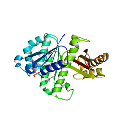 | | Synthetic Gene Encoded Bacillus Subtilis FtsZ NCS Dimer with Bound GDP and GTP-gamma-S | | 分子名称: | 5'-GUANOSINE-DIPHOSPHATE-MONOTHIOPHOSPHATE, Cell Division Protein ftsZ, GUANOSINE-5'-DIPHOSPHATE | | 著者 | Lovell, S, Halloran, Z, Hjerrild, K, Sheridan, D, Burgin, A, Stewart, L, Accelerated Technologies Center for Gene to 3D Structure (ATCG3D) | | 登録日 | 2007-10-09 | | 公開日 | 2008-10-21 | | 最終更新日 | 2024-02-21 | | 実験手法 | X-RAY DIFFRACTION (2.45 Å) | | 主引用文献 | Combined protein construct and synthetic gene engineering for heterologous protein expression and crystallization using Gene Composer.
BMC Biotechnol., 9, 2009
|
|
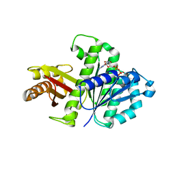
2RHH
 | | Synthetic Gene Encoded Bacillus Subtilis FtsZ with Bound Sulfate Ion | | 分子名称: | Cell Division Protein ftsZ, SULFATE ION | | 著者 | Lovell, S, Halloran, Z, Hjerrild, K, Sheridan, D, Burgin, A, Stewart, L, Accelerated Technologies Center for Gene to 3D Structure (ATCG3D) | | 登録日 | 2007-10-09 | | 公開日 | 2008-10-21 | | 最終更新日 | 2024-02-21 | | 実験手法 | X-RAY DIFFRACTION (2.001 Å) | | 主引用文献 | Combined protein construct and synthetic gene engineering for heterologous protein expression and crystallization using Gene Composer.
BMC Biotechnol., 9, 2009
|
|
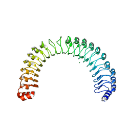
2RHL
 | | Synthetic Gene Encoded Bacillus Subtilis FtsZ NCS Dimer with Bound GDP | | 分子名称: | Cell Division Protein ftsZ, GUANOSINE-5'-DIPHOSPHATE | | 著者 | Lovell, S, Halloran, Z, Hjerrild, K, Sheridan, D, Burgin, A, Stewart, L, Accelerated Technologies Center for Gene to 3D Structure (ATCG3D) | | 登録日 | 2007-10-09 | | 公開日 | 2008-10-21 | | 最終更新日 | 2024-02-21 | | 実験手法 | X-RAY DIFFRACTION (2.45 Å) | | 主引用文献 | Combined protein construct and synthetic gene engineering for heterologous protein expression and crystallization using Gene Composer.
BMC Biotechnol., 9, 2009
|
|
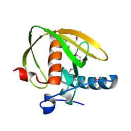
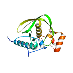
7T7A
 | | Crystal Structure of Human SHOC2: A Leucine-Rich Repeat Protein | | 分子名称: | Leucine-rich repeat protein SHOC-2, MAGNESIUM ION, NITRATE ION | | 著者 | Hajian, B, Lemke, C, Kwon, J, Bian, Y, Fuller, C, Aguirre, J. | | 登録日 | 2021-12-14 | | 公開日 | 2022-05-04 | | 最終更新日 | 2024-05-22 | | 実験手法 | X-RAY DIFFRACTION (1.79 Å) | | 主引用文献 | Structure-function analysis of the SHOC2-MRAS-PP1C holophosphatase complex.
Nature, 609, 2022
|
|
5IL7
 | |
3UWA
 | | Crystal structure of a probable peptide deformylase from synechococcus phage S-SSM7 | | 分子名称: | (4R)-2-METHYLPENTANE-2,4-DIOL, RIIA-RIIB membrane-associated protein, ZINC ION | | 著者 | Lorimer, D, Abendroth, J, Edwards, T.E, Burgin, A, Segall, A, Rohwer, F. | | 登録日 | 2011-12-01 | | 公開日 | 2013-01-09 | | 最終更新日 | 2013-06-19 | | 実験手法 | X-RAY DIFFRACTION (1.95 Å) | | 主引用文献 | Structure and function of a cyanophage-encoded peptide deformylase.
ISME J, 7, 2013
|
|
3UWB
 | | Crystal structure of a probable peptide deformylase from strucynechococcus phage S-SSM7 in complex with actinonin | | 分子名称: | 1,2-ETHANEDIOL, ACTINONIN, CHLORIDE ION, ... | | 著者 | Lorimer, D, Abendroth, J, Edwards, T.E, Burgin, A, Segall, A, Rohwer, F. | | 登録日 | 2011-12-01 | | 公開日 | 2013-01-09 | | 最終更新日 | 2023-12-06 | | 実験手法 | X-RAY DIFFRACTION (1.7 Å) | | 主引用文献 | Structure and function of a cyanophage-encoded peptide deformylase.
ISME J, 7, 2013
|
|
4DR8
 | | Crystal structure of a peptide deformylase from Synechococcus elongatus | | 分子名称: | 1,2-ETHANEDIOL, CHLORIDE ION, FORMIC ACID, ... | | 著者 | Lorimer, D, Abendroth, J, Craig, T, Burgin, A, Segall, A, Rohwer, F. | | 登録日 | 2012-02-17 | | 公開日 | 2013-03-06 | | 最終更新日 | 2023-09-13 | | 実験手法 | X-RAY DIFFRACTION (1.55 Å) | | 主引用文献 | Structure and function of a cyanophage-encoded peptide deformylase.
ISME J, 7, 2013
|
|
4DR9
 | | Crystal structure of a peptide deformylase from synechococcus elongatus in complex with actinonin | | 分子名称: | ACTINONIN, BROMIDE ION, Peptide deformylase, ... | | 著者 | Lorimer, D, Abendroth, J, Craig, T, Burgin, A, Segall, A, Rohwler, F. | | 登録日 | 2012-02-17 | | 公開日 | 2013-01-16 | | 最終更新日 | 2023-09-13 | | 実験手法 | X-RAY DIFFRACTION (1.9 Å) | | 主引用文献 | Structure and function of a cyanophage-encoded peptide deformylase.
ISME J, 7, 2013
|
|
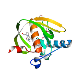
7UPI
 | | Cryo-EM structure of SHOC2-PP1c-MRAS holophosphatase complex | | 分子名称: | CHLORIDE ION, GUANOSINE-5'-TRIPHOSPHATE, Leucine-rich repeat protein SHOC-2, ... | | 著者 | Fuller, J.R, Hajian, B, Lemke, C, Kwon, J, Bian, Y, Aguirre, A. | | 登録日 | 2022-04-15 | | 公開日 | 2022-05-04 | | 最終更新日 | 2024-06-12 | | 実験手法 | ELECTRON MICROSCOPY (2.89 Å) | | 主引用文献 | Structure-function analysis of the SHOC2-MRAS-PP1C holophosphatase complex.
Nature, 609, 2022
|
|
6OP0
 | | Asymmetric hTNF-alpha | | 分子名称: | (R)-{1-[(2,5-dimethylphenyl)methyl]-6-(1-methyl-1H-pyrazol-4-yl)-1H-benzimidazol-2-yl}(pyridin-4-yl)methanol, Tumor necrosis factor | | 著者 | Arakaki, T.L, Edwards, T.E, Fairman, J.W, Davies, D.R, Foley, A, Ceska, T. | | 登録日 | 2019-04-23 | | 公開日 | 2019-12-25 | | 最終更新日 | 2024-04-03 | | 実験手法 | X-RAY DIFFRACTION (2.55 Å) | | 主引用文献 | Small molecules that inhibit TNF signalling by stabilising an asymmetric form of the trimer.
Nat Commun, 10, 2019
|
|
6OOY
 | | Asymmetric hTNF-alpha | | 分子名称: | (4R)-2-METHYLPENTANE-2,4-DIOL, Tumor necrosis factor, {1-[(2,5-dimethylphenyl)methyl]-1H-benzimidazol-2-yl}methanol | | 著者 | Arakaki, T.L, Edwards, T.E, Ceska, T. | | 登録日 | 2019-04-23 | | 公開日 | 2019-12-25 | | 最終更新日 | 2024-04-03 | | 実験手法 | X-RAY DIFFRACTION (2.5 Å) | | 主引用文献 | Small molecules that inhibit TNF signalling by stabilising an asymmetric form of the trimer.
Nat Commun, 10, 2019
|
|
6OOZ
 | | Asymmetric hTNF-alpha | | 分子名称: | (S)-{1-[(2,5-dimethylphenyl)methyl]-1H-benzimidazol-2-yl}(pyridin-4-yl)methanol, CHLORIDE ION, Tumor necrosis factor | | 著者 | Arakaki, T.L, Edwards, T.E, Fox III, D, Lecomte, F, Ceska, T. | | 登録日 | 2019-04-23 | | 公開日 | 2019-12-25 | | 最終更新日 | 2024-04-03 | | 実験手法 | X-RAY DIFFRACTION (2.8 Å) | | 主引用文献 | Small molecules that inhibit TNF signalling by stabilising an asymmetric form of the trimer.
Nat Commun, 10, 2019
|
|
2M7T
 | | Solution NMR Structure of Engineered Cystine Knot Protein 2.5D | | 分子名称: | Cystine Knot Protein 2.5D | | 著者 | Cochran, F.V, Das, R. | | 登録日 | 2013-04-30 | | 公開日 | 2014-05-07 | | 最終更新日 | 2023-06-14 | | 実験手法 | SOLUTION NMR | | 主引用文献 | Challenging the state of the art in protein structure prediction: Highlights of experimental target structures for the 10th Critical Assessment of Techniques for Protein Structure Prediction Experiment CASP10.
Proteins, 82 Suppl 2, 2014
|
|
4CSO
 | | The structure of OrfY from Thermoproteus tenax | | 分子名称: | ORFY PROTEIN, TRANSCRIPTION FACTOR | | 著者 | Zeth, K, Hagemann, A, Siebers, B, Martin, J, Lupas, A.N. | | 登録日 | 2014-03-09 | | 公開日 | 2014-03-19 | | 最終更新日 | 2024-05-08 | | 実験手法 | X-RAY DIFFRACTION (2.6 Å) | | 主引用文献 | Challenging the state of the art in protein structure prediction: Highlights of experimental target structures for the 10th Critical Assessment of Techniques for Protein Structure Prediction Experiment CASP10.
Proteins, 82 Suppl 2, 2014
|
|


