5KB3
 
 | |
5K1Z
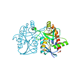
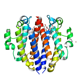
 | | Joint X-ray/neutron structure of MTAN complex with p-ClPh-Thio-DADMe-ImmA | | 分子名称: | (3R,4S)-1-[(4-amino-5H-pyrrolo[3,2-d]pyrimidin-7-yl)methyl]-4-{[(4-chlorophenyl)sulfanyl]methyl}pyrrolidin-3-ol, Aminodeoxyfutalosine nucleosidase | | 著者 | Banco, M.T, Kovalevsky, A.Y, Ronning, D.R. | | 登録日 | 2016-05-18 | | 公開日 | 2016-11-16 | | 最終更新日 | 2024-03-06 | | 実験手法 | NEUTRON DIFFRACTION (2.6 Å), X-RAY DIFFRACTION | | 主引用文献 | Neutron structures of the Helicobacter pylori 5'-methylthioadenosine nucleosidase highlight proton sharing and protonation states.
Proc. Natl. Acad. Sci. U.S.A., 113, 2016
|
|
5JPC
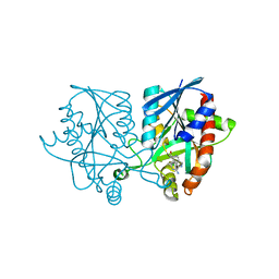
 | | Joint X-ray/neutron structure of MTAN complex with Formycin A | | 分子名称: | (1S)-1-(7-amino-1H-pyrazolo[4,3-d]pyrimidin-3-yl)-1,4-anhydro-D-ribitol, Aminodeoxyfutalosine nucleosidase | | 著者 | Banco, M.T, Kovalevsky, A.Y, Ronning, D.R. | | 登録日 | 2016-05-03 | | 公開日 | 2016-11-16 | | 最終更新日 | 2024-03-06 | | 実験手法 | NEUTRON DIFFRACTION (2.5 Å), X-RAY DIFFRACTION | | 主引用文献 | Neutron structures of the Helicobacter pylori 5'-methylthioadenosine nucleosidase highlight proton sharing and protonation states.
Proc. Natl. Acad. Sci. U.S.A., 113, 2016
|
|
5CCD
 | |
5CCE
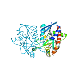
 | | Joint X-ray/neutron structure of wild type MTAN complexed with SRH and adenine | | 分子名称: | 5'-Methylthioadenosine Nucleosidase, ADENINE, S-ribosylhomocysteine, ... | | 著者 | Banco, M.T, Kovalevsky, A.Y, Ronning, D.R. | | 登録日 | 2015-07-02 | | 公開日 | 2016-11-16 | | 最終更新日 | 2023-09-27 | | 実験手法 | NEUTRON DIFFRACTION (2.5 Å), X-RAY DIFFRACTION | | 主引用文献 | Neutron structures of the Helicobacter pylori 5'-methylthioadenosine nucleosidase highlight proton sharing and protonation states.
Proc. Natl. Acad. Sci. U.S.A., 113, 2016
|
|
7SXP
 | |
8F5V
 | |
8FX9
 | |
8FI8
 | |
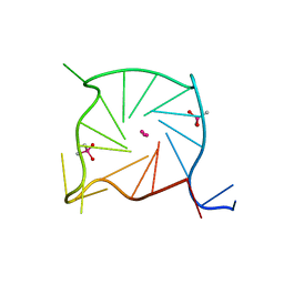
8FHV
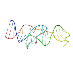
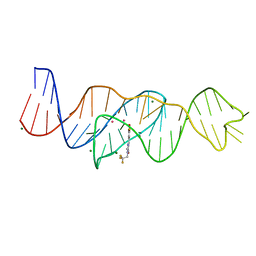 | | Structure of Lettuce aptamer bound to DFHBI-1T with thallium I ions | | 分子名称: | (5Z)-5-(3,5-difluoro-4-hydroxybenzylidene)-2-methyl-3-(2,2,2-trifluoroethyl)-3,5-dihydro-4H-imidazol-4-one, Lettuce DNA aptamer, MAGNESIUM ION, ... | | 著者 | Passalacqua, L.F.M, Ferre-D'Amare, A.R. | | 登録日 | 2022-12-15 | | 公開日 | 2023-05-10 | | 最終更新日 | 2024-05-22 | | 実験手法 | X-RAY DIFFRACTION (2.5 Å) | | 主引用文献 | Intricate 3D architecture of a DNA mimic of GFP.
Nature, 618, 2023
|
|
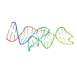
8FHZ
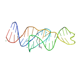
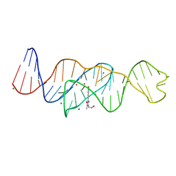 | | Structure of Lettuce aptamer bound to DFHO | | 分子名称: | (5Z)-5-[(3,5-difluoro-4-hydroxyphenyl)methylidene]-2-[(E)-(hydroxyimino)methyl]-3-methyl-3,5-dihydro-4H-imidazol-4-one, Lettuce DNA aptamer, MAGNESIUM ION, ... | | 著者 | Passalacqua, L.F.M, Ferre-D'Amare, A.R. | | 登録日 | 2022-12-15 | | 公開日 | 2023-05-10 | | 最終更新日 | 2023-10-25 | | 実験手法 | X-RAY DIFFRACTION (2.6 Å) | | 主引用文献 | Intricate 3D architecture of a DNA mimic of GFP.
Nature, 618, 2023
|
|
8FI2
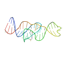
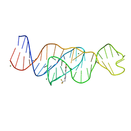 | | Structure of Lettuce C20T bound to DFHBI-1T | | 分子名称: | (5Z)-5-(3,5-difluoro-4-hydroxybenzylidene)-2-methyl-3-(2,2,2-trifluoroethyl)-3,5-dihydro-4H-imidazol-4-one, Lettuce DNA aptamer, MAGNESIUM ION, ... | | 著者 | Passalacqua, L.F.M, Ferre-D'Amare, A.R. | | 登録日 | 2022-12-15 | | 公開日 | 2023-05-10 | | 最終更新日 | 2023-10-25 | | 実験手法 | X-RAY DIFFRACTION (3 Å) | | 主引用文献 | Intricate 3D architecture of a DNA mimic of GFP.
Nature, 618, 2023
|
|
8FI1
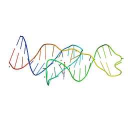 | | Structure of Lettuce C20G bound to DFHO | | 分子名称: | (5Z)-5-[(3,5-difluoro-4-hydroxyphenyl)methylidene]-2-[(E)-(hydroxyimino)methyl]-3-methyl-3,5-dihydro-4H-imidazol-4-one, Lettuce DNA aptamer, MAGNESIUM ION, ... | | 著者 | Passalacqua, L.F.M, Ferre-D'Amare, A.R. | | 登録日 | 2022-12-15 | | 公開日 | 2023-05-10 | | 最終更新日 | 2023-10-25 | | 実験手法 | X-RAY DIFFRACTION (2.6 Å) | | 主引用文献 | Intricate 3D architecture of a DNA mimic of GFP.
Nature, 618, 2023
|
|
8FI0
 | |
8FI7
 | | Structure of Lettuce C20T bound to DFHO | | 分子名称: | (5Z)-5-[(3,5-difluoro-4-hydroxyphenyl)methylidene]-2-[(E)-(hydroxyimino)methyl]-3-methyl-3,5-dihydro-4H-imidazol-4-one, Lettuce DNA aptamer, MAGNESIUM ION, ... | | 著者 | Passalacqua, L.F.M, Ferre-D'Amare, A.R. | | 登録日 | 2022-12-15 | | 公開日 | 2023-05-10 | | 最終更新日 | 2023-10-25 | | 実験手法 | X-RAY DIFFRACTION (2.9 Å) | | 主引用文献 | Intricate 3D architecture of a DNA mimic of GFP.
Nature, 618, 2023
|
|
8FHX
 | | Structure of Lettuce aptamer bound to DFHBI-1T | | 分子名称: | (5Z)-5-(3,5-difluoro-4-hydroxybenzylidene)-2-methyl-3-(2,2,2-trifluoroethyl)-3,5-dihydro-4H-imidazol-4-one, Lettuce DNA aptamer, MAGNESIUM ION, ... | | 著者 | Passalacqua, L.F.M, Ferre-D'Amare, A.R. | | 登録日 | 2022-12-15 | | 公開日 | 2023-05-10 | | 最終更新日 | 2023-10-25 | | 実験手法 | X-RAY DIFFRACTION (2.5 Å) | | 主引用文献 | Intricate 3D architecture of a DNA mimic of GFP.
Nature, 618, 2023
|
|
7SEX
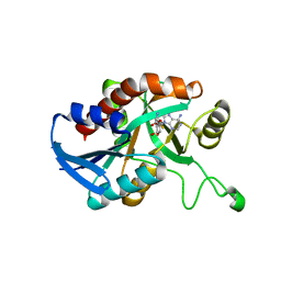
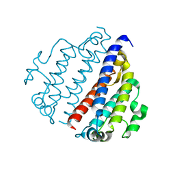
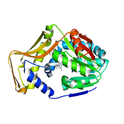 | | M. tb EgtD in complex with TGX221 | | 分子名称: | 7-methyl-2-morpholin-4-yl-9-[(1~{R})-1-phenylazanylethyl]-3~{H}-pyrido[1,2-a]pyrimidin-4-one, GLYCEROL, Histidine N-alpha-methyltransferase | | 著者 | Sudasinghe, T.D, Ronning, D.R. | | 登録日 | 2021-10-02 | | 公開日 | 2021-12-01 | | 最終更新日 | 2023-10-18 | | 実験手法 | X-RAY DIFFRACTION (2.2 Å) | | 主引用文献 | Inhibitors of Mycobacterium tuberculosis EgtD target both substrate binding sites to limit hercynine production.
Sci Rep, 11, 2021
|
|
7SEY
 | | M. tb EgtD in complex with SGH | | 分子名称: | 2-amino-1-[(3S)-3-methyl-4-(4-methylisoquinoline-5-sulfonyl)-1,4-diazepan-1-yl]ethan-1-one, Histidine N-alpha-methyltransferase | | 著者 | Sudasinghe, T.D, Ronning, D.R. | | 登録日 | 2021-10-02 | | 公開日 | 2021-12-01 | | 最終更新日 | 2023-10-18 | | 実験手法 | X-RAY DIFFRACTION (2.39 Å) | | 主引用文献 | Inhibitors of Mycobacterium tuberculosis EgtD target both substrate binding sites to limit hercynine production.
Sci Rep, 11, 2021
|
|
7SEW
 | | M. tb EgtD in complex with HD6 | | 分子名称: | 1,3-PROPANDIOL, GLYCEROL, Histidine N-alpha-methyltransferase, ... | | 著者 | Sudasinghe, T.D, Ronning, D.R. | | 登録日 | 2021-10-02 | | 公開日 | 2021-12-01 | | 最終更新日 | 2023-10-18 | | 実験手法 | X-RAY DIFFRACTION (1.72 Å) | | 主引用文献 | Inhibitors of Mycobacterium tuberculosis EgtD target both substrate binding sites to limit hercynine production.
Sci Rep, 11, 2021
|
|
7SF5
 | | M. tb EgtD in complex with HD3 | | 分子名称: | Histidine N-alpha-methyltransferase, N-(benzylcarbamothioyl)-L-histidine | | 著者 | Sudasinghe, T.D, Ronning, D.R. | | 登録日 | 2021-10-03 | | 公開日 | 2021-12-01 | | 最終更新日 | 2023-10-18 | | 実験手法 | X-RAY DIFFRACTION (2.52 Å) | | 主引用文献 | Inhibitors of Mycobacterium tuberculosis EgtD target both substrate binding sites to limit hercynine production.
Sci Rep, 11, 2021
|
|
7SF4
 | | M. tb EgtD in complex with imatinib | | 分子名称: | 1,2-ETHANEDIOL, 2-AMINO-4-PYRIDYL-PYRIMIDINE, GLYCEROL, ... | | 著者 | Sudasinghe, T.D, Ronning, D.R. | | 登録日 | 2021-10-03 | | 公開日 | 2021-12-01 | | 最終更新日 | 2023-10-18 | | 実験手法 | X-RAY DIFFRACTION (2.39 Å) | | 主引用文献 | Inhibitors of Mycobacterium tuberculosis EgtD target both substrate binding sites to limit hercynine production.
Sci Rep, 11, 2021
|
|
7SCF
 | | M. tb EgtD in complex with HD2 | | 分子名称: | (2S)-3-(1H-imidazol-5-yl)-2-(1H-pyrrol-1-yl)propanoic acid, DI(HYDROXYETHYL)ETHER, GLYCEROL, ... | | 著者 | Sudasinghe, T.D, Ronning, D.R. | | 登録日 | 2021-09-28 | | 公開日 | 2021-12-01 | | 最終更新日 | 2023-10-18 | | 実験手法 | X-RAY DIFFRACTION (2.67 Å) | | 主引用文献 | Inhibitors of Mycobacterium tuberculosis EgtD target both substrate binding sites to limit hercynine production.
Sci Rep, 11, 2021
|
|


