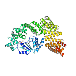
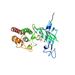
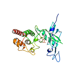
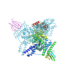
6I2P
 
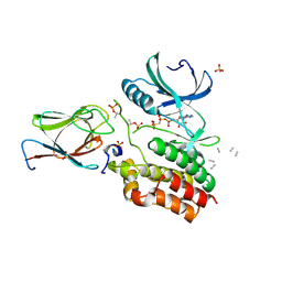 | | Crystal structure of the Mycobacterium tuberculosis PknB kinase domain (L33E mutant) in complex with its substrate GarA | | 分子名称: | Glycogen accumulation regulator GarA, MAGNESIUM ION, PHOSPHOMETHYLPHOSPHONIC ACID ADENYLATE ESTER, ... | | 著者 | Andre-Leroux, G, Hindie, V, Barilone, N, Bellinzoni, M, Alzari, P.M. | | 登録日 | 2018-11-01 | | 公開日 | 2019-05-22 | | 最終更新日 | 2024-01-24 | | 実験手法 | X-RAY DIFFRACTION (2.37 Å) | | 主引用文献 | Structural insights into the functional versatility of an FHA domain protein in mycobacterial signaling.
Sci.Signal., 12, 2019
|
|
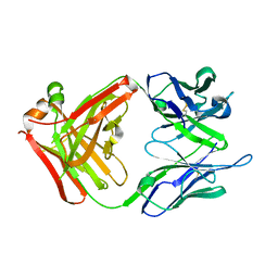
5NCR
 | | OH1 from the Orf virus: a tyrosine phosphatase that displays distinct structural features and triple substrate specificity | | 分子名称: | PHOSPHATE ION, SULFATE ION, tyrosine phosphatase | | 著者 | Segovia, D, Haouz, A, Berois, M, Villarino, A, Andre-Leroux, G. | | 登録日 | 2017-03-06 | | 公開日 | 2017-08-09 | | 最終更新日 | 2024-01-17 | | 実験手法 | X-RAY DIFFRACTION (1.89 Å) | | 主引用文献 | OH1 from Orf Virus: A New Tyrosine Phosphatase that Displays Distinct Structural Features and Triple Substrate Specificity.
J. Mol. Biol., 429, 2017
|
|
7QZN
 | | Amine Dehydrogenase from Cystobacter fuscus (CfusAmDH) W145A mutant with NAD+ | | 分子名称: | 2-AMINO-2-HYDROXYMETHYL-PROPANE-1,3-DIOL, Amine Dehydrogenase, NICOTINAMIDE-ADENINE-DINUCLEOTIDE | | 著者 | Bennett, M, Ducrot, L, Vaxelaire-Vergne, C, Grogan, G. | | 登録日 | 2022-01-31 | | 公開日 | 2022-12-07 | | 最終更新日 | 2024-01-31 | | 実験手法 | X-RAY DIFFRACTION (1.64 Å) | | 主引用文献 | Cover Feature: Expanding the Substrate Scope of Native Amine Dehydrogenases through In Silico Structural Exploration and Targeted Protein Engineering (ChemCatChem 22/2022)
Chemcatchem, 14, 2022
|
|
7QZL
 | | Amine Dehydrogenase from Cystobacter fuscus (CfusAmDH) W145A mutant with NADP+ and pentylamine | | 分子名称: | 2-AMINO-2-HYDROXYMETHYL-PROPANE-1,3-DIOL, AMYLAMINE, Amine Dehydrogenase, ... | | 著者 | Bennett, M, Ducrot, L, Vaxelaire-Vergne, C, Grogan, G. | | 登録日 | 2022-01-31 | | 公開日 | 2022-12-07 | | 最終更新日 | 2024-01-31 | | 実験手法 | X-RAY DIFFRACTION (1.5 Å) | | 主引用文献 | Cover Feature: Expanding the Substrate Scope of Native Amine Dehydrogenases through In Silico Structural Exploration and Targeted Protein Engineering (ChemCatChem 22/2022)
Chemcatchem, 14, 2022
|
|
3M4I
 | | Crystal structure of the second part of the Mycobacterium tuberculosis DNA gyrase reaction core: the TOPRIM domain at 1.95 A resolution | | 分子名称: | (4S)-2-METHYL-2,4-PENTANEDIOL, DNA gyrase subunit B | | 著者 | Piton, J, Petrella, S, Aubry, A, Mayer, C. | | 登録日 | 2010-03-11 | | 公開日 | 2010-09-29 | | 最終更新日 | 2023-11-01 | | 実験手法 | X-RAY DIFFRACTION (1.95 Å) | | 主引用文献 | Structural insights into the quinolone resistance mechanism of Mycobacterium tuberculosis DNA gyrase.
Plos One, 5, 2010
|
|
6MFV
 | | Crystal structure of the Signal Transduction ATPase with Numerous Domains (STAND) protein with a tetratricopeptide repeat sensor PH0952 from Pyrococcus horikoshii | | 分子名称: | ADENOSINE-5'-DIPHOSPHATE, tetratricopeptide repeat sensor PH0952 | | 著者 | Lisa, M.N, Alzari, P.M, Haouz, A, Danot, O. | | 登録日 | 2018-09-12 | | 公開日 | 2019-02-20 | | 最終更新日 | 2024-04-03 | | 実験手法 | X-RAY DIFFRACTION (3.4 Å) | | 主引用文献 | Double autoinhibition mechanism of signal transduction ATPases with numerous domains (STAND) with a tetratricopeptide repeat sensor.
Nucleic Acids Res., 47, 2019
|
|
2UYL
 | | Crystal structure of a monoclonal antibody directed against an antigenic determinant common to Ogawa and Inaba serotypes of Vibrio cholerae O1 | | 分子名称: | MONOCLONAL ANTIBODY F-22-30 | | 著者 | Ahmed, F, Haouz, A, Nato, F, Fournier, J.M, Alzari, P.M. | | 登録日 | 2007-04-10 | | 公開日 | 2008-05-13 | | 最終更新日 | 2024-05-01 | | 実験手法 | X-RAY DIFFRACTION (2.5 Å) | | 主引用文献 | Crystal Structure of a Monoclonal Antibody Directed Against an Antigenic Determinant Common to Ogawa and Inaba Serotypes of Vibrio Cholerae O1.
Proteins, 70, 2008
|
|
3IG0
 | |
3IFZ
 | | crystal structure of the first part of the Mycobacterium tuberculosis DNA gyrase reaction core: the breakage and reunion domain at 2.7 A resolution | | 分子名称: | (4S)-2-METHYL-2,4-PENTANEDIOL, DNA gyrase subunit A, SODIUM ION | | 著者 | Piton, J, Aubry, A, Delarue, M, Mayer, C. | | 登録日 | 2009-07-27 | | 公開日 | 2010-07-28 | | 最終更新日 | 2023-11-01 | | 実験手法 | X-RAY DIFFRACTION (2.7 Å) | | 主引用文献 | Structural insights into the quinolone resistance mechanism of Mycobacterium tuberculosis DNA gyrase.
Plos One, 5, 2010
|
|
4Y12
 | |
4Y0X
 | |
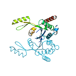
6I2S
 | | Crystal structure of the SucA domain of Mycobacterium smegmatis KGD (R802A) in complex with GarA, following 2-oxoglutarate soak | | 分子名称: | (4S)-4-{3-[(4-amino-2-methylpyrimidin-5-yl)methyl]-5-(2-{[(S)-hydroxy(phosphonooxy)phosphoryl]oxy}ethyl)-4-methyl-1,3lambda~5~-thiazol-2-yl}-4-hydroxybutanoic acid, CALCIUM ION, Glycogen accumulation regulator GarA, ... | | 著者 | Wagner, T, Bellinzoni, M, Alzari, P.M. | | 登録日 | 2018-11-01 | | 公開日 | 2019-05-22 | | 最終更新日 | 2024-01-24 | | 実験手法 | X-RAY DIFFRACTION (2.4 Å) | | 主引用文献 | Structural insights into the functional versatility of an FHA domain protein in mycobacterial signaling.
Sci.Signal., 12, 2019
|
|
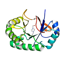
4PMO
 | | Crystal structure of the Mycobacterium tuberculosis Tat-secreted protein Rv2525c, monoclinic crystal form I | | 分子名称: | FORMIC ACID, GLYCEROL, SODIUM ION, ... | | 著者 | Bellinzoni, M, Haouz, A, Shepard, W, Alzari, P.M. | | 登録日 | 2014-05-22 | | 公開日 | 2014-10-08 | | 最終更新日 | 2023-12-20 | | 実験手法 | X-RAY DIFFRACTION (1.33 Å) | | 主引用文献 | Structural studies suggest a peptidoglycan hydrolase function for the Mycobacterium tuberculosis Tat-secreted protein Rv2525c.
J.Struct.Biol., 188, 2014
|
|
4PMN
 | | Crystal structure of the Mycobacterium tuberculosis Tat-secreted protein Rv2525c in complex with MES (monoclinic crystal form I) | | 分子名称: | 2-(N-MORPHOLINO)-ETHANESULFONIC ACID, GLYCEROL, SULFATE ION, ... | | 著者 | Bellinzoni, M, Haouz, A, Shepard, W, Alzari, P.M. | | 登録日 | 2014-05-22 | | 公開日 | 2014-10-08 | | 最終更新日 | 2014-12-10 | | 実験手法 | X-RAY DIFFRACTION (1.44 Å) | | 主引用文献 | Structural studies suggest a peptidoglycan hydrolase function for the Mycobacterium tuberculosis Tat-secreted protein Rv2525c.
J.Struct.Biol., 188, 2014
|
|
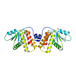
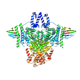
6I2Q
 | | Crystal structure of the wild-type SucA domain of Mycobacterium smegmatis KGD (alpha-ketoglutarate decarboxylase), in complex with GarA | | 分子名称: | CALCIUM ION, Glycogen accumulation regulator GarA, MAGNESIUM ION, ... | | 著者 | Wagner, T, Bellinzoni, M, Alzari, P.M. | | 登録日 | 2018-11-01 | | 公開日 | 2019-05-22 | | 最終更新日 | 2024-01-24 | | 実験手法 | X-RAY DIFFRACTION (2.15 Å) | | 主引用文献 | Structural insights into the functional versatility of an FHA domain protein in mycobacterial signaling.
Sci.Signal., 12, 2019
|
|
4PMQ
 | | Crystal structure of the Mycobacterium tuberculosis Tat-secreted protein Rv2525c in complex with L-tartrate (orthorhombic crystal form) | | 分子名称: | GLYCEROL, L(+)-TARTARIC ACID, Tat-secreted protein Rv2525c | | 著者 | Bellinzoni, M, Haouz, A, Shepard, W, Alzari, P.M. | | 登録日 | 2014-05-22 | | 公開日 | 2014-10-08 | | 最終更新日 | 2023-12-20 | | 実験手法 | X-RAY DIFFRACTION (1.61 Å) | | 主引用文献 | Structural studies suggest a peptidoglycan hydrolase function for the Mycobacterium tuberculosis Tat-secreted protein Rv2525c.
J.Struct.Biol., 188, 2014
|
|
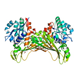
6I2R
 | | Crystal structure of the SucA domain of Mycobacterium smegmatis KGD (alpha-ketoglutarate decarboxylase), mutant R802A, in complex with GarA | | 分子名称: | CALCIUM ION, Glycogen accumulation regulator GarA, MAGNESIUM ION, ... | | 著者 | Wagner, T, Bellinzoni, M, Alzari, P.M. | | 登録日 | 2018-11-01 | | 公開日 | 2019-05-22 | | 最終更新日 | 2024-01-24 | | 実験手法 | X-RAY DIFFRACTION (2.2 Å) | | 主引用文献 | Structural insights into the functional versatility of an FHA domain protein in mycobacterial signaling.
Sci.Signal., 12, 2019
|
|
4PMR
 | | Crystal structure of the Mycobacterium tuberculosis Tat-secreted protein Rv2525c in complex with HEPES (monoclinic crystal form II) | | 分子名称: | 4-(2-HYDROXYETHYL)-1-PIPERAZINE ETHANESULFONIC ACID, SODIUM ION, Tat-secreted protein Rv2525c | | 著者 | Bellinzoni, M, Haouz, A, Shepard, W, Alzari, P.M. | | 登録日 | 2014-05-22 | | 公開日 | 2014-10-08 | | 最終更新日 | 2023-12-20 | | 実験手法 | X-RAY DIFFRACTION (1.81 Å) | | 主引用文献 | Structural studies suggest a peptidoglycan hydrolase function for the Mycobacterium tuberculosis Tat-secreted protein Rv2525c.
J.Struct.Biol., 188, 2014
|
|


