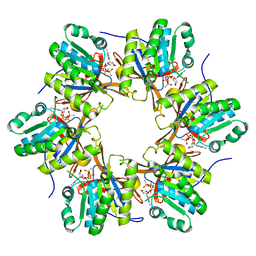
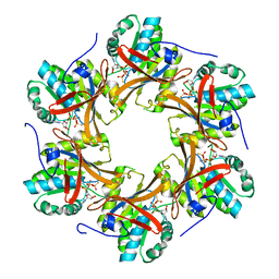
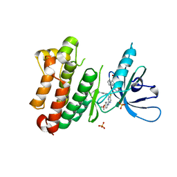
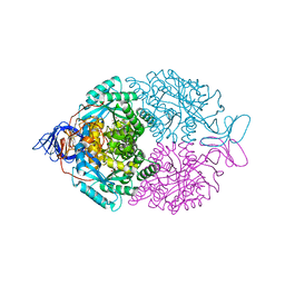
4TLE
 
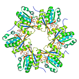 | | Crystal structure of N-terminal C1 domain of KaiC | | 分子名称: | CHLORIDE ION, Circadian clock protein kinase KaiC, MAGNESIUM ION, ... | | 著者 | Abe, J, Hiyama, T.B, Mukaiyama, A, Son, S, Akiyama, S. | | 登録日 | 2014-05-29 | | 公開日 | 2015-07-01 | | 最終更新日 | 2024-03-20 | | 実験手法 | X-RAY DIFFRACTION (1.936 Å) | | 主引用文献 | Atomic-scale origins of slowness in the cyanobacterial circadian clock
Science, 349, 2015
|
|
4TLC
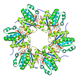 | | Crystal structure of N-terminal C1 domain of KaiC | | 分子名称: | CHLORIDE ION, Circadian clock protein kinase KaiC, MAGNESIUM ION, ... | | 著者 | Abe, J, Hiyama, T.B, Mukaiyama, A, Son, S, Akiyama, S. | | 登録日 | 2014-05-29 | | 公開日 | 2015-07-01 | | 最終更新日 | 2024-03-20 | | 実験手法 | X-RAY DIFFRACTION (2.09 Å) | | 主引用文献 | Atomic-scale origins of slowness in the cyanobacterial circadian clock
Science, 349, 2015
|
|
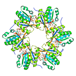
4TL7
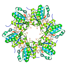 | | Crystal structure of N-terminal C1 domain of KaiC | | 分子名称: | ADENOSINE-5'-TRIPHOSPHATE, CHLORIDE ION, Circadian clock protein kinase KaiC, ... | | 著者 | Abe, J, Hiyama, T.B, Mukaiyama, A, Son, S, Akiyama, S. | | 登録日 | 2014-05-29 | | 公開日 | 2015-07-01 | | 最終更新日 | 2024-03-20 | | 実験手法 | X-RAY DIFFRACTION (1.936 Å) | | 主引用文献 | Circadian rhythms. Atomic-scale origins of slowness in the cyanobacterial circadian clock.
Science, 349, 2015
|
|
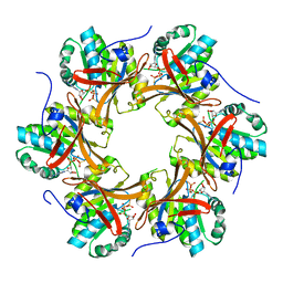
4TLA
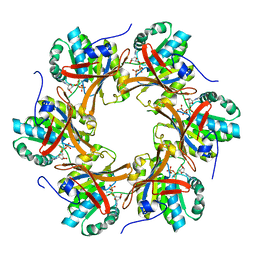 | | Crystal structure of N-terminal C1 domain of KaiC | | 分子名称: | ADENOSINE-5'-DIPHOSPHATE, CHLORIDE ION, Circadian clock protein kinase KaiC, ... | | 著者 | Abe, J, Hiyama, T.B, Mukaiyama, A, Son, S, Akiyama, S. | | 登録日 | 2014-05-29 | | 公開日 | 2015-07-01 | | 最終更新日 | 2024-03-20 | | 実験手法 | X-RAY DIFFRACTION (1.8 Å) | | 主引用文献 | Circadian rhythms. Atomic-scale origins of slowness in the cyanobacterial circadian clock.
Science, 349, 2015
|
|
4TLB
 | | Crystal structure of N-terminal C1 domain of KaiC | | 分子名称: | CHLORIDE ION, Circadian clock protein kinase KaiC, MAGNESIUM ION, ... | | 著者 | Abe, J, Hiyama, T.B, Mukaiyama, A, Son, S, Akiyama, S. | | 登録日 | 2014-05-29 | | 公開日 | 2015-07-01 | | 最終更新日 | 2024-03-20 | | 実験手法 | X-RAY DIFFRACTION (1.983 Å) | | 主引用文献 | Circadian rhythms. Atomic-scale origins of slowness in the cyanobacterial circadian clock.
Science, 349, 2015
|
|
4TL9
 | | Crystal structure of N-terminal C1 domain of KaiC | | 分子名称: | CHLORIDE ION, Circadian clock protein kinase KaiC, MAGNESIUM ION, ... | | 著者 | Abe, J, Hiyama, T.B, Mukaiyama, A, Son, S, Akiyama, S. | | 登録日 | 2014-05-29 | | 公開日 | 2015-07-01 | | 最終更新日 | 2024-03-20 | | 実験手法 | X-RAY DIFFRACTION (1.822 Å) | | 主引用文献 | Circadian rhythms. Atomic-scale origins of slowness in the cyanobacterial circadian clock.
Science, 349, 2015
|
|
4TL8
 | | Crystal structure of N-terminal C1 domain of KaiC | | 分子名称: | CHLORIDE ION, Circadian clock protein kinase KaiC, MAGNESIUM ION, ... | | 著者 | Abe, J, Hiyama, T.B, Mukaiyama, A, Son, S, Akiyama, S. | | 登録日 | 2014-05-29 | | 公開日 | 2015-07-01 | | 最終更新日 | 2023-11-08 | | 実験手法 | X-RAY DIFFRACTION (1.859 Å) | | 主引用文献 | Atomic-scale origins of slowness in the cyanobacterial circadian clock
Science, 349, 2015
|
|
4TL6
 | | Crystal structure of N-terminal domain of KaiC | | 分子名称: | CHLORIDE ION, Circadian clock protein kinase KaiC, MAGNESIUM ION, ... | | 著者 | Abe, J, Hiyama, T.B, Mukaiyama, A, Son, S, Akiyama, S. | | 登録日 | 2014-05-29 | | 公開日 | 2015-07-01 | | 最終更新日 | 2023-11-08 | | 実験手法 | X-RAY DIFFRACTION (1.763 Å) | | 主引用文献 | Circadian rhythms. Atomic-scale origins of slowness in the cyanobacterial circadian clock.
Science, 349, 2015
|
|
4TLD
 | | Crystal structure of N-terminal C1 domain of KaiC | | 分子名称: | CHLORIDE ION, Circadian clock protein kinase KaiC, MAGNESIUM ION, ... | | 著者 | Abe, J, Hiyama, T.B, Mukaiyama, A, Son, S, Akiyama, S. | | 登録日 | 2014-05-29 | | 公開日 | 2015-07-01 | | 最終更新日 | 2024-03-20 | | 実験手法 | X-RAY DIFFRACTION (1.949 Å) | | 主引用文献 | Atomic-scale origins of slowness in the cyanobacterial circadian clock
Science, 349, 2015
|
|
6Z4D
 | | Crystal Structure of EGFR-T790M/V948R in Complex with Mavelertinib and EAI001 | | 分子名称: | (2R)-2-(1-oxo-1,3-dihydro-2H-isoindol-2-yl)-2-phenyl-N-(1,3-thiazol-2-yl)acetamide, Epidermal growth factor receptor, N-[(3R,4R)-4-fluoro-1-{6-[(3-methoxy-1-methyl-1H-pyrazol-4-yl)amino]-9-methyl-9H-purin-2-yl}pyrrolidin-3-yl]propanamide, ... | | 著者 | Niggenaber, J, Mueller, M.P, Rauh, D. | | 登録日 | 2020-05-25 | | 公開日 | 2020-11-11 | | 最終更新日 | 2024-01-24 | | 実験手法 | X-RAY DIFFRACTION (2 Å) | | 主引用文献 | Complex Crystal Structures of EGFR with Third-Generation Kinase Inhibitors and Simultaneously Bound Allosteric Ligands.
Acs Med.Chem.Lett., 11, 2020
|
|
6Z4B
 | | Crystal Structure of EGFR-T790M/V948R in Complex with Osimertinib and EAI045 | | 分子名称: | (2R)-2-(5-fluoro-2-hydroxyphenyl)-2-(1-oxo-1,3-dihydro-2H-isoindol-2-yl)-N-(1,3-thiazol-2-yl)acetamide, Epidermal growth factor receptor, SULFATE ION, ... | | 著者 | Niggenaber, J, Mueller, M.P, Rauh, D. | | 登録日 | 2020-05-25 | | 公開日 | 2020-11-11 | | 最終更新日 | 2024-01-24 | | 実験手法 | X-RAY DIFFRACTION (2.5 Å) | | 主引用文献 | Complex Crystal Structures of EGFR with Third-Generation Kinase Inhibitors and Simultaneously Bound Allosteric Ligands.
Acs Med.Chem.Lett., 11, 2020
|
|
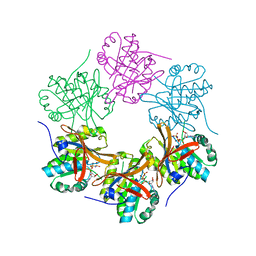
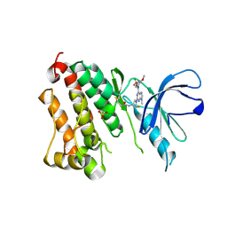
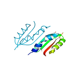
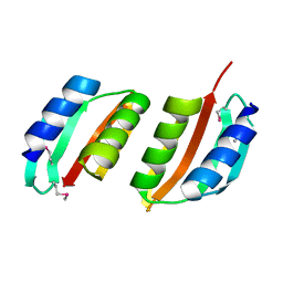
1YB3
 | | Conserved hypothetical protein Pfu-178653-001 from Pyrococcus furiosus | | 分子名称: | UNKNOWN ATOM OR ION, hypothetical protein | | 著者 | Habel, J, Zhou, W, Chang, J, Zhao, M, Xu, H, Chen, L, Lee, D, Nguyen, D, Chang, S.-H, Horanyi, P, Florence, Q, Tempel, W, Lin, D, Zhang, H, Praissman, J, Jenney Jr, F.E, Adams, M.W.W, Liu, Z.-J, Rose, J.P, Wang, B.-C, Southeast Collaboratory for Structural Genomics (SECSG) | | 登録日 | 2004-12-19 | | 公開日 | 2005-02-01 | | 最終更新日 | 2017-10-11 | | 実験手法 | X-RAY DIFFRACTION (1.6 Å) | | 主引用文献 | Conserved hypothetical protein Pfu-178653-001 from Pyrococcus furiosus
To be published
|
|
7A2A
 | | Crystal Structure of EGFR-T790M/V948R in Complex with Spebrutinib and EAI001 | | 分子名称: | (2R)-2-(1-oxo-1,3-dihydro-2H-isoindol-2-yl)-2-phenyl-N-(1,3-thiazol-2-yl)acetamide, CHLORIDE ION, Epidermal growth factor receptor, ... | | 著者 | Niggenaber, J, Mueller, M.P, Rauh, D. | | 登録日 | 2020-08-17 | | 公開日 | 2020-11-11 | | 最終更新日 | 2024-01-31 | | 実験手法 | X-RAY DIFFRACTION (1.9 Å) | | 主引用文献 | Complex Crystal Structures of EGFR with Third-Generation Kinase Inhibitors and Simultaneously Bound Allosteric Ligands.
Acs Med.Chem.Lett., 11, 2020
|
|
1YF5
 | | Cyto-Epsl: The Cytoplasmic Domain Of Epsl, An Inner Membrane Component Of The Type II Secretion System Of Vibrio Cholerae | | 分子名称: | General secretion pathway protein L | | 著者 | Abendroth, J, Murphy, P, Mushtaq, A, Sandkvist, M, Bagdasarian, M, Hol, W.G. | | 登録日 | 2004-12-30 | | 公開日 | 2005-05-03 | | 最終更新日 | 2023-11-15 | | 実験手法 | X-RAY DIFFRACTION (2.75 Å) | | 主引用文献 | The X-ray Structure of the Type II Secretion System Complex Formed by the N-terminal Domain of EpsE and the Cytoplasmic Domain of EpsL of Vibrio cholerae.
J.Mol.Biol., 348, 2005
|
|
2W7V
 | | periplasmic domain of EpsL from Vibrio parahaemolyticus | | 分子名称: | 1,2-ETHANEDIOL, GENERAL SECRETION PATHWAY PROTEIN L, PHOSPHATE ION | | 著者 | Abendroth, J, Kreger, A.C, Abendroth, H, Sandkvist, M, Hol, W.G.J. | | 登録日 | 2009-01-06 | | 公開日 | 2010-03-31 | | 最終更新日 | 2017-06-28 | | 実験手法 | X-RAY DIFFRACTION (2.3 Å) | | 主引用文献 | The Dimer Formed by the Periplasmic Domain of Epsl from the Type 2 Secretion System of Vibrio Parahaemolyticus.
J.Struct.Biol., 168, 2009
|
|
1W97
 | | cyto-EpsL: the cytoplasmic domain of EpsL, an inner membrane component of the type II secretion system of Vibrio cholerae | | 分子名称: | TYPE II SECRETION SYSTEM PROTEIN L | | 著者 | Abendroth, J, Bagdasarian, M, Sansdkvist, M, Hol, W.G.J. | | 登録日 | 2004-10-06 | | 公開日 | 2004-11-30 | | 最終更新日 | 2019-05-08 | | 実験手法 | X-RAY DIFFRACTION (2.7 Å) | | 主引用文献 | The Structure of the Cytoplasmic Domain of Epsl, an Inner Membrane Component of the Type II Secretion System of Vibrio Cholerae: An Unusual Member of the Actin-Like ATPase Superfamily
J.Mol.Biol., 344, 2004
|
|
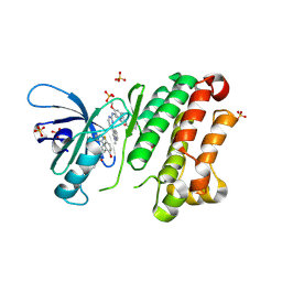
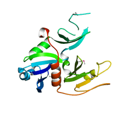
5UVM
 | | HIT family hydrolase from Clostridium thermocellum Cth-393 | | 分子名称: | ADENOSINE, Histidine triad (HIT) protein, UNKNOWN ATOM OR ION, ... | | 著者 | Habel, J, Tempel, W, Liu, Z.-J, Rose, J, Wang, B.-C, Southeast Collaboratory for Structural Genomics (SECSG) | | 登録日 | 2017-02-20 | | 公開日 | 2017-04-05 | | 最終更新日 | 2023-11-15 | | 実験手法 | X-RAY DIFFRACTION (2.3 Å) | | 主引用文献 | HIT family hydrolase from Clostridium thermocellum Cth-393
To Be Published
|
|
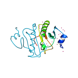
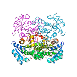
1GKP
 | | D-Hydantoinase (Dihydropyrimidinase) from Thermus sp. in space group C2221 | | 分子名称: | 4-(2-HYDROXYETHYL)-1-PIPERAZINE ETHANESULFONIC ACID, HYDANTOINASE, SULFATE ION, ... | | 著者 | Abendroth, J, Niefind, K, Schomburg, D. | | 登録日 | 2001-08-20 | | 公開日 | 2002-06-27 | | 最終更新日 | 2019-07-24 | | 実験手法 | X-RAY DIFFRACTION (1.295 Å) | | 主引用文献 | X-Ray Structure of a Dihydropyrimidinase from Thermus Sp. At 1.3 A Resolution
J.Mol.Biol., 320, 2002
|
|
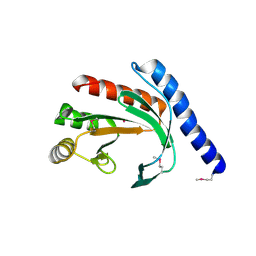
1UV7
 | | periplasmic domain of EpsM from Vibrio cholerae | | 分子名称: | GENERAL SECRETION PATHWAY PROTEIN M | | 著者 | Abendroth, J, Hol, W.G.J. | | 登録日 | 2004-01-15 | | 公開日 | 2004-04-23 | | 最終更新日 | 2019-05-15 | | 実験手法 | X-RAY DIFFRACTION (1.7 Å) | | 主引用文献 | The Crystal Structure of the Periplasmic Domain of the Type II Secretion System Protein Epsm from Vibrio Cholerae: The Simplest Version of the Ferredoxin Fold
J.Mol.Biol., 338, 2004
|
|
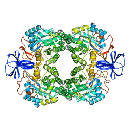
2VMA
 | | The three-dimensional structure of the cytoplasmic domains of EpsF from the Type 2 Secretion System of Vibrio cholerae | | 分子名称: | CALCIUM ION, GENERAL SECRETION PATHWAY PROTEIN F, IODIDE ION | | 著者 | Abendroth, J, Korotkov, K.V, Mitchell, D.D, Kreger, A, Hol, W.G.J. | | 登録日 | 2008-01-25 | | 公開日 | 2009-02-10 | | 最終更新日 | 2017-06-28 | | 実験手法 | X-RAY DIFFRACTION (1.9 Å) | | 主引用文献 | The Three-Dimensional Structure of the Cytoplasmic Domains of Epsf from the Type 2 Secretion System of Vibrio Cholerae.
J.Struct.Biol., 166, 2009
|
|
2VMB
 | | The three-dimensional structure of the cytoplasmic domains of EpsF from the Type 2 Secretion System of Vibrio cholerae | | 分子名称: | CALCIUM ION, GENERAL SECRETION PATHWAY PROTEIN F | | 著者 | Abendroth, J, Korotkov, K.V, Mitchell, D.D, Kreger, A, Hol, W.G.J. | | 登録日 | 2008-01-25 | | 公開日 | 2009-02-10 | | 最終更新日 | 2024-05-08 | | 実験手法 | X-RAY DIFFRACTION (1.95 Å) | | 主引用文献 | The Three-Dimensional Structure of the Cytoplasmic Domains of Epsf from the Type 2 Secretion System of Vibrio Cholerae.
J.Struct.Biol., 166, 2009
|
|
3K2E
 | |
3C1Q
 | | The three-dimensional structure of the cytoplasmic domains of EpsF from the Type 2 Secretion System of Vibrio cholerae | | 分子名称: | 3,6,9,12,15,18,21,24-OCTAOXAHEXACOSAN-1-OL, CALCIUM ION, CHLORIDE ION, ... | | 著者 | Abendroth, J, Mitchell, D.D, Korotkov, K.V, Kreeger, A, Hol, W.G.J. | | 登録日 | 2008-01-24 | | 公開日 | 2009-02-03 | | 最終更新日 | 2023-11-15 | | 実験手法 | X-RAY DIFFRACTION (1.7 Å) | | 主引用文献 | The three-dimensional structure of the cytoplasmic domains of EpsF from the type 2 secretion system of Vibrio cholerae
J.Struct.Biol., 166, 2009
|
|
1GKQ
 | |
1GKR
 | |


