Loading
PDBj
メニューPDBj@FacebookPDBj@X(formerly Twitter)PDBj@BlueSkyPDBj@YouTubewwPDB FoundationwwPDBDonate
RCSB PDBPDBeBMRBAdv. SearchSearch help
Search by PDB author
6QO9
DownloadVisualize
BU of 6qo9 by Molmil
Crystal structure of ribonucleotide reductase NrdF from Bacillus anthracis soaked with manganese ions
分子名称: 2-[BIS-(2-HYDROXY-ETHYL)-AMINO]-2-HYDROXYMETHYL-PROPANE-1,3-DIOL, MANGANESE (II) ION, Ribonucleoside-diphosphate reductase subunit beta, ...
著者Grave, K, Hogbom, M.
登録日2019-02-12
公開日2019-08-21
最終更新日2024-01-24
実験手法X-RAY DIFFRACTION (1.299 Å)
主引用文献Redox-induced structural changes in the di-iron and di-manganese forms of Bacillus anthracis ribonucleotide reductase subunit NrdF suggest a mechanism for gating of radical access.
J.Biol.Inorg.Chem., 24, 2019
6QO7
DownloadVisualize
BU of 6qo7 by Molmil
Crystal structure of ribonucleotide reductase NrdF from Bacillus anthracis aerobically soaked with ferrous ions (photo-reduced)
分子名称: CHLORIDE ION, FE (II) ION, Ribonucleoside-diphosphate reductase subunit beta
著者Grave, K, Hogbom, M.
登録日2019-02-12
公開日2019-08-21
最終更新日2024-05-15
実験手法X-RAY DIFFRACTION (1.628 Å)
主引用文献Redox-induced structural changes in the di-iron and di-manganese forms of Bacillus anthracis ribonucleotide reductase subunit NrdF suggest a mechanism for gating of radical access.
J.Biol.Inorg.Chem., 24, 2019
6QO5
DownloadVisualize
BU of 6qo5 by Molmil
Crystal structure of apo (metal-free) ribonucleotide reductase NrdF from Bacillus anthracis
分子名称: CHLORIDE ION, Ribonucleoside-diphosphate reductase subunit beta, SULFATE ION
著者Grave, K, Hogbom, M.
登録日2019-02-12
公開日2019-08-21
最終更新日2024-01-24
実験手法X-RAY DIFFRACTION (1.511 Å)
主引用文献Redox-induced structural changes in the di-iron and di-manganese forms of Bacillus anthracis ribonucleotide reductase subunit NrdF suggest a mechanism for gating of radical access.
J.Biol.Inorg.Chem., 24, 2019
6QOB
DownloadVisualize
BU of 6qob by Molmil
Crystal structure of ribonucleotide reductase NrdF from Bacillus anthracis with partially oxidised di-iron metallocofactor
分子名称: 2-[BIS-(2-HYDROXY-ETHYL)-AMINO]-2-HYDROXYMETHYL-PROPANE-1,3-DIOL, FE (III) ION, Ribonucleoside-diphosphate reductase subunit beta, ...
著者Grave, K, Hogbom, M.
登録日2019-02-12
公開日2019-08-21
最終更新日2024-01-24
実験手法X-RAY DIFFRACTION (1.457 Å)
主引用文献Redox-induced structural changes in the di-iron and di-manganese forms of Bacillus anthracis ribonucleotide reductase subunit NrdF suggest a mechanism for gating of radical access.
J.Biol.Inorg.Chem., 24, 2019
2RDO
DownloadVisualize
BU of 2rdo by Molmil
50S subunit with EF-G(GDPNP) and RRF bound
分子名称: 23S RIBOSOMAL RNA, 50S ribosomal protein L1, 50S ribosomal protein L11, ...
著者Gao, N, Zavialov, A.V, Ehrenberg, M, Frank, J.
登録日2007-09-24
公開日2008-03-04
最終更新日2024-02-21
実験手法ELECTRON MICROSCOPY (9.1 Å)
主引用文献Specific interaction between EF-G and RRF and its implication for GTP-dependent ribosome splitting into subunits.
J.Mol.Biol., 374, 2007
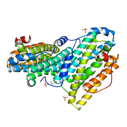
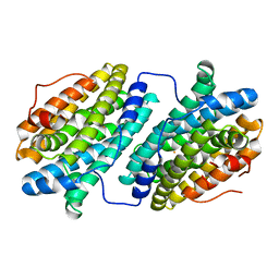
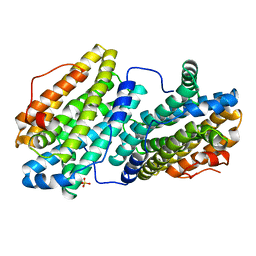
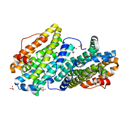
6ZBA
DownloadVisualize
BU of 6zba by Molmil
Crystal structure of PDE4D2 in complex with inhibitor LEO39652
分子名称: 1,2-ETHANEDIOL, 2-methylpropyl 1-[8-methoxy-5-(1-oxidanylidene-3~{H}-2-benzofuran-5-yl)-[1,2,4]triazolo[1,5-a]pyridin-2-yl]cyclopropane-1-carboxylate, DIMETHYL SULFOXIDE, ...
著者Akutsu, M, Hakansson, M, Welin, M, Svensson, A, Logan, D.T, Sorensen, M.D.
登録日2020-06-08
公開日2020-09-23
最終更新日2024-01-24
実験手法X-RAY DIFFRACTION (1.6 Å)
主引用文献Discovery and Early Clinical Development of Isobutyl 1-[8-Methoxy-5-(1-oxo-3 H -isobenzofuran-5-yl)-[1,2,4]triazolo[1,5- a ]pyridin-2-yl]cyclopropanecarboxylate (LEO 39652), a Novel "Dual-Soft" PDE4 Inhibitor for Topical Treatment of Atopic Dermatitis.
J.Med.Chem., 63, 2020

238582

件を2025-07-09に公開中

PDB statisticsPDBj update infoContact PDBjnumon