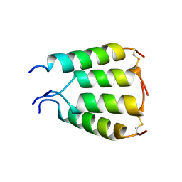
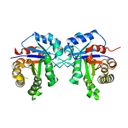
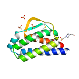
6QIZ
 
 | | CI-2, conformation 2 | | 分子名称: | Subtilisin-chymotrypsin inhibitor-2A | | 著者 | Romero, A, Ruiz, F.M. | | 登録日 | 2019-01-21 | | 公開日 | 2019-12-25 | | 最終更新日 | 2024-01-24 | | 実験手法 | X-RAY DIFFRACTION (1.65 Å) | | 主引用文献 | Engineering protein assemblies with allosteric control via monomer fold-switching.
Nat Commun, 10, 2019
|
|
8VJ7
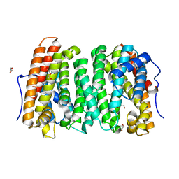
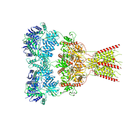 | | GluA2 bound to GYKI-52466 and Glutamate, Inhibited State 2 | | 分子名称: | 4-[(5S,8R)-8-methyl-6,7,8,9-tetrahydro-2H,5H-[1,3]dioxolo[4,5-h][2,3]benzodiazepin-5-yl]aniline, GLUTAMIC ACID, Isoform Flip of Glutamate receptor 2 | | 著者 | Hale, W.D, Montano Romero, A, Huganir, R.L, Twomey, E.C. | | 登録日 | 2024-01-05 | | 公開日 | 2024-06-05 | | 実験手法 | ELECTRON MICROSCOPY (4.85 Å) | | 主引用文献 | Allosteric Competition and Inhibition in AMPA Receptors.
Nat.Struct.Mol.Biol., 2024
|
|
8VJ6
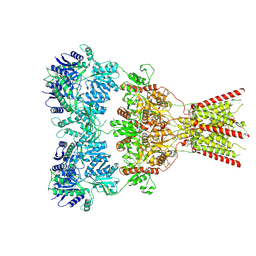 | | GluA2 bound to GYKI-52466 and Glutamate, Inhibited State 1 | | 分子名称: | 4-[(5S,8R)-8-methyl-6,7,8,9-tetrahydro-2H,5H-[1,3]dioxolo[4,5-h][2,3]benzodiazepin-5-yl]aniline, Isoform Flip of Glutamate receptor 2 | | 著者 | Hale, W.D, Montano Romero, A, Huganir, R.L, Twomey, E.C. | | 登録日 | 2024-01-05 | | 公開日 | 2024-06-05 | | 実験手法 | ELECTRON MICROSCOPY (3.5 Å) | | 主引用文献 | Allosteric Competition and Inhibition in AMPA Receptors.
Nat.Struct.Mol.Biol., 2024
|
|
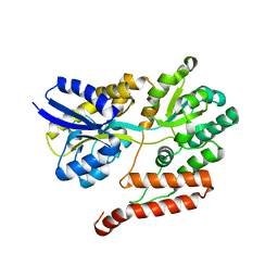
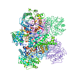
6Y91
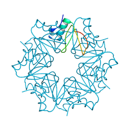
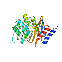
 | | Crystal structure of malate dehydrogenase from Plasmodium Falciparum in complex with NADH | | 分子名称: | Malate dehydrogenase, NICOTINAMIDE-ADENINE-DINUCLEOTIDE | | 著者 | Romero, A.R, Calderone, V, Gentili, M, Lunev, S, Groves, M, Popowicz, G, Domling, A, Sattler, M. | | 登録日 | 2020-03-06 | | 公開日 | 2021-03-31 | | 最終更新日 | 2024-01-24 | | 実験手法 | X-RAY DIFFRACTION (2.5 Å) | | 主引用文献 | A fragment-based approach identifies an allosteric pocket that impacts malate dehydrogenase activity.
Commun Biol, 4, 2021
|
|
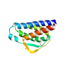
6YQY
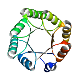 | | Crystal structure of sTIM11noCys, a de novo designed TIM barrel | | 分子名称: | de novo designed TIM barrel sTIM11noCys | | 著者 | Romero-Romero, S, Wiese, G.J, Kordes, S, Shanmugaratnam, S, Fernandez-Velasco, D.A, Hocker, B. | | 登録日 | 2020-04-18 | | 公開日 | 2021-07-21 | | 最終更新日 | 2024-01-24 | | 実験手法 | X-RAY DIFFRACTION (1.876 Å) | | 主引用文献 | The Stability Landscape of de novo TIM Barrels Explored by a Modular Design Approach.
J.Mol.Biol., 433, 2021
|
|
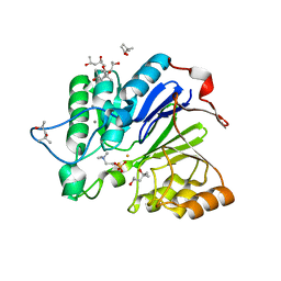
6YQX
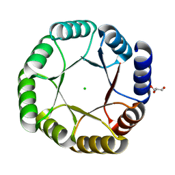 | | Crystal structure of DeNovoTIM13, a de novo designed TIM barrel | | 分子名称: | CHLORIDE ION, GLYCEROL, de novo designed TIM barrel DeNovoTIM13 | | 著者 | Romero-Romero, S, Kordes, S, Shanmugaratnam, S, Fernandez-Velasco, D.A, Hocker, B. | | 登録日 | 2020-04-18 | | 公開日 | 2021-07-21 | | 最終更新日 | 2024-05-01 | | 実験手法 | X-RAY DIFFRACTION (1.638 Å) | | 主引用文献 | The Stability Landscape of de novo TIM Barrels Explored by a Modular Design Approach.
J.Mol.Biol., 433, 2021
|
|
2FT6
 | |
2FT8
 | |
2FT7
 | |
2FTA
 | |
2W0K
 | |
2W0L
 | |
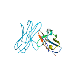
1XKM
 | | NMR structure of antimicrobial peptide distinctin in water | | 分子名称: | Distinctin chain A, Distinctin chain B | | 著者 | Amodeo, P, Raimondo, D, Andreotti, G, Motta, A, Scaloni, A. | | 登録日 | 2004-09-29 | | 公開日 | 2005-04-05 | | 最終更新日 | 2022-03-02 | | 実験手法 | SOLUTION NMR | | 主引用文献 | A folding-dependent mechanism of antimicrobial peptide resistance to degradation unveiled by solution structure of distinctin.
Proc.Natl.Acad.Sci.Usa, 102, 2005
|
|
3MBZ
 | | OXA-24 beta-lactamase complex soaked with 10mM SA4-17 inhibitor for 15min | | 分子名称: | (2S,3R)-2-[(7-aminocarbonyl-2-methanoyl-indolizin-3-yl)amino]-4-aminocarbonyloxy-3-methyl-3-sulfino-butanoic acid, Betalactamase OXA24, SULFATE ION | | 著者 | Sampson, J, van den Akker, F. | | 登録日 | 2010-03-26 | | 公開日 | 2011-03-16 | | 最終更新日 | 2011-07-13 | | 実験手法 | X-RAY DIFFRACTION (2.6 Å) | | 主引用文献 | Design, synthesis, and crystal structures of 6-alkylidene-2'-substituted penicillanic acid sulfones as potent inhibitors of Acinetobacter baumannii OXA-24 carbapenemase
J.Am.Chem.Soc., 132, 2010
|
|
6OH8
 | |
6OH6
 | |
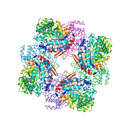
3TH6
 | | Crystal structure of Triosephosphate isomerase from Rhipicephalus (Boophilus) microplus. | | 分子名称: | Triosephosphate isomerase | | 著者 | Arreola, R, Rodriguez-Romero, A, Moraes, J, Gomez-Puyou, A, Perez-Montfort, R, Logullo, C. | | 登録日 | 2011-08-18 | | 公開日 | 2011-08-31 | | 最終更新日 | 2023-09-13 | | 実験手法 | X-RAY DIFFRACTION (2.4 Å) | | 主引用文献 | Structural and biochemical characterization of a recombinant triosephosphate isomerase from Rhipicephalus (Boophilus) microplus.
Insect Biochem.Mol.Biol., 41, 2011
|
|
3UOR
 | |
1WRA
 | |
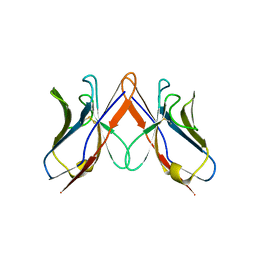
2ERM
 | | Solution structure of a biologically active human FGF-1 monomer, complexed to a hexasaccharide heparin-analogue | | 分子名称: | 2-deoxy-2-(sulfoamino)-alpha-D-glucopyranose-(1-4)-2-O-sulfo-alpha-L-idopyranuronic acid-(1-4)-2-acetamido-2-deoxy-6-O-sulfo-alpha-D-glucopyranose-(1-4)-alpha-L-idopyranuronic acid-(1-4)-2-deoxy-2-(sulfoamino)-alpha-D-glucopyranose-(1-4)-2-O-sulfo-alpha-L-idopyranuronic acid, Heparin-binding growth factor 1, ISOPROPYL ALCOHOL | | 著者 | Canales, A, Lozano, R, Nieto, P.M, Martin-Lomas, M, Gimenez-Gallego, G, Jimenez-Barbero, J. | | 登録日 | 2005-10-25 | | 公開日 | 2006-10-03 | | 最終更新日 | 2024-05-01 | | 実験手法 | SOLUTION NMR | | 主引用文献 | Solution NMR structure of a human FGF-1 monomer, activated by a hexasaccharide heparin-analogue.
Febs J., 273, 2006
|
|
1HEV
 | |
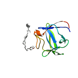
7RA9
 | | Designed StabIL-2 seq1 | | 分子名称: | 4-(2-HYDROXYETHYL)-1-PIPERAZINE ETHANESULFONIC ACID, Interleukin-2, PHOSPHATE ION | | 著者 | Jude, K.M, Chu, A.E, Huang, P.-S, Garcia, K.C. | | 登録日 | 2021-06-30 | | 公開日 | 2022-03-16 | | 最終更新日 | 2023-10-25 | | 実験手法 | X-RAY DIFFRACTION (2.2 Å) | | 主引用文献 | Interleukin-2 superkines by computational design.
Proc.Natl.Acad.Sci.USA, 119, 2022
|
|
7RAA
 | | Designed StabIL-2 seq15 | | 分子名称: | Interleukin-2, MAGNESIUM ION | | 著者 | Jude, K.M, Chu, A.E, Huang, P.-S, Garcia, K.C. | | 登録日 | 2021-06-30 | | 公開日 | 2022-03-16 | | 最終更新日 | 2024-04-03 | | 実験手法 | X-RAY DIFFRACTION (2.69 Å) | | 主引用文献 | Interleukin-2 superkines by computational design.
Proc.Natl.Acad.Sci.USA, 119, 2022
|
|
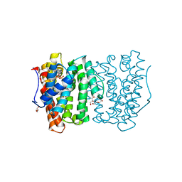
1BGG
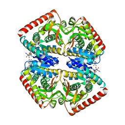
 | | GLUCOSIDASE A FROM BACILLUS POLYMYXA COMPLEXED WITH GLUCONATE | | 分子名称: | BETA-GLUCOSIDASE A, D-gluconic acid | | 著者 | Sanz-Aparicio, J, Hermoso, J, Martinez-Ripoll, M, Polaina, J. | | 登録日 | 1997-05-12 | | 公開日 | 1998-05-27 | | 最終更新日 | 2024-05-22 | | 実験手法 | X-RAY DIFFRACTION (2.3 Å) | | 主引用文献 | Crystal structure of beta-glucosidase A from Bacillus polymyxa: insights into the catalytic activity in family 1 glycosyl hydrolases.
J.Mol.Biol., 275, 1998
|
|
1BGA
 | |


