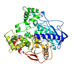
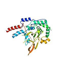
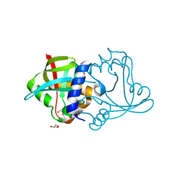
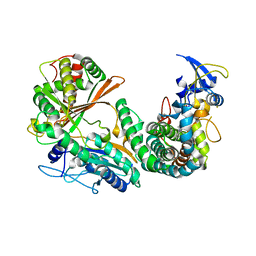
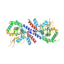
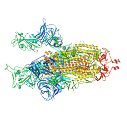
5C8V
 
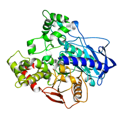 | |
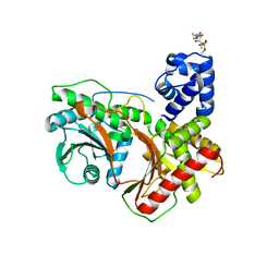
5CH3
 | | E3 alpha-esterase-7 carboxylesterase | | 分子名称: | Carboxylic ester hydrolase | | 著者 | Correy, G, Mabbitt, P, Jackson, C.J. | | 登録日 | 2015-07-10 | | 公開日 | 2016-06-15 | | 最終更新日 | 2023-09-27 | | 実験手法 | X-RAY DIFFRACTION (1.71 Å) | | 主引用文献 | Mapping the Accessible Conformational Landscape of an Insect Carboxylesterase Using Conformational Ensemble Analysis and Kinetic Crystallography.
Structure, 24, 2016
|
|
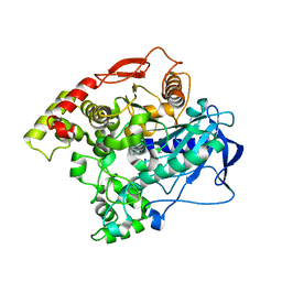
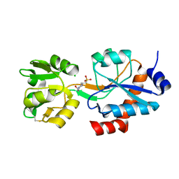
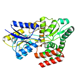
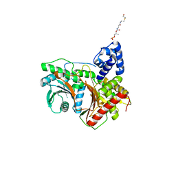
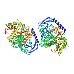
5CH5
 | | E3 alpha-esterase-7 carboxylesterase | | 分子名称: | Carboxylic ester hydrolase, DIETHYL HYDROGEN PHOSPHATE | | 著者 | Correy, G, Mabbitt, P, Jackson, C.J. | | 登録日 | 2015-07-10 | | 公開日 | 2016-06-15 | | 最終更新日 | 2023-09-27 | | 実験手法 | X-RAY DIFFRACTION (1.53 Å) | | 主引用文献 | Mapping the Accessible Conformational Landscape of an Insect Carboxylesterase Using Conformational Ensemble Analysis and Kinetic Crystallography.
Structure, 24, 2016
|
|
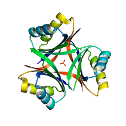
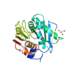
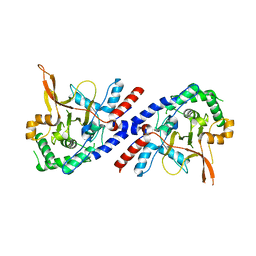
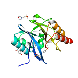
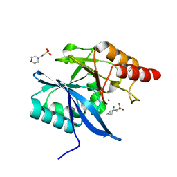
5EH9
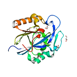 | | Indirect contributions of mutations underlie optimization of new enzyme function | | 分子名称: | 2-HYDROXYETHYL DISULFIDE, GLYCEROL, N-acyl homoserine lactonase AiiA, ... | | 著者 | Hong, N.-S, Jackson, C.J, Tokuriki, N, Yang, G, Baier, F. | | 登録日 | 2015-10-28 | | 公開日 | 2016-09-07 | | 最終更新日 | 2023-09-27 | | 実験手法 | X-RAY DIFFRACTION (1.29 Å) | | 主引用文献 | Conformational Tinkering Drives Evolution of a Promiscuous Activity through Indirect Mutational Effects.
Biochemistry, 55, 2016
|
|
5DQ6
 | |
6WUP
 | |
6N4A
 | |
6NTB
 | |
8D4W
 | | Asymmetric ene-reduction of alpha,beta-unsaturated compounds using MSMEG_2850 | | 分子名称: | Cell entry (Mce) related family protein, DI(HYDROXYETHYL)ETHER, GLYCEROL | | 著者 | Kang, S.W, Frkic, R.L, Jackson, C. | | 登録日 | 2022-06-02 | | 公開日 | 2023-01-25 | | 最終更新日 | 2023-10-25 | | 実験手法 | X-RAY DIFFRACTION (1.35 Å) | | 主引用文献 | Asymmetric Ene-Reduction of alpha , beta-Unsaturated Compounds by F 420 -Dependent Oxidoreductases A Enzymes from Mycobacterium smegmatis .
Biochemistry, 62, 2023
|
|
8KD0
 | |
8CRU
 | | PETase Ancestral Sequence Reconstruction 008 | | 分子名称: | CITRIC ACID, Poly(ethylene terephthalate) hydrolase | | 著者 | Joho, Y, Royan, S, Caputo, A.T, Ardevol Grau, A, Jackson, C. | | 登録日 | 2022-05-11 | | 公開日 | 2022-09-21 | | 最終更新日 | 2023-10-25 | | 実験手法 | X-RAY DIFFRACTION (1.3 Å) | | 主引用文献 | Ancestral Sequence Reconstruction Identifies Structural Changes Underlying the Evolution of Ideonella sakaiensis PETase and Variants with Improved Stability and Activity.
Biochemistry, 62, 2023
|
|
7OQ6
 | | Crystal structure of cytochrome P450 Sas16 from Streptomyces asterosporus | | 分子名称: | Cytochrome P450, PROTOPORPHYRIN IX CONTAINING FE, THIOCYANATE ION | | 著者 | Zhang, L, Zhang, S, Bechthold, A, Einsle, O. | | 登録日 | 2021-06-02 | | 公開日 | 2022-06-22 | | 最終更新日 | 2023-09-13 | | 実験手法 | X-RAY DIFFRACTION (2 Å) | | 主引用文献 | P450-mediated dehydrotyrosine formation during WS9326 biosynthesis proceeds via dehydrogenation of a specific acylated dipeptide substrate.
Acta Pharm Sin B, 13, 2023
|
|
6M7L
 | | Complex of OxyA with the X-domain from GPA biosynthesis | | 分子名称: | PROTOPORPHYRIN IX CONTAINING FE, Putative cytochrome P450 hydroxylase, Putative non-ribosomal peptide synthetase | | 著者 | Greule, A, Izore, T, Tailhades, J, Peschke, M, Schoppet, M, Ahmed, I, Kulik, A, Adamek, M, Ziemert, N, De Voss, J, Stegmann, E, Cryle, M.J. | | 登録日 | 2018-08-20 | | 公開日 | 2019-05-22 | | 最終更新日 | 2023-10-11 | | 実験手法 | X-RAY DIFFRACTION (2.648297 Å) | | 主引用文献 | Kistamicin biosynthesis reveals the biosynthetic requirements for production of highly crosslinked glycopeptide antibiotics.
Nat Commun, 10, 2019
|
|
8FX7
 | |
8G3I
 | |
8G3J
 | |
8FX6
 | |
5V3B
 | |
5V3P
 | |
5IKX
 | |
5K4M
 | |
5JQJ
 | |
8BON
 | |
3IIV
 | |
3IIO
 | |


