3WR2
 
 | |
3WXT
 | |
3WXU
 | |
2Z5Q
 | | Apo-Fr with intermediate content of Pd ion | | 分子名称: | CADMIUM ION, Ferritin light chain, GLYCEROL, ... | | 著者 | Ueno, T, Hirata, K, Abe, M, Suzuki, M, Abe, S, Shimizu, N, Yamamoto, M, Takata, M, Watanabe, Y. | | 登録日 | 2007-07-16 | | 公開日 | 2008-07-29 | | 最終更新日 | 2023-11-01 | | 実験手法 | X-RAY DIFFRACTION (2.1 Å) | | 主引用文献 | Process of accumulation of metal ions on the interior surface of apo-ferritin: crystal structures of a series of apo-ferritins containing variable quantities of Pd(II) ions.
J.Am.Chem.Soc., 131, 2009
|
|
2Z5R
 | | Apo-Fr with high content of Pd ions | | 分子名称: | CADMIUM ION, Ferritin light chain, PALLADIUM ION | | 著者 | Ueno, T, Hirata, K, Abe, M, Suzuki, M, Abe, S, Shimizu, N, Yamamoto, M, Takata, M, Watanabe, Y. | | 登録日 | 2007-07-16 | | 公開日 | 2008-07-29 | | 最終更新日 | 2023-11-01 | | 実験手法 | X-RAY DIFFRACTION (2.5 Å) | | 主引用文献 | Process of accumulation of metal ions on the interior surface of apo-ferritin: crystal structures of a series of apo-ferritins containing variable quantities of Pd(II) ions.
J.Am.Chem.Soc., 131, 2009
|
|
2Z4P
 | | Crystal structure of FFRP-DM1 | | 分子名称: | 75aa long hypothetical regulatory protein AsnC, ISOLEUCINE | | 著者 | Yamada, M, Koike, H, Kudo, N, Suzuki, M. | | 登録日 | 2007-06-21 | | 公開日 | 2007-09-18 | | 最終更新日 | 2024-03-13 | | 実験手法 | X-RAY DIFFRACTION (1.95 Å) | | 主引用文献 | A Structural Code for Discriminating between Transcription Signals Revealed by the Feast/Famine Regulatory Protein DM1 in Complex with Ligands
Structure, 15, 2007
|
|
3ASX
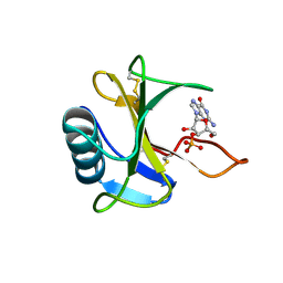
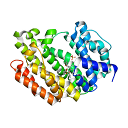 | | Human Squalene synthase in complex with 1-{4-[{4-chloro-2-[(2-chlorophenyl)(hydroxy)methyl]phenyl}(2,2-dimethylpropyl)amino]-4-oxobutanoyl}piperidine-3-carboxylic acid | | 分子名称: | (3R)-1-{4-[{4-chloro-2-[(S)-(2-chlorophenyl)(hydroxy)methyl]phenyl}(2,2-dimethylpropyl)amino]-4-oxobutanoyl}piperidine-3-carboxylic acid, PHOSPHATE ION, Squalene synthase | | 著者 | Shimizu, H, Suzuki, M, Katakura, S, Yamazaki, K, Higashihashi, N, Ichikawa, M, Yokomizo, A, Itoh, M, Sugita, K, Usui, H. | | 登録日 | 2010-12-22 | | 公開日 | 2011-12-21 | | 最終更新日 | 2023-11-01 | | 実験手法 | X-RAY DIFFRACTION (2 Å) | | 主引用文献 | Discovery of a new 2-aminobenzhydrol template for highly potent squalene synthase inhibitors
Bioorg.Med.Chem., 19, 2011
|
|
3VQG
 | | Crystal Structure Analysis of the PDZ Domain Derived from the Tight Junction Regulating Protein | | 分子名称: | C-terminal peptide from Immunoglobulin superfamily member 5, E3 ubiquitin-protein ligase LNX, SULFATE ION | | 著者 | Akiyoshi, Y, Hamada, D, Goda, N, Tenno, T, Narita, H, Nakagawa, A, Furuse, M, Suzuki, M, Hiroaki, H. | | 登録日 | 2012-03-23 | | 公開日 | 2013-03-27 | | 最終更新日 | 2023-11-08 | | 実験手法 | X-RAY DIFFRACTION (1.35 Å) | | 主引用文献 | Structural basis for down regulation of tight junction by PDZ-domain containing E3-Ubiquitin ligase
To be Published
|
|
2ZNY
 | | Crystal structure of the FFRP | | 分子名称: | ARGININE, Uncharacterized HTH-type transcriptional regulator PH1519 | | 著者 | Yamada, M, Suzuki, M. | | 登録日 | 2008-05-02 | | 公開日 | 2009-03-17 | | 最終更新日 | 2023-11-01 | | 実験手法 | X-RAY DIFFRACTION (2.59 Å) | | 主引用文献 | Interactions between the archaeal transcription repressor FL11 and its coregulators lysine and arginine.
Proteins, 74, 2009
|
|
3WUN
 | |
3AWE
 | | Crystal structure of Pten-like domain of Ci-VSP (248-576) | | 分子名称: | ACETIC ACID, SODIUM ION, SULFATE ION, ... | | 著者 | Matsuda, M, Sakata, S, Takeshita, K, Suzuki, M, Yamashita, E, Okamura, Y, Nakagawa, A. | | 登録日 | 2011-03-19 | | 公開日 | 2011-05-04 | | 最終更新日 | 2023-11-01 | | 実験手法 | X-RAY DIFFRACTION (2.77 Å) | | 主引用文献 | Crystal structure of the cytoplasmic phosphatase and tensin homolog (PTEN)-like region of Ciona intestinalis voltage-sensing phosphatase provides insight into substrate specificity and redox regulation of the phosphoinositide phosphatase activity
J.Biol.Chem., 286, 2011
|
|
2ZNZ
 | | Crystal structure of FFRP | | 分子名称: | LYSINE, Uncharacterized HTH-type transcriptional regulator PH1519 | | 著者 | Yamada, M, Suzuki, M. | | 登録日 | 2008-05-02 | | 公開日 | 2009-03-17 | | 最終更新日 | 2023-11-01 | | 実験手法 | X-RAY DIFFRACTION (2.39 Å) | | 主引用文献 | Interactions between the archaeal transcription repressor FL11 and its coregulators lysine and arginine.
Proteins, 74, 2009
|
|
3AWF
 | | Crystal structure of Pten-like domain of Ci-VSP (236-576) | | 分子名称: | GLYCEROL, SULFATE ION, Voltage-sensor containing phosphatase | | 著者 | Matsuda, M, Sakata, S, Takeshita, K, Suzuki, M, Yamashita, E, Okamura, Y, Nakagawa, A. | | 登録日 | 2011-03-19 | | 公開日 | 2011-05-04 | | 最終更新日 | 2023-11-01 | | 実験手法 | X-RAY DIFFRACTION (1.99 Å) | | 主引用文献 | Crystal structure of the cytoplasmic phosphatase and tensin homolog (PTEN)-like region of Ciona intestinalis voltage-sensing phosphatase provides insight into substrate specificity and redox regulation of the phosphoinositide phosphatase activity
J.Biol.Chem., 286, 2011
|
|
3AWG
 | | Crystal structure of Pten-like domain of Ci-VSP G356A mutant (248-576) | | 分子名称: | SULFATE ION, Voltage-sensor containing phosphatase | | 著者 | Matsuda, M, Sakata, S, Takeshita, K, Suzuki, M, Yamashita, E, Okamura, Y, Nakagawa, A. | | 登録日 | 2011-03-19 | | 公開日 | 2011-05-04 | | 最終更新日 | 2023-11-01 | | 実験手法 | X-RAY DIFFRACTION (2.39 Å) | | 主引用文献 | Crystal structure of the cytoplasmic phosphatase and tensin homolog (PTEN)-like region of Ciona intestinalis voltage-sensing phosphatase provides insight into substrate specificity and redox regulation of the phosphoinositide phosphatase activity
J.Biol.Chem., 286, 2011
|
|
3VRR
 | | Crystal structure of the tyrosine kinase binding domain of Cbl-c (PL mutant) in complex with phospho-EGFR peptide | | 分子名称: | CALCIUM ION, Epidermal growth factor receptor, Signal transduction protein CBL-C | | 著者 | Takeshita, K, Tezuka, T, Isozaki, Y, Yamashita, E, Suzuki, M, Yamanashi, Y, Yamamoto, T, Nakagawa, A. | | 登録日 | 2012-04-13 | | 公開日 | 2013-03-06 | | 実験手法 | X-RAY DIFFRACTION (2 Å) | | 主引用文献 | Structural flexibility regulates phosphopeptide-binding activity of the tyrosine kinase binding domain of Cbl-c.
J.Biochem., 152, 2012
|
|
3VRO
 | | Crystal structure of the tyrosine kinase binding domain of Cbl-c in complex with phospho-Src peptide | | 分子名称: | CALCIUM ION, Proto-oncogene tyrosine-protein kinase Src, Signal transduction protein CBL-C | | 著者 | Takeshita, K, Tezuka, T, Isozaki, Y, Yamashita, E, Suzuki, M, Yamanashi, Y, Yamamoto, T, Nakagawa, A. | | 登録日 | 2012-04-13 | | 公開日 | 2013-03-06 | | 最終更新日 | 2017-11-22 | | 実験手法 | X-RAY DIFFRACTION (1.8 Å) | | 主引用文献 | Structural flexibility regulates phosphopeptide-binding activity of the tyrosine kinase binding domain of Cbl-c.
J.Biochem., 152, 2012
|
|
3VRQ
 | | Crystal structure of the tyrosine kinase binding domain of Cbl-c (PL mutant) | | 分子名称: | CALCIUM ION, Signal transduction protein CBL-C | | 著者 | Takeshita, K, Tezuka, T, Isozaki, Y, Yamashita, E, Suzuki, M, Yamanashi, Y, Yamamoto, T, Nakagawa, A. | | 登録日 | 2012-04-13 | | 公開日 | 2013-03-06 | | 最終更新日 | 2024-03-20 | | 実験手法 | X-RAY DIFFRACTION (2.39 Å) | | 主引用文献 | Structural flexibility regulates phosphopeptide-binding activity of the tyrosine kinase binding domain of Cbl-c.
J.Biochem., 152, 2012
|
|
2ZUQ
 | | Crystal structure of DsbB-Fab complex | | 分子名称: | Disulfide bond formation protein B, Fab fragment heavy chain, Fab fragment light chain, ... | | 著者 | Inaba, K, Suzuki, M, Murakami, S. | | 登録日 | 2008-10-28 | | 公開日 | 2009-04-14 | | 最終更新日 | 2023-11-01 | | 実験手法 | X-RAY DIFFRACTION (3.3 Å) | | 主引用文献 | Dynamic nature of disulphide bond formation catalysts revealed by crystal structures of DsbB
Embo J., 28, 2009
|
|
3APQ
 | |
3APO
 | |
3APS
 | |
2ZIB
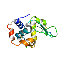
 | | Crystal structure analysis of calcium-independent type II antifreeze protein | | 分子名称: | SULFATE ION, Type II antifreeze protein | | 著者 | Nishimiya, Y, Sato, R, Kondo, H, Noro, N, Sugimoto, H, Suzuki, M, Tsuda, S. | | 登録日 | 2008-02-14 | | 公開日 | 2008-08-19 | | 最終更新日 | 2011-07-13 | | 実験手法 | X-RAY DIFFRACTION (1.34 Å) | | 主引用文献 | Crystal structure and mutational analysis of Ca2+-independent type II antifreeze protein from longsnout poacher, Brachyopsis rostratus
J.Mol.Biol., 382, 2008
|
|
2ZUP
 | | Updated crystal structure of DsbB-DsbA complex from E. coli | | 分子名称: | Disulfide bond formation protein B, Thiol:disulfide interchange protein dsbA, UBIQUINONE-1, ... | | 著者 | Inaba, K, Suzuki, M, Murakami, S, Nakagawa, A. | | 登録日 | 2008-10-28 | | 公開日 | 2009-04-14 | | 最終更新日 | 2023-11-01 | | 実験手法 | X-RAY DIFFRACTION (3.7 Å) | | 主引用文献 | Dynamic nature of disulphide bond formation catalysts revealed by crystal structures of DsbB
Embo J., 28, 2009
|
|
3AXA
 | | Crystal structure of afadin PDZ domain in complex with the C-terminal peptide from nectin-3 | | 分子名称: | Afadin, Nectin-3 | | 著者 | Fujiwara, Y, Goda, N, Narita, H, Satomura, K, Nakagawa, A, Sakisaka, T, Suzuki, M, Hiroaki, H. | | 登録日 | 2011-03-31 | | 公開日 | 2012-04-25 | | 最終更新日 | 2023-11-01 | | 実験手法 | X-RAY DIFFRACTION (2.78 Å) | | 主引用文献 | Crystal structure of afadin PDZ domain-nectin-3 complex shows the structural plasticity of the ligand-binding site.
Protein Sci., 24, 2015
|
|
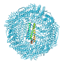
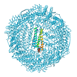
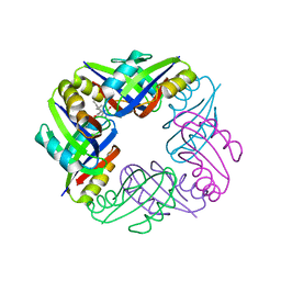
3VJJ
 | | Crystal Structure Analysis of the P9-1 | | 分子名称: | P9-1 | | 著者 | Akita, F, Higashiura, A, Suzuki, M, Tsukihara, T, Nakagawa, A, Omura, T. | | 登録日 | 2011-10-24 | | 公開日 | 2011-12-21 | | 最終更新日 | 2024-03-20 | | 実験手法 | X-RAY DIFFRACTION (3 Å) | | 主引用文献 | Crystallographic analysis reveals octamerization of viroplasm matrix protein P9-1 of Rice black streaked dwarf virus
J.Virol., 86, 2012
|
|


