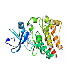
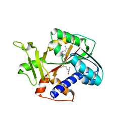
6T2B
 
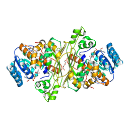 | | Glycoside hydrolase family 109 from Akkermansia muciniphila in complex with GalNAc and NAD+. | | 分子名称: | 2-acetamido-2-deoxy-alpha-D-galactopyranose, Glycosyl hydrolase family 109 protein 2, NICOTINAMIDE-ADENINE-DINUCLEOTIDE, ... | | 著者 | Chaberski, E.K, Fredslund, F, Teze, D, Shuoker, B, Kunstmann, S, Karlsson, E.N, Hachem, M.A, Welner, D.H. | | 登録日 | 2019-10-08 | | 公開日 | 2020-02-26 | | 最終更新日 | 2024-01-24 | | 実験手法 | X-RAY DIFFRACTION (2.13 Å) | | 主引用文献 | The Catalytic Acid-Base in GH109 Resides in a Conserved GGHGG Loop and Allows for Comparable alpha-Retaining and beta-Inverting Activity in an N-Acetylgalactosaminidase from Akkermansia muciniphila
Acs Catalysis, 2020
|
|
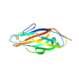
3I5R
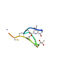
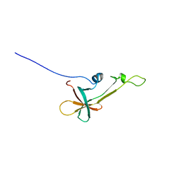
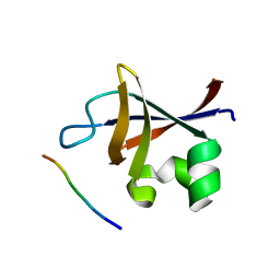 | | PI3K SH3 domain in complex with a peptide ligand | | 分子名称: | Peptide ligand, Phosphatidylinositol 3-kinase regulatory subunit alpha | | 著者 | Batra-Safferling, R, Granzin, J, Modder, S, Hoffmann, S, Willbold, D. | | 登録日 | 2009-07-06 | | 公開日 | 2010-03-02 | | 最終更新日 | 2023-09-06 | | 実験手法 | X-RAY DIFFRACTION (1.7 Å) | | 主引用文献 | Structural studies of the phosphatidylinositol 3-kinase (PI3K) SH3 domain in complex with a peptide ligand: role of the anchor residue in ligand binding.
Biol.Chem., 391, 2010
|
|
3I5S
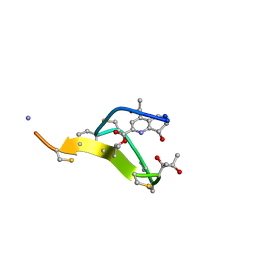
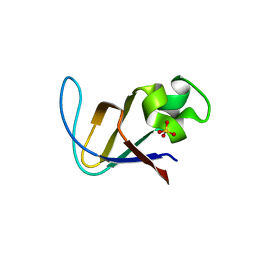 | | Crystal structure of PI3K SH3 | | 分子名称: | Phosphatidylinositol 3-kinase regulatory subunit alpha, SULFATE ION | | 著者 | Batra-Safferling, R, Granzin, J, Modder, S, Hoffmann, S, Willbold, D. | | 登録日 | 2009-07-06 | | 公開日 | 2010-03-02 | | 最終更新日 | 2023-09-06 | | 実験手法 | X-RAY DIFFRACTION (3 Å) | | 主引用文献 | Structural studies of the phosphatidylinositol 3-kinase (PI3K) SH3 domain in complex with a peptide ligand: role of the anchor residue in ligand binding.
Biol.Chem., 391, 2010
|
|
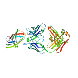
1YG9
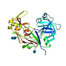 | | The structure of mutant (N93Q) of bla g 2 | | 分子名称: | 2-acetamido-2-deoxy-beta-D-glucopyranose, 2-acetamido-2-deoxy-beta-D-glucopyranose-(1-4)-2-acetamido-2-deoxy-beta-D-glucopyranose, Aspartic protease Bla g 2, ... | | 著者 | Li, M, Gustchina, A, Wuenschmann, S, Pomes, A, Wlodawer, A. | | 登録日 | 2005-01-04 | | 公開日 | 2005-03-22 | | 最終更新日 | 2021-10-20 | | 実験手法 | X-RAY DIFFRACTION (1.3 Å) | | 主引用文献 | Crystal structure of cockroach allergen Bla g 2, an unusual zinc binding aspartic protease with a novel mode of self-inhibition.
J.Mol.Biol., 348, 2005
|
|
6TFP
 | | BTK in complex with LOU064, a potent and highly selective covalent inhibitor | | 分子名称: | SODIUM ION, Tyrosine-protein kinase BTK, ~{N}-[3-[6-azanyl-5-[2-[methyl(propanoyl)amino]ethoxy]pyrimidin-4-yl]-5-fluoranyl-2-methyl-phenyl]-4-cyclopropyl-2-fluoranyl-benzamide | | 著者 | Scheufler, C, Hinniger, A, Gutmann, S. | | 登録日 | 2019-11-14 | | 公開日 | 2020-03-04 | | 最終更新日 | 2024-05-01 | | 実験手法 | X-RAY DIFFRACTION (2 Å) | | 主引用文献 | Discovery of LOU064 (Remibrutinib), a Potent and Highly Selective Covalent Inhibitor of Bruton's Tyrosine Kinase.
J.Med.Chem., 63, 2020
|
|
6OY4
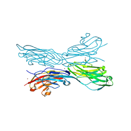
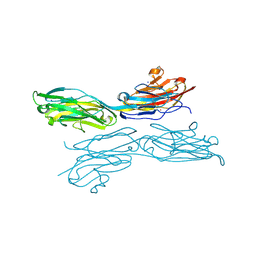
 | | Crystal structure of complex between recombinant Der p 2.0103 and Fab fragment of 7A1 | | 分子名称: | Der p 2 variant 3, Fab fragment of IgG, HEAVY CHAIN, ... | | 著者 | Kapingidza, A.B, Offermann, L.R, Glesner, J, Wunschmann, S, Vailes, L.D, Chapman, M.D.C, Pomes, A, Chruszcz, M. | | 登録日 | 2019-05-14 | | 公開日 | 2019-08-28 | | 最終更新日 | 2023-10-11 | | 実験手法 | X-RAY DIFFRACTION (2.45 Å) | | 主引用文献 | A Human IgE Antibody Binding Site on Der p 2 for the Design of a Recombinant Allergen for Immunotherapy.
J Immunol., 203, 2019
|
|
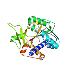
1RI1
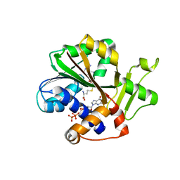 | | Structure and mechanism of mRNA cap (guanine N-7) methyltransferase | | 分子名称: | 7-METHYL-GUANOSINE-5'-TRIPHOSPHATE-5'-GUANOSINE, S-ADENOSYL-L-HOMOCYSTEINE, mRNA CAPPING ENZYME | | 著者 | Fabrega, C, Hausmann, S, Shen, V, Shuman, S, Lima, C.D. | | 登録日 | 2003-11-16 | | 公開日 | 2004-02-03 | | 最終更新日 | 2024-02-14 | | 実験手法 | X-RAY DIFFRACTION (2.5 Å) | | 主引用文献 | Structure and mechanism of mRNA cap (Guanine-n7) methyltransferase
Mol.Cell, 13, 2004
|
|
4J7Z
 | | Thermus thermophilus DNAJ J- and G/F-DOMAINS | | 分子名称: | Chaperone protein DnaJ 2, GLYCEROL | | 著者 | Barends, T.R.M, Brosi, R.W, Steinmetz, A, Scherer, A, Hartmann, E, Eschenbach, J, Lorenz, T, Seidel, R, Shoeman, R, Zimmermann, S, Bittl, R, Schlichting, I, Reinstein, J. | | 登録日 | 2013-02-14 | | 公開日 | 2013-07-31 | | 最終更新日 | 2024-02-28 | | 実験手法 | EPR (1.64 Å), X-RAY DIFFRACTION | | 主引用文献 | Combining crystallography and EPR: crystal and solution structures of the multidomain cochaperone DnaJ.
Acta Crystallogr.,Sect.D, 69, 2013
|
|
4J80
 | | Thermus thermophilus DnaJ | | 分子名称: | Chaperone protein DnaJ 2 | | 著者 | Barends, T.R.M, Brosi, R.W, Steinmetz, A, Scherer, A, Hartmann, E, Eschenbach, J, Lorenz, T, Seidel, R, Shoeman, R, Zimmermann, S, Bittl, R, Schlichting, I, Reinstein, J. | | 登録日 | 2013-02-14 | | 公開日 | 2013-07-31 | | 最終更新日 | 2013-10-09 | | 実験手法 | EPR (2.9 Å), X-RAY DIFFRACTION | | 主引用文献 | Combining crystallography and EPR: crystal and solution structures of the multidomain cochaperone DnaJ.
Acta Crystallogr.,Sect.D, 69, 2013
|
|
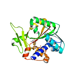
1RI3
 | | Structure and mechanism of mRNA cap (guanine N-7) methyltransferase | | 分子名称: | S-ADENOSYL-L-HOMOCYSTEINE, mRNA CAPPING ENZYME | | 著者 | Fabrega, C, Hausmann, S, Shen, V, Shuman, S, Lima, C.D. | | 登録日 | 2003-11-16 | | 公開日 | 2004-02-03 | | 最終更新日 | 2024-02-14 | | 実験手法 | X-RAY DIFFRACTION (2.5 Å) | | 主引用文献 | Structure and mechanism of mRNA cap (Guanine-n7) methyltransferase
Mol.Cell, 13, 2004
|
|
1RI2
 | | Structure and mechanism of mRNA cap (guanine N-7) methyltransferase | | 分子名称: | 7-METHYL-GUANOSINE-5'-TRIPHOSPHATE-5'-GUANOSINE, mRNA CAPPING ENZYME | | 著者 | Fabrega, C, Hausmann, S, Shen, V, Shuman, S, Lima, C.D. | | 登録日 | 2003-11-16 | | 公開日 | 2004-02-03 | | 最終更新日 | 2024-02-14 | | 実験手法 | X-RAY DIFFRACTION (2.7 Å) | | 主引用文献 | Structure and mechanism of mRNA cap (Guanine-n7) methyltransferase
Mol.Cell, 13, 2004
|
|
1RI5
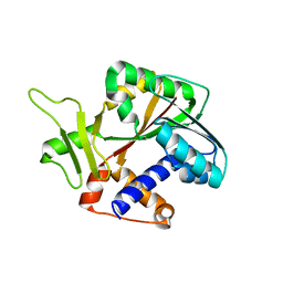 | | Structure and mechanism of mRNA cap (guanine N-7) methyltransferase | | 分子名称: | mRNA CAPPING ENZYME | | 著者 | Fabrega, C, Hausmann, S, Shen, V, Shuman, S, Lima, C.D, Burley, S.K, New York SGX Research Center for Structural Genomics (NYSGXRC) | | 登録日 | 2003-11-16 | | 公開日 | 2004-02-03 | | 最終更新日 | 2024-02-14 | | 実験手法 | X-RAY DIFFRACTION (2.1 Å) | | 主引用文献 | Structure and mechanism of mRNA cap (Guanine-n7) methyltransferase
Mol.Cell, 13, 2004
|
|
1RI4
 | | Structure and mechanism of mRNA cap (guanine N-7) methyltransferase | | 分子名称: | S-ADENOSYLMETHIONINE, mRNA CAPPING ENZYME | | 著者 | Fabrega, C, Hausmann, S, Shen, V, Shuman, S, Lima, C.D. | | 登録日 | 2003-11-16 | | 公開日 | 2004-02-03 | | 最終更新日 | 2024-02-14 | | 実験手法 | X-RAY DIFFRACTION (2.4 Å) | | 主引用文献 | Structure and mechanism of mRNA cap (Guanine-n7) methyltransferase
Mol.Cell, 13, 2004
|
|
1TR7
 | | FimH adhesin receptor binding domain from uropathogenic E. coli | | 分子名称: | (4S)-2-METHYL-2,4-PENTANEDIOL, CACODYLATE ION, FimH protein, ... | | 著者 | Bouckaert, J, Berglund, J, Schembri, M, De Genst, E, Cools, L, Wuhrer, M, Hung, C.S, Pinkner, J, Slattegard, R, Zavialov, A, Choudhury, D, Langermann, S, Hultgren, S.J, Wyns, L, Klemm, P, Oscarson, S, Knight, S.D, De Greve, H. | | 登録日 | 2004-06-21 | | 公開日 | 2005-05-03 | | 最終更新日 | 2023-10-25 | | 実験手法 | X-RAY DIFFRACTION (2.1 Å) | | 主引用文献 | Receptor binding studies disclose a novel class of high-affinity inhibitors of the Escherichia coli FimH adhesin
Mol.Microbiol., 55, 2005
|
|
5CVS
 | | GlgE isoform 1 from Streptomyces coelicolor E423A mutant soaked in maltoheptaose | | 分子名称: | Alpha-1,4-glucan:maltose-1-phosphate maltosyltransferase 1, alpha-D-glucopyranose-(1-4)-alpha-D-glucopyranose-(1-4)-alpha-D-glucopyranose-(1-4)-alpha-D-glucopyranose-(1-4)-alpha-D-glucopyranose, alpha-D-glucopyranose-(1-4)-alpha-D-glucopyranose-(1-4)-alpha-D-glucopyranose-(1-4)-alpha-D-glucopyranose-(1-4)-alpha-D-glucopyranose-(1-4)-alpha-D-glucopyranose-(1-4)-alpha-D-glucopyranose | | 著者 | Rashid, A.M, Syson, K, Koliwer-Brandl, H, van de Weerd, R, Stevenson, C.E.M, Batey, S.F.D, Miah, F, Alber, M, Ioerger, T.R, Chandra, G, Appelmelk, B.J, Nartowski, K.P, Khimyak, Y.Z, Lawson, D.M, Jacobs, W.R, Geurtsen, J, Kalscheuer, R, Bornemann, S. | | 登録日 | 2015-07-27 | | 公開日 | 2016-08-17 | | 最終更新日 | 2024-01-10 | | 実験手法 | X-RAY DIFFRACTION (2.3 Å) | | 主引用文献 | Ligand-bound structures and site-directed mutagenesis identify the acceptor and secondary binding sites of Streptomyces coelicolor maltosyltransferase GlgE.
J.Biol.Chem., 291, 2016
|
|
1R3O
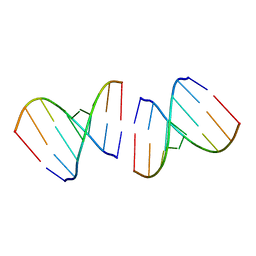 | | Crystal structure of the first RNA duplex in L-conformation at 1.9A resolution | | 分子名称: | L-RNA | | 著者 | Vallazza, M, Perbandt, M, Klussmann, S, Rypniewski, W, Erdmann, V.A, Betzel, C. | | 登録日 | 2003-10-02 | | 公開日 | 2003-12-23 | | 最終更新日 | 2024-02-14 | | 実験手法 | X-RAY DIFFRACTION (1.9 Å) | | 主引用文献 | First look at RNA in L-configuration.
Acta Crystallogr.,Sect.D, 60, 2004
|
|
2LBU
 | | HADDOCK calculated model of Congo red bound to the HET-s amyloid | | 分子名称: | Small s protein, sodium 3,3'-(1E,1'E)-biphenyl-4,4'-diylbis(diazene-2,1-diyl)bis(4-aminonaphthalene-1-sulfonate) | | 著者 | Schutz, A.K, Soragni, A, Hornemann, S, Aguzzi, A, Ernst, M, Bockmann, A, Meier, B.H. | | 登録日 | 2011-04-07 | | 公開日 | 2011-06-01 | | 最終更新日 | 2024-05-01 | | 実験手法 | SOLID-STATE NMR | | 主引用文献 | The Amyloid-Congo Red Interface at Atomic Resolution.
Angew.Chem.Int.Ed.Engl., 2011
|
|
2L2Y
 | | Thiostrepton, epimer form of residue 9 | | 分子名称: | Thiostrepton | | 著者 | Jonker, H.R.A, Baumann, S, Wolf, A, Schoof, S, Hiller, F, Schulte, K.W, Kirschner, K.N, Schwalbe, H, Arndt, H.-D. | | 登録日 | 2010-08-27 | | 公開日 | 2011-02-02 | | 最終更新日 | 2013-06-26 | | 実験手法 | SOLUTION NMR | | 主引用文献 | NMR structures of thiostrepton derivatives for characterization of the ribosomal binding site.
Angew.Chem.Int.Ed.Engl., 50, 2011
|
|
2L2X
 | | Thiostrepton, oxidized at CA-CB bond of residue 9 | | 分子名称: | Thiostrepton | | 著者 | Jonker, H.R.A, Baumann, S, Wolf, A, Schoof, S, Hiller, F, Schulte, K.W, Kirschner, K.N, Schwalbe, H, Arndt, H.-D. | | 登録日 | 2010-08-27 | | 公開日 | 2011-02-02 | | 最終更新日 | 2013-06-26 | | 実験手法 | SOLUTION NMR | | 主引用文献 | NMR structures of thiostrepton derivatives for characterization of the ribosomal binding site.
Angew.Chem.Int.Ed.Engl., 50, 2011
|
|
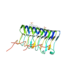
5K6V
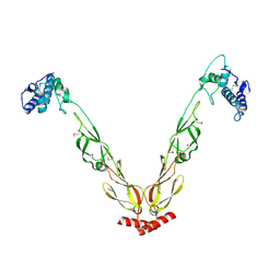
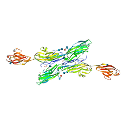
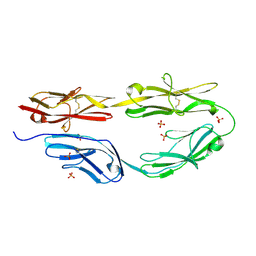
 | | Sidekick-1 immunoglobulin domains 1-4, crystal form 2 | | 分子名称: | 2-acetamido-2-deoxy-beta-D-glucopyranose, 2-acetamido-2-deoxy-beta-D-glucopyranose-(1-4)-2-acetamido-2-deoxy-beta-D-glucopyranose, Protein sidekick-1, ... | | 著者 | Jin, X, Goodman, K.M, Mannepalli, S, Honig, B, Shapiro, L. | | 登録日 | 2016-05-25 | | 公開日 | 2016-09-28 | | 最終更新日 | 2024-04-03 | | 実験手法 | X-RAY DIFFRACTION (3.208 Å) | | 主引用文献 | Molecular basis of sidekick-mediated cell-cell adhesion and specificity.
Elife, 5, 2016
|
|
5K6W
 | | Sidekick-1 immunoglobulin domains 1-5 | | 分子名称: | 2-acetamido-2-deoxy-beta-D-glucopyranose, 2-acetamido-2-deoxy-beta-D-glucopyranose-(1-4)-2-acetamido-2-deoxy-beta-D-glucopyranose, GLYCEROL, ... | | 著者 | Goodman, K.M, Mannepalli, S, Honig, B, Shapiro, L. | | 登録日 | 2016-05-25 | | 公開日 | 2016-09-28 | | 最終更新日 | 2020-07-29 | | 実験手法 | X-RAY DIFFRACTION (3.5 Å) | | 主引用文献 | Molecular basis of sidekick-mediated cell-cell adhesion and specificity.
Elife, 5, 2016
|
|
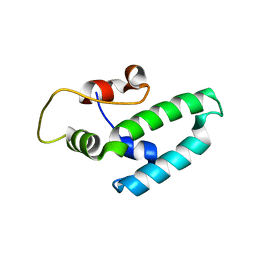
2LVL
 | | NMR Structure the lantibiotic immunity protein SpaI | | 分子名称: | SpaI | | 著者 | Christ, N, Bochmann, S, Gottstein, D, Duchardt-Ferner, E, Hellmich, U.A, Duesterhus, S, Koetter, P, Guentert, P, Entian, K, Woehnert, J. | | 登録日 | 2012-07-06 | | 公開日 | 2012-08-29 | | 最終更新日 | 2024-05-15 | | 実験手法 | SOLUTION NMR | | 主引用文献 | The First Structure of a Lantibiotic Immunity Protein, SpaI from Bacillus subtilis, Reveals a Novel Fold.
J.Biol.Chem., 287, 2012
|
|
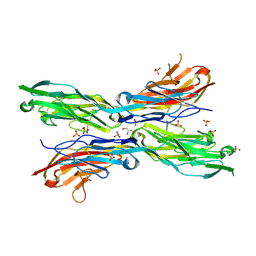
5K70
 | | Sidekick-2 immunoglobulin domains 1-4 H18R/N22S mutant | | 分子名称: | 2-acetamido-2-deoxy-beta-D-glucopyranose, 2-acetamido-2-deoxy-beta-D-glucopyranose-(1-4)-[alpha-L-fucopyranose-(1-6)]2-acetamido-2-deoxy-beta-D-glucopyranose, PHOSPHATE ION, ... | | 著者 | Goodman, K.M, Mannepalli, S, Honig, B, Shapiro, L. | | 登録日 | 2016-05-25 | | 公開日 | 2016-09-28 | | 最終更新日 | 2024-04-03 | | 実験手法 | X-RAY DIFFRACTION (2.7 Å) | | 主引用文献 | Molecular basis of sidekick-mediated cell-cell adhesion and specificity.
Elife, 5, 2016
|
|
5K6U
 | | Sidekick-1 immunoglobulin domains 1-4, crystal form 1 | | 分子名称: | 2-acetamido-2-deoxy-beta-D-glucopyranose-(1-4)-2-acetamido-2-deoxy-beta-D-glucopyranose, CESIUM ION, IODIDE ION, ... | | 著者 | Jin, X, Goodman, K.M, Mannepalli, S, Honig, B, Shapiro, L. | | 登録日 | 2016-05-25 | | 公開日 | 2016-09-28 | | 最終更新日 | 2020-07-29 | | 実験手法 | X-RAY DIFFRACTION (2.201 Å) | | 主引用文献 | Molecular basis of sidekick-mediated cell-cell adhesion and specificity.
Elife, 5, 2016
|
|
5K6X
 | | Sidekick-2 immunoglobulin domains 1-4, crystal form 1 | | 分子名称: | 2-acetamido-2-deoxy-beta-D-glucopyranose, 2-acetamido-2-deoxy-beta-D-glucopyranose-(1-4)-[alpha-L-fucopyranose-(1-6)]2-acetamido-2-deoxy-beta-D-glucopyranose, GLYCEROL, ... | | 著者 | Goodman, K.M, Mannepalli, S, Honig, B, Shapiro, L. | | 登録日 | 2016-05-25 | | 公開日 | 2016-09-28 | | 最終更新日 | 2024-04-03 | | 実験手法 | X-RAY DIFFRACTION (2.7 Å) | | 主引用文献 | Molecular basis of sidekick-mediated cell-cell adhesion and specificity.
Elife, 5, 2016
|
|


