6DO5
 
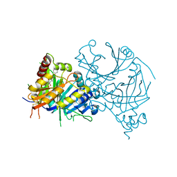
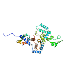
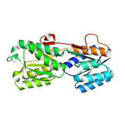
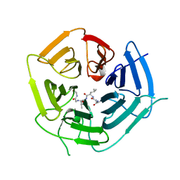 | | KLHDC2 ubiquitin ligase in complex with USP1 C-end degron | | 分子名称: | Kelch domain-containing protein 2, USP1 C-END DEGRON | | 著者 | Rusnac, D.V, Lin, H.C, Yen, H.C.S, Zheng, N. | | 登録日 | 2018-06-08 | | 公開日 | 2018-12-19 | | 最終更新日 | 2023-10-11 | | 実験手法 | X-RAY DIFFRACTION (2.5 Å) | | 主引用文献 | Recognition of the Diglycine C-End Degron by CRL2KLHDC2Ubiquitin Ligase.
Mol. Cell, 72, 2018
|
|
6DO4
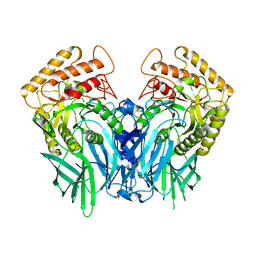
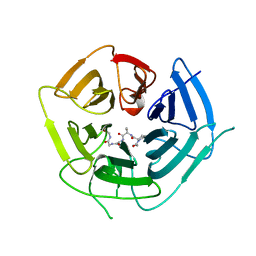 | | KLHDC2 ubiquitin ligase in complex with SelS C-end degron | | 分子名称: | Kelch domain-containing protein 2, SELS C-END DEGRON | | 著者 | Rusnac, D.V, Lin, H.C, Yen, H.C.S, Zheng, N. | | 登録日 | 2018-06-08 | | 公開日 | 2018-12-19 | | 最終更新日 | 2023-10-11 | | 実験手法 | X-RAY DIFFRACTION (2.2 Å) | | 主引用文献 | Recognition of the Diglycine C-End Degron by CRL2KLHDC2Ubiquitin Ligase.
Mol. Cell, 72, 2018
|
|
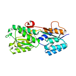
1JWA
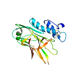
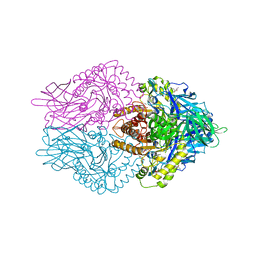
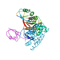 | | Structure of the ATP-bound MoeB-MoaD Protein Complex | | 分子名称: | ADENOSINE-5'-TRIPHOSPHATE, MOLYBDOPTERIN BIOSYNTHESIS MOEB PROTEIN, MOLYBDOPTERIN [MPT] CONVERTING FACTOR, ... | | 著者 | Lake, M.W, Wuebbens, M.M, Rajagopalan, K.V, Schindelin, H. | | 登録日 | 2001-09-03 | | 公開日 | 2001-11-21 | | 最終更新日 | 2024-02-07 | | 実験手法 | X-RAY DIFFRACTION (2.9 Å) | | 主引用文献 | Mechanism of ubiquitin activation revealed by the structure of a bacterial MoeB-MoaD complex.
Nature, 414, 2001
|
|
1JWB
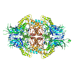
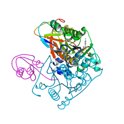 | | Structure of the Covalent Acyl-Adenylate Form of the MoeB-MoaD Protein Complex | | 分子名称: | ADENOSINE MONOPHOSPHATE, MOLYBDOPTERIN BIOSYNTHESIS MOEB PROTEIN, MOLYBDOPTERIN [MPT] CONVERTING FACTOR, ... | | 著者 | Lake, M.W, Wuebbens, M.M, Rajagopalan, K.V, Schindelin, H. | | 登録日 | 2001-09-03 | | 公開日 | 2001-11-21 | | 最終更新日 | 2017-10-04 | | 実験手法 | X-RAY DIFFRACTION (2.1 Å) | | 主引用文献 | Mechanism of ubiquitin activation revealed by the structure of a bacterial MoeB-MoaD complex.
Nature, 414, 2001
|
|
8GWD
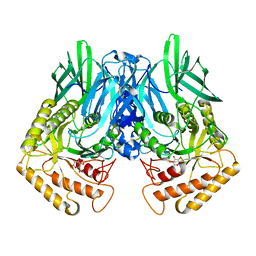
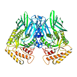
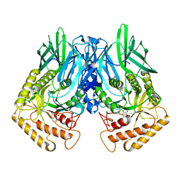
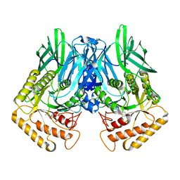
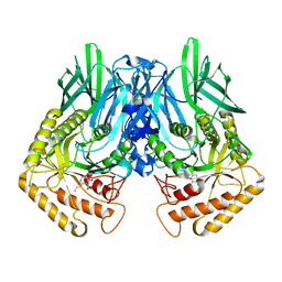
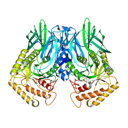
 | | Crystal structure of AtHPPD-Y18734 complex | | 分子名称: | 1,5-dimethyl-6-(2-oxidanyl-6-oxidanylidene-cyclohexen-1-yl)carbonyl-3-[(1S)-1-phenylethyl]quinazoline-2,4-dione, 4-hydroxyphenylpyruvate dioxygenase, COBALT (II) ION | | 著者 | Yang, G.-F, Lin, H.-Y, Dong, J. | | 登録日 | 2022-09-16 | | 公開日 | 2023-09-20 | | 実験手法 | X-RAY DIFFRACTION (1.889 Å) | | 主引用文献 | Crystal structure of AtHPPD-Y18734 complex
To Be Published
|
|
5Z19
 | |
7QU8
 | | ADGRG3/GPR97 Extracellular Region | | 分子名称: | Adhesion G protein-coupled receptor G3 | | 著者 | Zheng-Gerard, C, Chu, T.Y, El Omari, K, Lin, H.H, Seiradake, E. | | 登録日 | 2022-01-17 | | 公開日 | 2022-09-28 | | 最終更新日 | 2024-01-31 | | 実験手法 | X-RAY DIFFRACTION (3.37 Å) | | 主引用文献 | GPR97-mediated PAR2 transactivation via a mPR3-associated macromolecular complex induces inflammatory activation of human neutrophils
Nat Commun, 2022
|
|
5Z1B
 | |
3LZD
 | | Crystal structure of Dph2 from Pyrococcus horikoshii with 4Fe-4S cluster | | 分子名称: | Dph2, IRON/SULFUR CLUSTER, SULFATE ION | | 著者 | Torelli, A.T, Zhang, Y, Zhu, X, Lee, M, Dzikovski, B, Koralewski, R.M, Wang, E, Freed, J, Krebs, C, Lin, H, Ealick, S.E. | | 登録日 | 2010-03-01 | | 公開日 | 2010-07-14 | | 最終更新日 | 2023-09-06 | | 実験手法 | X-RAY DIFFRACTION (2.1 Å) | | 主引用文献 | Diphthamide biosynthesis requires an organic radical generated by an iron-sulphur enzyme.
Nature, 465, 2010
|
|
5Z18
 | |
5Z1A
 | |
3ESW
 | | Complex of yeast PNGase with GlcNAc2-IAc. | | 分子名称: | 2-acetamido-2-deoxy-beta-D-glucopyranose-(1-4)-2-acetamido-2-deoxy-beta-D-glucopyranose, Peptide-N(4)-(N-acetyl-beta-glucosaminyl)asparagine amidase, UV excision repair protein RAD23, ... | | 著者 | Zhao, G, Zhou, X, Lennarz, W.J, Schindelin, H. | | 登録日 | 2008-10-06 | | 公開日 | 2008-11-11 | | 最終更新日 | 2023-09-06 | | 実験手法 | X-RAY DIFFRACTION (3.4 Å) | | 主引用文献 | Structural and mutational studies on the importance of oligosaccharide binding for the activity of yeast PNGase.
Glycobiology, 19, 2009
|
|
2IPM
 | |
2IPN
 | |
3LZC
 | | Crystal structure of Dph2 from Pyrococcus horikoshii | | 分子名称: | Dph2 | | 著者 | Zhang, Y, Zhu, X, Torelli, A.T, Lee, M, Dzikovski, B, Koralewski, R.M, Wang, E, Freed, J, Krebs, C, Lin, H, Ealick, S.E. | | 登録日 | 2010-03-01 | | 公開日 | 2010-06-23 | | 最終更新日 | 2024-02-21 | | 実験手法 | X-RAY DIFFRACTION (2.261 Å) | | 主引用文献 | Diphthamide biosynthesis requires an organic radical generated by an iron-sulphur enzyme.
Nature, 465, 2010
|
|
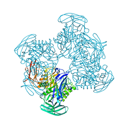
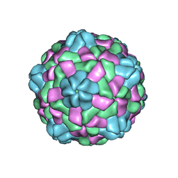
6JJC
 | | Cryo-EM structure of giant freshwater prawn Macrobrachium rosenbergii nodavirus (MrNV) semi-empty VLP | | 分子名称: | CALCIUM ION, Capsid protein | | 著者 | Chang, W.H, Wang, C.H, Lin, H.H, Lin, S.Y, Chong, S.C, Wu, Y.Y. | | 登録日 | 2019-02-25 | | 公開日 | 2019-05-01 | | 最終更新日 | 2024-03-27 | | 実験手法 | ELECTRON MICROSCOPY (2.92 Å) | | 主引用文献 | Cryo-EM structure of giant freshwater prawn Macrobrachium rosenbergii nodavirus (MrNV) semi-empty VLP
To Be Published
|
|
6JJA
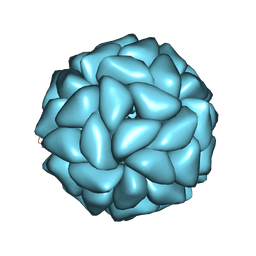 | | Cryo-EM structure of giant freshwater prawn Macrobrachium rosenbergii extra small virus (XSV) VLP | | 分子名称: | CALCIUM ION, Nucleocapsid protein CP17 | | 著者 | Chang, W.H, Wang, C.H, Lin, H.H, Lin, S.Y, Chong, S.C, Wu, Y.Y. | | 登録日 | 2019-02-25 | | 公開日 | 2019-07-17 | | 最終更新日 | 2024-03-27 | | 実験手法 | ELECTRON MICROSCOPY (2.91 Å) | | 主引用文献 | Cryo-EM structure of giant freshwater prawn Macrobrachium rosenbergii extra small virus (XSV) VLP
To Be Published
|
|
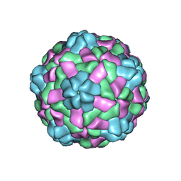
6JJD
 | | Cryo-EM structure of giant freshwater prawn Macrobrachium rosenbergii nodavirus (MrNV) full VLP | | 分子名称: | CALCIUM ION, Capsid protein | | 著者 | Chang, W.H, Wang, C.H, Lin, H.H, Lin, S.Y, Chong, S.C, Wu, Y.Y. | | 登録日 | 2019-02-25 | | 公開日 | 2019-05-01 | | 最終更新日 | 2024-03-27 | | 実験手法 | ELECTRON MICROSCOPY (3.21 Å) | | 主引用文献 | Cryo-EM structure of giant freshwater prawn Macrobrachium rosenbergii nodavirus (MrNV) full VLP
To Be Published
|
|
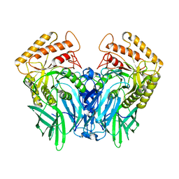
6JZ8
 | | b-glucuronidase from Ruminococcus gnavus in complex with D-glucaro 1,5-lactone | | 分子名称: | (2S,3S,4S,5R)-3,4,5-trihydroxy-6-oxo-oxane-2-carboxylic acid, Beta-glucuronidase | | 著者 | Dashnyam, P, Lin, H.Y. | | 登録日 | 2019-04-30 | | 公開日 | 2020-06-10 | | 最終更新日 | 2023-11-22 | | 実験手法 | X-RAY DIFFRACTION (1.583 Å) | | 主引用文献 | Substituent Position of Iminocyclitols Determines the Potency and Selectivity for Gut Microbial Xenobiotic-Reactivating Enzymes.
J.Med.Chem., 63, 2020
|
|
6JZ5
 | |
6JZ3
 | | b-glucuronidase from Ruminococcus gnavus in complex with uronic deoxynojirimycin | | 分子名称: | (2~{S},3~{R},4~{R},5~{S})-3,4,5-tris(oxidanyl)piperidine-2-carboxylic acid, (4R)-2-METHYLPENTANE-2,4-DIOL, Beta-glucuronidase | | 著者 | Dashnyam, P, Lin, H.Y. | | 登録日 | 2019-04-30 | | 公開日 | 2020-05-13 | | 最終更新日 | 2024-03-27 | | 実験手法 | X-RAY DIFFRACTION (1.502 Å) | | 主引用文献 | Substituent Position of Iminocyclitols Determines the Potency and Selectivity for Gut Microbial Xenobiotic-Reactivating Enzymes.
J.Med.Chem., 63, 2020
|
|
6JZ7
 | |
6JZ6
 | | b-glucuronidase from Ruminococcus gnavus in complex with C6-substituted uronic isofagomine | | 分子名称: | (2~{S},3~{S},4~{R},5~{R})-4,5-bis(oxidanyl)-2-propyl-piperidine-3-carboxylic acid, Beta-glucuronidase | | 著者 | Dashnyam, P, Lin, H.Y. | | 登録日 | 2019-04-30 | | 公開日 | 2020-06-10 | | 最終更新日 | 2023-11-22 | | 実験手法 | X-RAY DIFFRACTION (1.605 Å) | | 主引用文献 | Substituent Position of Iminocyclitols Determines the Potency and Selectivity for Gut Microbial Xenobiotic-Reactivating Enzymes.
J.Med.Chem., 63, 2020
|
|
6JZ2
 | | b-glucuronidase from Ruminococcus gnavus in complex with uronic isofagomine at 1.3 Angstroms resolution | | 分子名称: | (3S,4R,5R)-4,5-dihydroxypiperidine-3-carboxylic acid, (4R)-2-METHYLPENTANE-2,4-DIOL, (4S)-2-METHYL-2,4-PENTANEDIOL, ... | | 著者 | Dashnyam, P, Lin, H.Y. | | 登録日 | 2019-04-30 | | 公開日 | 2020-06-10 | | 最終更新日 | 2023-11-22 | | 実験手法 | X-RAY DIFFRACTION (1.29 Å) | | 主引用文献 | Substituent Position of Iminocyclitols Determines the Potency and Selectivity for Gut Microbial Xenobiotic-Reactivating Enzymes.
J.Med.Chem., 63, 2020
|
|
6JZ4
 | | b-glucuronidase from Ruminococcus gnavus in complex with D-glucaro-d-lactam | | 分子名称: | (2S,3R,4S,5R)-3,4,5-trihydroxy-6-oxopiperidine-2-carboxylic acid, (4R)-2-METHYLPENTANE-2,4-DIOL, Beta-glucuronidase | | 著者 | Dashnyam, P, Lin, H.Y. | | 登録日 | 2019-04-30 | | 公開日 | 2020-06-03 | | 最終更新日 | 2024-03-27 | | 実験手法 | X-RAY DIFFRACTION (1.712 Å) | | 主引用文献 | Substituent Position of Iminocyclitols Determines the Potency and Selectivity for Gut Microbial Xenobiotic-Reactivating Enzymes.
J.Med.Chem., 63, 2020
|
|


