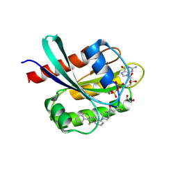
3UFF
 
 | |
3UGI
 | | Structural and functional characterization of an anesthetic binding site in the second cysteine-rich domain of protein kinase C delta | | 分子名称: | (methoxymethyl)cyclopropane, PHOSPHATE ION, Protein kinase C delta type, ... | | 著者 | Shanmugasundararaj, S, Stehle, T, Miller, K.W. | | 登録日 | 2011-11-02 | | 公開日 | 2012-12-12 | | 最終更新日 | 2023-09-13 | | 実験手法 | X-RAY DIFFRACTION (1.361 Å) | | 主引用文献 | Structural and Functional Characterization of an Anesthetic Binding Site in the Second Cysteine-Rich Domain of Protein Kinase Cdelta
Biophys.J., 103, 2012
|
|
6FXB
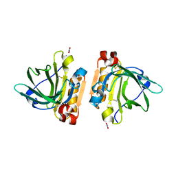 | | Bovine beta-lactoglobulin variant A at pH 4.0 | | 分子名称: | DI(HYDROXYETHYL)ETHER, Major allergen beta-lactoglobulin, NITRATE ION | | 著者 | Khan, S, Ipsen, R, Almdal, K, Svensson, B, Harris, P. | | 登録日 | 2018-03-08 | | 公開日 | 2018-05-23 | | 最終更新日 | 2019-02-20 | | 実験手法 | X-RAY DIFFRACTION (2 Å) | | 主引用文献 | Revealing the Dimeric Crystal and Solution Structure of beta-Lactoglobulin at pH 4 and Its pH and Salt Dependent Monomer-Dimer Equilibrium.
Biomacromolecules, 19, 2018
|
|
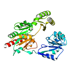
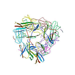
5CDI
 | | Chloroplast chaperonin 60b1 of Chlamydomonas | | 分子名称: | Chaperonin 60B1 | | 著者 | Zhang, S, Zhou, H, Yu, F, Gao, F, He, J, Liu, C. | | 登録日 | 2015-07-04 | | 公開日 | 2016-05-11 | | 最終更新日 | 2023-11-08 | | 実験手法 | X-RAY DIFFRACTION (3.807 Å) | | 主引用文献 | Structural insight into the cooperation of chloroplast chaperonin subunits
Bmc Biol., 14, 2016
|
|
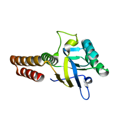
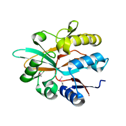
5CDJ
 | | apical domain of chloroplast chaperonin 60a | | 分子名称: | RuBisCO large subunit-binding protein subunit alpha, chloroplastic | | 著者 | Zhang, S, Yu, F, Liu, C, Gao, F. | | 登録日 | 2015-07-04 | | 公開日 | 2016-07-13 | | 最終更新日 | 2024-03-20 | | 実験手法 | X-RAY DIFFRACTION (1.75 Å) | | 主引用文献 | Functional Partition of Cpn60 alpha and Cpn60 beta Subunits in Substrate Recognition and Cooperation with Co-chaperonins
Mol Plant, 9, 2016
|
|
8QSN
 | |
8QSM
 | |
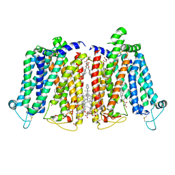
8QSL
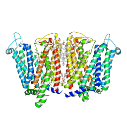 | | Cryo-EM structure of human SLC15A4 dimer in outward open state in LMNG | | 分子名称: | (1S)-2-{[{[(2R)-2,3-DIHYDROXYPROPYL]OXY}(HYDROXY)PHOSPHORYL]OXY}-1-[(PALMITOYLOXY)METHYL]ETHYL STEARATE, 2-acetamido-2-deoxy-beta-D-glucopyranose, CHOLESTEROL HEMISUCCINATE, ... | | 著者 | Rehan, S, Matsuoka, R, Jazayeri, A, Duerr, K.L. | | 登録日 | 2023-10-10 | | 公開日 | 2023-10-25 | | 実験手法 | ELECTRON MICROSCOPY (2.81 Å) | | 主引用文献 | Cryo-EM structure of human SLC15A4 dimer in outward open state in LMNG
To Be Published
|
|
8QSK
 | | Cryo-EM structure of human SLC15A4 dimer in outward open state in MSP1D1 nanodisc | | 分子名称: | (1S)-2-{[{[(2R)-2,3-DIHYDROXYPROPYL]OXY}(HYDROXY)PHOSPHORYL]OXY}-1-[(PALMITOYLOXY)METHYL]ETHYL STEARATE, 2-acetamido-2-deoxy-beta-D-glucopyranose, CHOLESTEROL HEMISUCCINATE, ... | | 著者 | Rehan, S, Matsuoka, R, Jazayeri, A, Duerr, K.L. | | 登録日 | 2023-10-10 | | 公開日 | 2023-11-01 | | 実験手法 | ELECTRON MICROSCOPY (3.3 Å) | | 主引用文献 | Cryo-EM structure of human SLC15A4 dimer in outward open state in MSP1D1 nanodisc
To Be Published
|
|
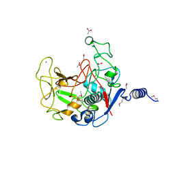
5GVX
 | | Structural insight into dephosphorylation by Trehalose 6-phosphate Phosphatase (OtsB2) from Mycobacterium Tuberculosis | | 分子名称: | MAGNESIUM ION, Trehalose-phosphate phosphatase | | 著者 | Shan, S, Min, H, Liu, T, Jiang, D, Rao, Z. | | 登録日 | 2016-09-07 | | 公開日 | 2017-09-27 | | 最終更新日 | 2024-03-20 | | 実験手法 | X-RAY DIFFRACTION (2.596 Å) | | 主引用文献 | Structural insight into dephosphorylation by trehalose 6-phosphate phosphatase (OtsB2) from Mycobacterium tuberculosis.
FASEB J., 30, 2016
|
|
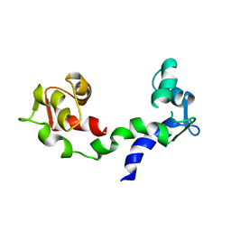
5CDK
 | |
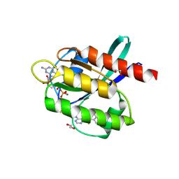
3UGD
 | | Structural and functional characterization of an anesthetic binding site in the second cysteine-rich domain of protein kinase C delta | | 分子名称: | 1,2-ETHANEDIOL, PHOSPHATE ION, Protein kinase C delta type, ... | | 著者 | Shanmugasundararaj, S, Stehle, T, Miller, K.W. | | 登録日 | 2011-11-02 | | 公開日 | 2012-12-12 | | 最終更新日 | 2023-09-13 | | 実験手法 | X-RAY DIFFRACTION (1.45 Å) | | 主引用文献 | Structural and functional characterization of an anesthetic binding site in the second cysteine-rich domain of protein kinase C delta
Biophys.J., 103, 2012
|
|
5VF4
 | |
5YXZ
 | | Co-crystal Structure of KRAS (G12C) covalently bound with Quinazoline based inhibitor JBI484 | | 分子名称: | 1,2-ETHANEDIOL, 1-[4-[6-chloranyl-8-fluoranyl-7-(2-fluorophenyl)quinazolin-4-yl]piperazin-1-yl]propan-1-one, GTPase KRas, ... | | 著者 | Swaminathan, S, Thakur, M.K, Kandan, S, Gautam, A, Kanavalli, M, Simhadri, P, Gosu, R. | | 登録日 | 2017-12-07 | | 公開日 | 2018-04-18 | | 最終更新日 | 2023-11-22 | | 実験手法 | X-RAY DIFFRACTION (1.7 Å) | | 主引用文献 | Co-crystal Structure of KRAS (G12C) covalently bound with Quinazoline based inhibitor JBI484
To Be Published
|
|
5YY1
 | | Co-crystal Structure of KRAS (G12C) covalently bound with Quinazoline based inhibitor JBI739 | | 分子名称: | 1,2-ETHANEDIOL, 1-[4-[6-chloranyl-8-fluoranyl-7-[2-(trifluoromethyl)phenyl]quinazolin-4-yl]piperazin-1-yl]propan-1-one, GTPase KRas, ... | | 著者 | Swaminathan, S, Thakur, M.K, Kandan, S, Gautam, A, Kanavalli, M, Simhadri, P, Gosu, R. | | 登録日 | 2017-12-07 | | 公開日 | 2018-04-18 | | 最終更新日 | 2023-11-22 | | 実験手法 | X-RAY DIFFRACTION (1.69 Å) | | 主引用文献 | Co-crystal Structure of KRAS (G12C) covalently bound with Quinazoline based inhibitor JBI739
To Be Published
|
|
5ZS3
 | | Small heat shock protein from M. marinum:Form-1 | | 分子名称: | CHLORIDE ION, GLY-ARG-LEU-LEU-PRO, Molecular chaperone (Small heat shock protein), ... | | 著者 | Bhandari, S, Suguna, K. | | 登録日 | 2018-04-27 | | 公開日 | 2019-01-30 | | 最終更新日 | 2019-04-17 | | 実験手法 | X-RAY DIFFRACTION (2.001 Å) | | 主引用文献 | Dodecameric structure of a small heat shock protein from Mycobacterium marinum M.
Proteins, 87, 2019
|
|
5ZS6
 | |
5ZUL
 | |
1FEX
 | |
5IOO
 | | Accommodation of massive sequence variation in Nanoarchaeota by the C-type lectin fold | | 分子名称: | AvpA, MAGNESIUM ION | | 著者 | Handa, S, Paul, B, Miller, J, Valentine, D, Ghosh, P. | | 登録日 | 2016-03-08 | | 公開日 | 2016-11-09 | | 最終更新日 | 2019-12-11 | | 実験手法 | X-RAY DIFFRACTION (2.521 Å) | | 主引用文献 | Conservation of the C-type lectin fold for accommodating massive sequence variation in archaeal diversity-generating retroelements.
Bmc Struct.Biol., 16, 2016
|
|
1DEG
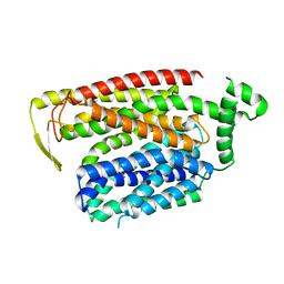
 | | THE LINKER OF DES-GLU84 CALMODULIN IS BENT AS SEEN IN THE CRYSTAL STRUCTURE | | 分子名称: | CALCIUM ION, CALMODULIN | | 著者 | Raghunathan, S, Chandross, R, Cheng, B.P, Persechini, A, Sobottk, S.E, Kretsinger, R.H. | | 登録日 | 1993-06-07 | | 公開日 | 1994-05-31 | | 最終更新日 | 2024-02-07 | | 実験手法 | X-RAY DIFFRACTION (2.9 Å) | | 主引用文献 | The linker of des-Glu84-calmodulin is bent.
Proc.Natl.Acad.Sci.Usa, 90, 1993
|
|
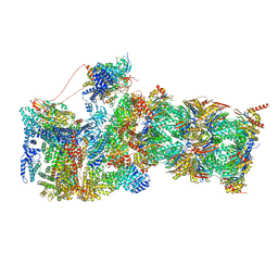
7W3B
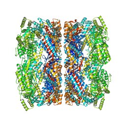
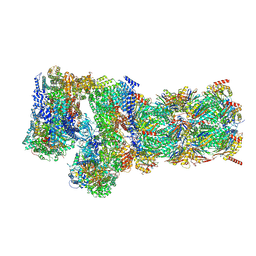 | | Structure of USP14-bound human 26S proteasome in substrate-engaged state ED5_USP14 | | 分子名称: | 26S protease regulatory subunit 4, 26S protease regulatory subunit 6A, 26S protease regulatory subunit 6B, ... | | 著者 | Zhang, S, Zou, S, Yin, D, Wu, Z, Mao, Y. | | 登録日 | 2021-11-25 | | 公開日 | 2022-05-04 | | 最終更新日 | 2022-06-01 | | 実験手法 | ELECTRON MICROSCOPY (3.6 Å) | | 主引用文献 | USP14-regulated allostery of the human proteasome by time-resolved cryo-EM.
Nature, 605, 2022
|
|
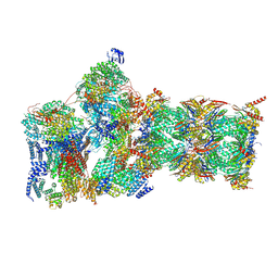
7W37
 | | Structure of USP14-bound human 26S proteasome in state EA1_UBL | | 分子名称: | 26S protease regulatory subunit 4, 26S protease regulatory subunit 6A, 26S protease regulatory subunit 6B, ... | | 著者 | Zhang, S, Zou, S, Yin, D, Wu, Z, Mao, Y. | | 登録日 | 2021-11-25 | | 公開日 | 2022-05-04 | | 最終更新日 | 2022-06-01 | | 実験手法 | ELECTRON MICROSCOPY (3 Å) | | 主引用文献 | USP14-regulated allostery of the human proteasome by time-resolved cryo-EM.
Nature, 605, 2022
|
|
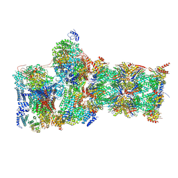
7W3G
 | | Structure of USP14-bound human 26S proteasome in substrate-engaged state ED2.0_USP14 | | 分子名称: | 26S protease regulatory subunit 4, 26S protease regulatory subunit 6A, 26S protease regulatory subunit 6B, ... | | 著者 | Zhang, S, Zou, S, Yin, D, Wu, Z, Mao, Y. | | 登録日 | 2021-11-25 | | 公開日 | 2022-05-04 | | 最終更新日 | 2022-06-01 | | 実験手法 | ELECTRON MICROSCOPY (3.2 Å) | | 主引用文献 | USP14-regulated allostery of the human proteasome by time-resolved cryo-EM.
Nature, 605, 2022
|
|
7W3K
 | | Structure of USP14-bound human 26S proteasome in substrate-inhibited state SD4_USP14 | | 分子名称: | 26S protease regulatory subunit 4, 26S protease regulatory subunit 6A, 26S protease regulatory subunit 6B, ... | | 著者 | Zhang, S, Zou, S, Yin, D, Wu, Z, Mao, Y. | | 登録日 | 2021-11-25 | | 公開日 | 2022-05-04 | | 最終更新日 | 2022-06-01 | | 実験手法 | ELECTRON MICROSCOPY (3.6 Å) | | 主引用文献 | USP14-regulated allostery of the human proteasome by time-resolved cryo-EM.
Nature, 605, 2022
|
|


