Loading
PDBj
メニューPDBj@FacebookPDBj@TwitterPDBj@YouTubewwPDB FoundationwwPDB
RCSB PDBPDBeBMRBAdv. SearchSearch help
Search by PDB author
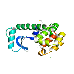
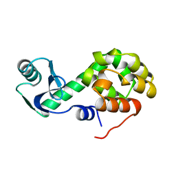
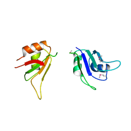
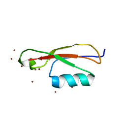
5E0Z
DownloadVisualize
BU of 5e0z by Molmil
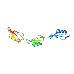
Crystal Structure of PASTA Domains 3 and 4 of Mycobacterium tuberculosis Protein Kinase B
分子名称: Serine/threonine-protein kinase PknB
著者Prigozhin, D.M, TB Structural Genomics Consortium (TBSGC)
登録日2015-09-29
公開日2016-09-14
最終更新日2024-03-06
実験手法X-RAY DIFFRACTION (2 Å)
主引用文献Structural and Genetic Analyses of the Mycobacterium tuberculosis Protein Kinase B Sensor Domain Identify a Potential Ligand-binding Site.
J.Biol.Chem., 291, 2016
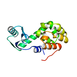
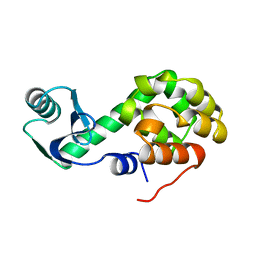
6CAH
DownloadVisualize
BU of 6cah by Molmil
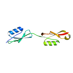
NMR-based structure of the FHA-2 domain from Mycobacterium tuberculosis ABC transporter Rv1747
分子名称: ABC transporter ATP-binding/permease protein Rv1747
著者Heinkel, F, Okon, M, Gsponer, J, McIntosh, L.P.
登録日2018-01-30
公開日2018-06-20
最終更新日2024-05-15
実験手法SOLUTION NMR
主引用文献Biophysical Characterization of the Tandem FHA Domain Regulatory Module from the Mycobacterium tuberculosis ABC Transporter Rv1747.
Structure, 26, 2018
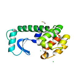
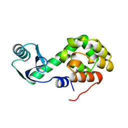
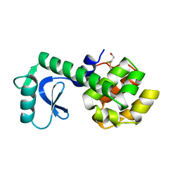
5E0Y
DownloadVisualize
BU of 5e0y by Molmil
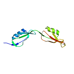
Crystal Structure of PASTA Domain 4 of Mycobacterium tuberculosis Protein Kinase B
分子名称: Serine/threonine-protein kinase PknB, ZINC ION
著者Prigozhin, D.M, TB Structural Genomics Consortium (TBSGC)
登録日2015-09-29
公開日2016-09-14
最終更新日2024-03-06
実験手法X-RAY DIFFRACTION (2.001 Å)
主引用文献Structural and Genetic Analyses of the Mycobacterium tuberculosis Protein Kinase B Sensor Domain Identify a Potential Ligand-binding Site.
J.Biol.Chem., 291, 2016
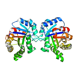
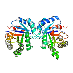
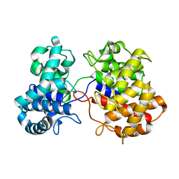
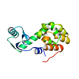
4P0M
DownloadVisualize
BU of 4p0m by Molmil
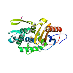
Crystal structure of an evolved putative penicillin-binding protein homolog, Rv2911, from Mycobacterium tuberculosis
分子名称: D-alanyl-D-alanine carboxypeptidase
著者Krieger, I, Yu, M, Bursey, E, Hung, L.-W, Terwilliger, T.C, TB Structural Genomics Consortium (TBSGC)
登録日2014-02-21
公開日2014-03-12
最終更新日2023-12-27
実験手法X-RAY DIFFRACTION (2 Å)
主引用文献Subfamily-Specific Adaptations in the Structures of Two Penicillin-Binding Proteins from Mycobacterium tuberculosis.
Plos One, 9, 2014
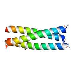
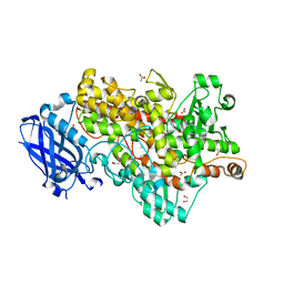
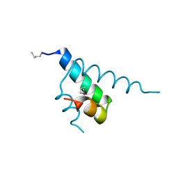
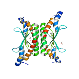
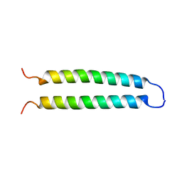
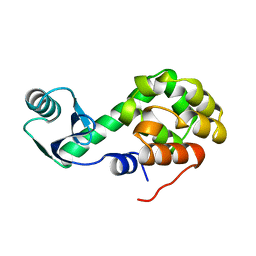
3HXA
DownloadVisualize
BU of 3hxa by Molmil
Crystal Structure of DCoH1Thr51Ser
分子名称: GLYCEROL, Pterin-4-alpha-carbinolamine dehydratase, SULFATE ION
著者Rho, H.Y, Jones, C.N, Rose, R.B.
登録日2009-06-20
公開日2010-08-25
最終更新日2024-02-21
実験手法X-RAY DIFFRACTION (1.8 Å)
主引用文献Crystal Structure of DCoH1Thr51Ser
To be Published
6DHO
DownloadVisualize
BU of 6dho by Molmil
RT XFEL structure of the two-flash state of Photosystem II (2F, S3-rich) at 2.07 Angstrom resolution
分子名称: 1,2-DI-O-ACYL-3-O-[6-DEOXY-6-SULFO-ALPHA-D-GLUCOPYRANOSYL]-SN-GLYCEROL, 1,2-DIPALMITOYL-PHOSPHATIDYL-GLYCEROLE, 1,2-DISTEAROYL-MONOGALACTOSYL-DIGLYCERIDE, ...
著者Kern, J, Chatterjee, R, Young, I.D, Fuller, F.D, Lassalle, L, Ibrahim, M, Gul, S, Fransson, T, Brewster, A.S, Alonso-Mori, R, Hussein, R, Zhang, M, Douthit, L, de Lichtenberg, C, Cheah, M.H, Shevela, D, Wersig, J, Seufert, I, Sokaras, D, Pastor, E, Weninger, C, Kroll, T, Sierra, R.G, Aller, P, Butryn, A, Orville, A.M, Liang, M, Batyuk, A, Koglin, J.E, Carbajo, S, Boutet, S, Moriarty, N.W, Holton, J.M, Dobbek, H, Adams, P.D, Bergmann, U, Sauter, N.K, Zouni, A, Messinger, J, Yano, J, Yachandra, V.K.
登録日2018-05-20
公開日2018-11-21
最終更新日2024-03-13
実験手法X-RAY DIFFRACTION (2.07 Å)
主引用文献Structures of the intermediates of Kok's photosynthetic water oxidation clock.
Nature, 563, 2018
6DHG
DownloadVisualize
BU of 6dhg by Molmil
RT XFEL structure of Photosystem II 150 microseconds after the second illumination at 2.5 Angstrom resolution
分子名称: 1,2-DI-O-ACYL-3-O-[6-DEOXY-6-SULFO-ALPHA-D-GLUCOPYRANOSYL]-SN-GLYCEROL, 1,2-DIPALMITOYL-PHOSPHATIDYL-GLYCEROLE, 1,2-DISTEAROYL-MONOGALACTOSYL-DIGLYCERIDE, ...
著者Kern, J, Chatterjee, R, Young, I.D, Fuller, F.D, Lassalle, L, Ibrahim, M, Gul, S, Fransson, T, Brewster, A.S, Alonso-Mori, R, Hussein, R, Zhang, M, Douthit, L, de Lichtenberg, C, Cheah, M.H, Shevela, D, Wersig, J, Seufert, I, Sokaras, D, Pastor, E, Weninger, C, Kroll, T, Sierra, R.G, Aller, P, Butryn, A, Orville, A.M, Liang, M, Batyuk, A, Koglin, J.E, Carbajo, S, Boutet, S, Moriarty, N.W, Holton, J.M, Dobbek, H, Adams, P.D, Bergmann, U, Sauter, N.K, Zouni, A, Messinger, J, Yano, J, Yachandra, V.K.
登録日2018-05-20
公開日2018-11-21
最終更新日2024-03-13
実験手法X-RAY DIFFRACTION (2.5 Å)
主引用文献Structures of the intermediates of Kok's photosynthetic water oxidation clock.
Nature, 563, 2018
2KLN
DownloadVisualize
BU of 2kln by Molmil
Solution Structure of STAS domain of RV1739c from M. tuberculosis
分子名称: PROBABLE SULPHATE-TRANSPORT TRANSMEMBRANE PROTEIN, COG0659
著者Sharma, A.K, Ye, L, Zolotarev, A.S, Alper, S.L, Rigby, A.C.
登録日2009-07-06
公開日2010-12-15
最終更新日2024-05-01
実験手法SOLUTION NMR
主引用文献Solution Structure of the Guanine Nucleotide-binding STAS Domain of SLC26-related SulP Protein Rv1739c from Mycobacterium tuberculosis.
J.Biol.Chem., 286, 2011
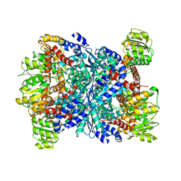
3SBO
DownloadVisualize
BU of 3sbo by Molmil
Structure of E.coli GDH from native source
分子名称: CHLORIDE ION, NADP-specific glutamate dehydrogenase
著者Gee, C.L, Zubieta, C, Echols, N, Totir, M.
登録日2011-06-06
公開日2012-03-21
最終更新日2024-02-28
実験手法X-RAY DIFFRACTION (3.204 Å)
主引用文献Macro-to-Micro Structural Proteomics: Native Source Proteins for High-Throughput Crystallization.
Plos One, 7, 2012
3SR0
DownloadVisualize
BU of 3sr0 by Molmil
Crystal Structure of the Phosphoryl Transfer Transition State Mimic in the Adenylate Kinase: ADP/AlF4/AMP in the active site
分子名称: ADENOSINE MONOPHOSPHATE, ADENOSINE-5'-DIPHOSPHATE, Adenylate kinase, ...
著者Cho, Y.-J, Kern, D.
登録日2011-07-06
公開日2012-10-10
最終更新日2024-02-28
実験手法X-RAY DIFFRACTION (1.565 Å)
主引用文献The energy landscape of adenylate kinase during catalysis.
Nat.Struct.Mol.Biol., 22, 2015
4CF7
DownloadVisualize
BU of 4cf7 by Molmil
Crystal structure of adenylate kinase from Aquifex aeolicus with MgADP bound
分子名称: ADENOSINE MONOPHOSPHATE, ADENOSINE-5'-DIPHOSPHATE, ADENYLATE KINASE, ...
著者Kerns, S.J, Agafonov, R.V, Cho, Y.-J, Pontiggia, F, Otten, R, Pachov, D.V, Kutter, S, Phung, L.A, Murphy, P.N, Thai, V, Hagan, M.F, Kern, D.
登録日2013-11-13
公開日2014-12-03
最終更新日2023-12-20
実験手法X-RAY DIFFRACTION (1.594 Å)
主引用文献The Energy Landscape of Adenylate Kinase During Catalysis.
Nat.Struct.Mol.Biol., 22, 2015
4JLP
DownloadVisualize
BU of 4jlp by Molmil
Crystal structure of R150K aquifex adenylate kinase mutant
分子名称: ADENOSINE-5'-DIPHOSPHATE, Adenylate kinase
著者Cho, Y.-J, Phung, L, Kern, D.
登録日2013-03-12
公開日2014-06-25
最終更新日2024-02-28
実験手法X-RAY DIFFRACTION (1.433 Å)
主引用文献The energy landscape of adenylate kinase during catalysis.
Nat.Struct.Mol.Biol., 22, 2015
4JLO
DownloadVisualize
BU of 4jlo by Molmil
Crystal structure of Aquifex adenylate kinase R150K mutant
分子名称: ADENOSINE-5'-DIPHOSPHATE, Adenylate kinase
著者Cho, Y.-J, Phung, L, Kern, D.
登録日2013-03-12
公開日2014-06-25
最終更新日2024-02-28
実験手法X-RAY DIFFRACTION (1.73 Å)
主引用文献The energy landscape of adenylate kinase during catalysis.
Nat.Struct.Mol.Biol., 22, 2015
4JL6
DownloadVisualize
BU of 4jl6 by Molmil
Crystal Structure of Adenylate kinase with 2 ADP's
分子名称: ADENOSINE-5'-DIPHOSPHATE, Adenylate kinase
著者Cho, Y.-J, Kern, D.
登録日2013-03-12
公開日2014-06-25
最終更新日2024-02-28
実験手法X-RAY DIFFRACTION (1.65 Å)
主引用文献The energy landscape of adenylate kinase during catalysis.
Nat.Struct.Mol.Biol., 22, 2015
4JL5
DownloadVisualize
BU of 4jl5 by Molmil
A high resolution structure of Aquifex Adenylate kinase with 2 ADP's
分子名称: ADENOSINE-5'-DIPHOSPHATE, Adenylate kinase
著者Cho, Y.-J, Kern, D.
登録日2013-03-12
公開日2014-06-25
最終更新日2024-02-28
実験手法X-RAY DIFFRACTION (1.24 Å)
主引用文献The energy landscape of adenylate kinase during catalysis.
Nat.Struct.Mol.Biol., 22, 2015
5TR0
DownloadVisualize
BU of 5tr0 by Molmil
Lipoxygenase-1 (soybean) L754A mutant at 293K
分子名称: FE (II) ION, Seed linoleate 13S-lipoxygenase-1
著者Poss, E.M, Fraser, J.S.
登録日2016-10-24
公開日2017-11-01
最終更新日2023-10-04
実験手法X-RAY DIFFRACTION (1.85 Å)
主引用文献Biophysical Characterization of a Disabled Double Mutant of Soybean Lipoxygenase: The "Undoing" of Precise Substrate Positioning Relative to Metal Cofactor and an Identified Dynamical Network.
J.Am.Chem.Soc., 141, 2019
5TQP
DownloadVisualize
BU of 5tqp by Molmil
LIPOXYGENASE-1 (SOYBEAN) I553G MUTANT AT 300K
分子名称: FE (III) ION, Seed linoleate 13S-lipoxygenase-1
著者Poss, E.M, Fraser, J.S.
登録日2016-10-24
公開日2017-11-01
最終更新日2023-10-04
実験手法X-RAY DIFFRACTION (1.7 Å)
主引用文献Hydrogen-Deuterium Exchange of Lipoxygenase Uncovers a Relationship between Distal, Solvent Exposed Protein Motions and the Thermal Activation Barrier for Catalytic Proton-Coupled Electron Tunneling.
ACS Cent Sci, 3, 2017
5TQO
DownloadVisualize
BU of 5tqo by Molmil
Lipoxygenase-1 (soybean) L546A/L754A mutant at 300K
分子名称: FE (III) ION, Seed linoleate 13S-lipoxygenase-1
著者Poss, E.M, Fraser, J.S, Gee, C.
登録日2016-10-24
公開日2017-11-01
最終更新日2023-10-04
実験手法X-RAY DIFFRACTION (1.7 Å)
主引用文献Biophysical Characterization of a Disabled Double Mutant of Soybean Lipoxygenase: The "Undoing" of Precise Substrate Positioning Relative to Metal Cofactor and an Identified Dynamical Network.
J.Am.Chem.Soc., 141, 2019
5TQN
DownloadVisualize
BU of 5tqn by Molmil
Lipoxygenase-1 (soybean) L546A mutant at 293K
分子名称: FE (II) ION, Seed linoleate 13S-lipoxygenase-1
著者Poss, E.M, Fraser, J.S, Gee, C.
登録日2016-10-24
公開日2017-11-01
最終更新日2023-10-04
実験手法X-RAY DIFFRACTION (1.8 Å)
主引用文献Biophysical Characterization of a Disabled Double Mutant of Soybean Lipoxygenase: The "Undoing" of Precise Substrate Positioning Relative to Metal Cofactor and an Identified Dynamical Network.
J.Am.Chem.Soc., 141, 2019
5T5V
DownloadVisualize
BU of 5t5v by Molmil
LIPOXYGENASE-1 (SOYBEAN) AT 293K
分子名称: FE (III) ION, Seed linoleate 13S-lipoxygenase-1
著者Poss, E.M, Fraser, J.S.
登録日2016-08-31
公開日2017-09-06
最終更新日2023-10-04
実験手法X-RAY DIFFRACTION (1.8 Å)
主引用文献Hydrogen-Deuterium Exchange of Lipoxygenase Uncovers a Relationship between Distal, Solvent Exposed Protein Motions and the Thermal Activation Barrier for Catalytic Proton-Coupled Electron Tunneling.
ACS Cent Sci, 3, 2017
4JL8
DownloadVisualize
BU of 4jl8 by Molmil
A various kinds of ADP conformations in the Adenylate kinase active site
分子名称: ADENOSINE-5'-DIPHOSPHATE, Adenylate kinase
著者Cho, Y.-J, Kern, D.
登録日2013-03-12
公開日2014-06-25
最終更新日2024-02-28
実験手法X-RAY DIFFRACTION (1.79 Å)
主引用文献The energy landscape of adenylate kinase during catalysis.
Nat.Struct.Mol.Biol., 22, 2015
4JLD
DownloadVisualize
BU of 4jld by Molmil
Crystal Structures of adenylate kinase with 2 ADP's
分子名称: ADENOSINE-5'-DIPHOSPHATE, Adenylate kinase
著者Cho, Y.-J, Kern, D.
登録日2013-03-12
公開日2014-06-25
最終更新日2024-02-28
実験手法X-RAY DIFFRACTION (1.552 Å)
主引用文献The energy landscape of adenylate kinase during catalysis.
Nat.Struct.Mol.Biol., 22, 2015
4JLA
DownloadVisualize
BU of 4jla by Molmil
Crystal Structure of Adenylate kinase with 2 ADP's in the active site
分子名称: ADENOSINE-5'-DIPHOSPHATE, Adenylate kinase
著者Cho, Y.-J, Kern, D.
登録日2013-03-12
公開日2014-06-25
最終更新日2024-02-28
実験手法X-RAY DIFFRACTION (2.12 Å)
主引用文献The energy landscape of adenylate kinase during catalysis.
Nat.Struct.Mol.Biol., 22, 2015
4JKY
DownloadVisualize
BU of 4jky by Molmil
Cobalt between two ADP's in the active site of adenylate kinase
分子名称: ADENOSINE-5'-DIPHOSPHATE, Adenylate kinase, COBALT (II) ION
著者Cho, Y.-J, Kern, D.
登録日2013-03-12
公開日2014-06-25
最終更新日2024-02-28
実験手法X-RAY DIFFRACTION (2.373 Å)
主引用文献The energy landscape of adenylate kinase during catalysis.
Nat.Struct.Mol.Biol., 22, 2015
4JLB
DownloadVisualize
BU of 4jlb by Molmil
Crystal Structures of Adenylate kinase with 2ADP's
分子名称: ADENOSINE-5'-DIPHOSPHATE, Adenylate kinase
著者Cho, Y.-J, Kern, D.
登録日2013-03-12
公開日2014-06-25
最終更新日2024-02-28
実験手法X-RAY DIFFRACTION (1.528 Å)
主引用文献The energy landscape of adenylate kinase during catalysis.
Nat.Struct.Mol.Biol., 22, 2015

222415

件を2024-07-10に公開中

PDB statisticsPDBj update infoContact PDBjnumon