7C4C
 
 | |
7C4B
 | |
7C45
 | |
7C47
 | |
7C42
 | |
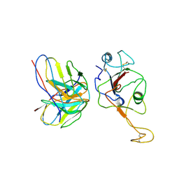
7F7G
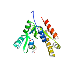 | | a linear Peptide Inhibitors in complex with GK domain | | 分子名称: | DLG4 GK domain, UNK-ARG-ILE-ARG-ARG-ASP-GLU-TYR-LEU-LYS-ALA-ILE-GLN-UNK | | 著者 | Shang, Y, Huang, X, Li, X, Zhang, M. | | 登録日 | 2021-06-29 | | 公開日 | 2022-02-23 | | 最終更新日 | 2023-11-29 | | 実験手法 | X-RAY DIFFRACTION (2.446 Å) | | 主引用文献 | Entropy of stapled peptide inhibitors in free state is the major contributor to the improvement of binding affinity with the GK domain.
Rsc Chem Biol, 2, 2021
|
|
7E9Q
 | |
7E9O
 | |
7E9P
 | |
7E9N
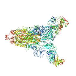 | |
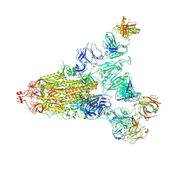
7ENF
 | | Cryo-EM structure of the SARS-CoV-2 S-6P in complex with Fab30 | | 分子名称: | 2-acetamido-2-deoxy-beta-D-glucopyranose, 2-acetamido-2-deoxy-beta-D-glucopyranose-(1-4)-2-acetamido-2-deoxy-beta-D-glucopyranose, Heavy chain of Fab30, ... | | 著者 | Wang, X.F, Zhu, Y.Q. | | 登録日 | 2021-04-16 | | 公開日 | 2022-04-06 | | 最終更新日 | 2022-09-21 | | 実験手法 | ELECTRON MICROSCOPY (2.76 Å) | | 主引用文献 | A potent human monoclonal antibody with pan-neutralizing activities directly dislocates S trimer of SARS-CoV-2 through binding both up and down forms of RBD
Signal Transduct Target Ther, 7, 2022
|
|
7ENG
 | |
5YKS
 | |
5WHZ
 | | PGDM1400-10E8v4 CODV Fab | | 分子名称: | Anti-HIV CODV-Fab Heavy chain, Anti-HIV CODV-Fab Light chain | | 著者 | Lord, D.M, Wei, R.R. | | 登録日 | 2017-07-18 | | 公開日 | 2017-10-11 | | 最終更新日 | 2023-10-04 | | 実験手法 | X-RAY DIFFRACTION (3.549 Å) | | 主引用文献 | Trispecific broadly neutralizing HIV antibodies mediate potent SHIV protection in macaques.
Science, 358, 2017
|
|
5XMC
 | |
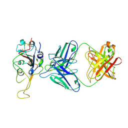
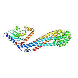
5WUJ
 | | Crystal structure of FliF-FliG complex from H. pylori | | 分子名称: | Flagellar M-ring protein, Flagellar motor switch protein FliG, GLYCEROL | | 著者 | Au, S.W, Xue, C, Lam, K.H, Lee, S.H. | | 登録日 | 2016-12-19 | | 公開日 | 2017-12-20 | | 最終更新日 | 2024-03-20 | | 実験手法 | X-RAY DIFFRACTION (2.3 Å) | | 主引用文献 | Crystal structure of the FliF-FliG complex from Helicobacter pylori yields insight into the assembly of the motor MS-C ring in the bacterial flagellum
J. Biol. Chem., 293, 2018
|
|
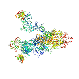
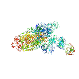
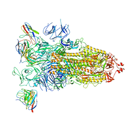
7F46
 | | Cryo-EM structure of the SARS-CoV-2 S-6P in complex with 35B5 Fab (state1, local refinement of the RBD, NTD and 35B5 Fab) | | 分子名称: | 2-acetamido-2-deoxy-beta-D-glucopyranose, Heavy chain of 35B5 Fab, Light chain of 35B5 Fab, ... | | 著者 | Wang, X.F, Zhu, Y.Q. | | 登録日 | 2021-06-17 | | 公開日 | 2022-03-23 | | 最終更新日 | 2022-09-21 | | 実験手法 | ELECTRON MICROSCOPY (4.79 Å) | | 主引用文献 | A potent human monoclonal antibody with pan-neutralizing activities directly dislocates S trimer of SARS-CoV-2 through binding both up and down forms of RBD
Signal Transduct Target Ther, 7, 2022
|
|
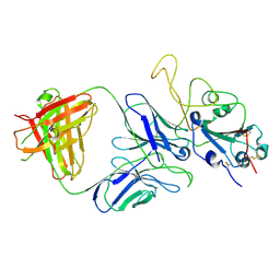
7CKG
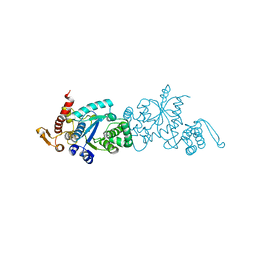 | | Crystal structure of TMSiPheRS complexed with TMSiPhe | | 分子名称: | 4-(trimethylsilyl)-L-phenylalanine, Tyrosine--tRNA ligase | | 著者 | Sun, J.P, Wang, J.Y, Zhu, Z.L, He, Q.T, Xiao, P. | | 登録日 | 2020-07-17 | | 公開日 | 2021-03-31 | | 最終更新日 | 2023-11-29 | | 実験手法 | X-RAY DIFFRACTION (2.053 Å) | | 主引用文献 | DeSiphering receptor core-induced and ligand-dependent conformational changes in arrestin via genetic encoded trimethylsilyl 1 H-NMR probe.
Nat Commun, 11, 2020
|
|
7CKH
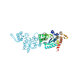 | | Crystal structure of TMSiPheRS | | 分子名称: | Tyrosine--tRNA ligase | | 著者 | Sun, J.P, Wang, J.Y, Zhu, Z.L, He, Q.T, Xiao, P. | | 登録日 | 2020-07-17 | | 公開日 | 2021-03-31 | | 最終更新日 | 2023-11-29 | | 実験手法 | X-RAY DIFFRACTION (1.79492676 Å) | | 主引用文献 | DeSiphering receptor core-induced and ligand-dependent conformational changes in arrestin via genetic encoded trimethylsilyl 1 H-NMR probe.
Nat Commun, 11, 2020
|
|
5WDF
 | |
5Z33
 | |
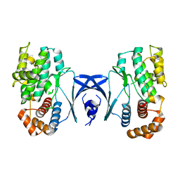
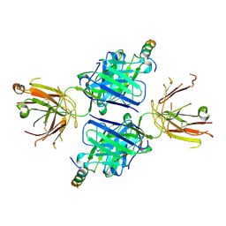
7V97
 | | Arsenic-bound p53 DNA-binding domain mutant V272M | | 分子名称: | ARSENIC, Cellular tumor antigen p53, ZINC ION | | 著者 | Lu, M, Xing, Y.F, Wang, Z.Y, Ni, Y, Song, H.X. | | 登録日 | 2021-08-24 | | 公開日 | 2022-08-31 | | 最終更新日 | 2023-11-29 | | 実験手法 | X-RAY DIFFRACTION (2.02 Å) | | 主引用文献 | Diverse rescue potencies of p53 mutations to ATO are predetermined by intrinsic mutational properties.
Sci Transl Med, 15, 2023
|
|
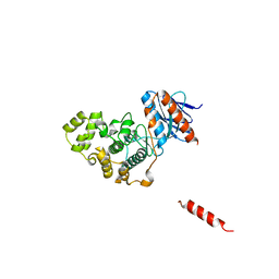
7WB5
 | | local structure of hu33 and spike | | 分子名称: | Surface glycoprotein, beta-D-mannopyranose-(1-4)-2-acetamido-2-deoxy-beta-D-glucopyranose-(1-4)-2-acetamido-2-deoxy-beta-D-glucopyranose, hu33 heavy chain, ... | | 著者 | Pulan, L. | | 登録日 | 2021-12-15 | | 公開日 | 2022-10-26 | | 実験手法 | ELECTRON MICROSCOPY (3.7 Å) | | 主引用文献 | A non-ACE2-blocking neutralizing antibody against Omicron-included SARS-CoV-2 variants.
Signal Transduct Target Ther, 7, 2022
|
|
7WBH
 | | overall structure of hu33 and spike | | 分子名称: | 2-acetamido-2-deoxy-beta-D-glucopyranose, 2-acetamido-2-deoxy-beta-D-glucopyranose-(1-4)-2-acetamido-2-deoxy-beta-D-glucopyranose, Spike glycoprotein, ... | | 著者 | Pulan, L. | | 登録日 | 2021-12-16 | | 公開日 | 2022-10-26 | | 実験手法 | ELECTRON MICROSCOPY (3.7 Å) | | 主引用文献 | A non-ACE2-blocking neutralizing antibody against Omicron-included SARS-CoV-2 variants.
Signal Transduct Target Ther, 7, 2022
|
|
7W75
 | |


