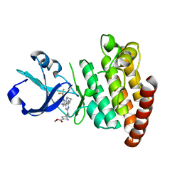
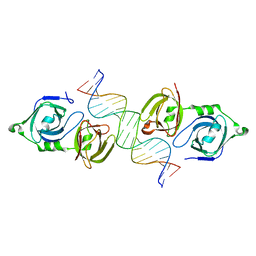
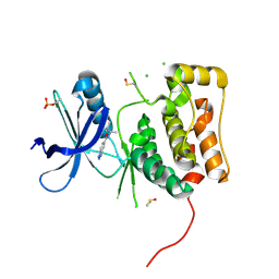
8H5E
 
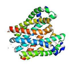 | | Crystal structure of the MgtE TM domain in complex with Ca2+ ions at 2.5 angstrom resolution | | 分子名称: | (2R)-2,3-dihydroxypropyl (9Z)-octadec-9-enoate, CALCIUM ION, Magnesium transporter MgtE | | 著者 | Teng, X, Sheng, D, Hattori, M. | | 登録日 | 2022-10-13 | | 公開日 | 2022-12-07 | | 最終更新日 | 2023-11-29 | | 実験手法 | X-RAY DIFFRACTION (2.5 Å) | | 主引用文献 | Ion selectivity mechanism of the MgtE channel for Mg 2+ over Ca 2 .
Iscience, 25, 2022
|
|
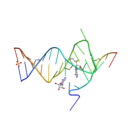
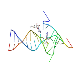
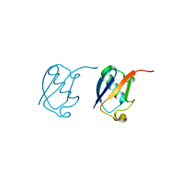
6YMJ
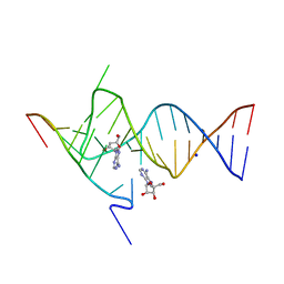 | | Crystal structure of the SAM-SAH riboswitch with adenosine. | | 分子名称: | 5-BROMOCYTIDINE 5'-(DIHYDROGEN PHOSPHATE), ADENOSINE, Chains: A,C,F,I,M,O, ... | | 著者 | Huang, L, Lilley, D.M.J. | | 登録日 | 2020-04-08 | | 公開日 | 2020-07-22 | | 最終更新日 | 2024-02-07 | | 実験手法 | X-RAY DIFFRACTION (2.04 Å) | | 主引用文献 | Crystal structure and ligand-induced folding of the SAM/SAH riboswitch.
Nucleic Acids Res., 48, 2020
|
|
6YFZ
 | |
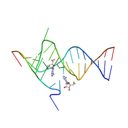
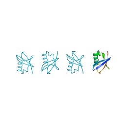
6YLB
 | | Crystal structure of the SAM-SAH riboswitch with SAM | | 分子名称: | Chains: A,C,F,I,M,O, Chains: B,D,G,J,N,P, S-ADENOSYLMETHIONINE | | 著者 | Huang, L, Lilley, D.M.J. | | 登録日 | 2020-04-07 | | 公開日 | 2020-07-22 | | 最終更新日 | 2024-01-24 | | 実験手法 | X-RAY DIFFRACTION (2.12 Å) | | 主引用文献 | Crystal structure and ligand-induced folding of the SAM/SAH riboswitch.
Nucleic Acids Res., 2020
|
|
6YML
 | |
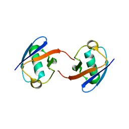
6YMK
 | | Crystal structure of the SAM-SAH riboswitch with AMP | | 分子名称: | 5'-DEOXY-5'-METHYLTHIOADENOSINE, Chains: A,C,F,I,M,O, Chains: B,D,G,J,N,P, ... | | 著者 | Huang, L, Lilley, D.M.J. | | 登録日 | 2020-04-08 | | 公開日 | 2020-07-22 | | 最終更新日 | 2024-01-24 | | 実験手法 | X-RAY DIFFRACTION (2.03 Å) | | 主引用文献 | Crystal structure and ligand-induced folding of the SAM/SAH riboswitch.
Nucleic Acids Res., 48, 2020
|
|
6YG4
 | |
6YG2
 | | Crystal structure of MKK7 (MAP2K7) in complex with ibrutnib, with covalent and allosteric binding modes | | 分子名称: | 1,2-ETHANEDIOL, 1-[(3~{R})-3-[4-azanyl-3-(4-phenoxyphenyl)pyrazolo[3,4-d]pyrimidin-1-yl]piperidin-1-yl]propan-1-one, 1-{(3R)-3-[4-amino-3-(4-phenoxyphenyl)-1H-pyrazolo[3,4-d]pyrimidin-1-yl]piperidin-1-yl}prop-2-en-1-one, ... | | 著者 | Chaikuad, A, Knapp, S, Structural Genomics Consortium (SGC) | | 登録日 | 2020-03-27 | | 公開日 | 2020-08-12 | | 最終更新日 | 2024-01-24 | | 実験手法 | X-RAY DIFFRACTION (2 Å) | | 主引用文献 | Catalytic Domain Plasticity of MKK7 Reveals Structural Mechanisms of Allosteric Activation and Diverse Targeting Opportunities.
Cell Chem Biol, 27, 2020
|
|
6YMM
 | |
6YG0
 | |
6YG5
 | |
6YZ4
 | | Crystal structure of MKK7 (MAP2K7) with ibrutinib bound at allosteric site | | 分子名称: | 1,2-ETHANEDIOL, 1-{(3R)-3-[4-amino-3-(4-phenoxyphenyl)-1H-pyrazolo[3,4-d]pyrimidin-1-yl]piperidin-1-yl}prop-2-en-1-one, DIMETHYL SULFOXIDE, ... | | 著者 | Chaikuad, A, Knapp, S, Structural Genomics Consortium (SGC) | | 登録日 | 2020-05-06 | | 公開日 | 2020-08-12 | | 最終更新日 | 2024-01-24 | | 実験手法 | X-RAY DIFFRACTION (1.7 Å) | | 主引用文献 | Catalytic Domain Plasticity of MKK7 Reveals Structural Mechanisms of Allosteric Activation and Diverse Targeting Opportunities.
Cell Chem Biol, 27, 2020
|
|
8DEG
 | | Crystal structure of DLK in complex with inhibitor DN0011197 | | 分子名称: | Mitogen-activated protein kinase kinase kinase 12, methyl (1S,4S)-5-{(4P)-4-[5-amino-6-(difluoromethoxy)pyrazin-2-yl]-6-[(1R,4R)-2-azabicyclo[2.1.1]hexan-2-yl]pyridin-2-yl}-2,5-diazabicyclo[2.2.1]heptane-2-carboxylate | | 著者 | Srivastava, A, Lexa, K, de Vicente, J. | | 登録日 | 2022-06-20 | | 公開日 | 2022-12-14 | | 最終更新日 | 2024-04-03 | | 実験手法 | X-RAY DIFFRACTION (2.79 Å) | | 主引用文献 | Discovery of Potent and Selective Dual Leucine Zipper Kinase/Leucine Zipper-Bearing Kinase Inhibitors with Neuroprotective Properties in In Vitro and In Vivo Models of Amyotrophic Lateral Sclerosis.
J.Med.Chem., 65, 2022
|
|
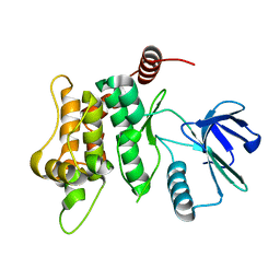
5HYX
 | | Plant peptide hormone receptor RGFR1 in complex with RGF1 | | 分子名称: | 2-acetamido-2-deoxy-beta-D-glucopyranose, 2-acetamido-2-deoxy-beta-D-glucopyranose-(1-4)-2-acetamido-2-deoxy-beta-D-glucopyranose, PTR-SER-ASN-PRO-GLY-HIS-HIS-PRO-HYP-ARG-HIS-ASN, ... | | 著者 | Song, W, Han, Z, Chai, J. | | 登録日 | 2016-02-02 | | 公開日 | 2017-01-11 | | 最終更新日 | 2020-07-29 | | 実験手法 | X-RAY DIFFRACTION (2.596 Å) | | 主引用文献 | Signature motif-guided identification of receptors for peptide hormones essential for root meristem growth
Cell Res., 26, 2016
|
|
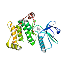
8SQF
 | | OXA-48 bound to inhibitor CDD-2725 | | 分子名称: | (1M)-3'-(benzyloxy)-5-hydroxy[1,1'-biphenyl]-3,4'-dicarboxylic acid, BICARBONATE ION, Beta-lactamase | | 著者 | Park, S, Judge, A, Fan, J, Sankaran, B, Prasad, B.V.V, Palzkill, T. | | 登録日 | 2023-05-04 | | 公開日 | 2024-01-03 | | 最終更新日 | 2024-01-17 | | 実験手法 | X-RAY DIFFRACTION (2.3 Å) | | 主引用文献 | Exploiting the Carboxylate-Binding Pocket of beta-Lactamase Enzymes Using a Focused DNA-Encoded Chemical Library.
J.Med.Chem., 67, 2024
|
|
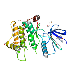
8SQG
 | | OXA-48 bound to inhibitor CDD-2801 | | 分子名称: | (1M)-3'-(benzyloxy)-5-[2-(methylamino)-2-oxoethoxy][1,1'-biphenyl]-3,4'-dicarboxylic acid, BICARBONATE ION, Beta-lactamase | | 著者 | Park, S, Judge, A, Fan, J, Sankaran, B, Palzkill, T. | | 登録日 | 2023-05-04 | | 公開日 | 2024-01-03 | | 最終更新日 | 2024-01-17 | | 実験手法 | X-RAY DIFFRACTION (2.03 Å) | | 主引用文献 | Exploiting the Carboxylate-Binding Pocket of beta-Lactamase Enzymes Using a Focused DNA-Encoded Chemical Library.
J.Med.Chem., 67, 2024
|
|
4LNQ
 | |
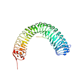
4HZU
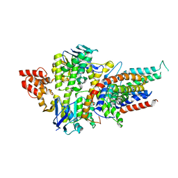 | | Structure of a bacterial energy-coupling factor transporter | | 分子名称: | Energy-coupling factor transporter ATP-binding protein EcfA 1, Energy-coupling factor transporter ATP-binding protein EcfA 2, Energy-coupling factor transporter transmembrane protein EcfT, ... | | 著者 | Wang, T.L, Fu, G.B, Pan, X.J, Shi, Y.G. | | 登録日 | 2012-11-15 | | 公開日 | 2013-04-17 | | 最終更新日 | 2024-02-28 | | 実験手法 | X-RAY DIFFRACTION (3.53 Å) | | 主引用文献 | Structure of a bacterial energy-coupling factor transporter.
Nature, 497, 2013
|
|
5GOG
 | | Lys29-linked di-ubiquitin | | 分子名称: | D-ubiquitin, Ubiquitin | | 著者 | Gao, S, Pan, M, Zheng, Y. | | 登録日 | 2016-07-27 | | 公開日 | 2016-11-02 | | 最終更新日 | 2023-11-15 | | 実験手法 | X-RAY DIFFRACTION (1.977 Å) | | 主引用文献 | Monomer/Oligomer Quasi-Racemic Protein Crystallography
J.Am.Chem.Soc., 138, 2016
|
|
5GOK
 | | K11/K63-branched tri-Ubiquitin | | 分子名称: | D-ubiquitin, Ubiquitin | | 著者 | Gao, S, Pan, M, Zheng, Y, Liu, L. | | 登録日 | 2016-07-27 | | 公開日 | 2016-11-02 | | 最終更新日 | 2023-11-15 | | 実験手法 | X-RAY DIFFRACTION (1.84 Å) | | 主引用文献 | Monomer/Oligomer Quasi-Racemic Protein Crystallography
J.Am.Chem.Soc., 138, 2016
|
|
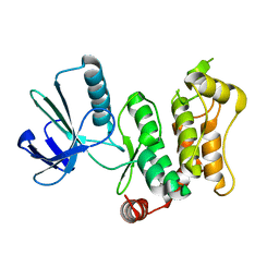
6MNH
 | | ULK1 Unc-51 like autophagy activating kinase in complex with inhibitor BTC | | 分子名称: | CHLORIDE ION, DIMETHYL SULFOXIDE, N-[(2R)-3-methylbutan-2-yl]-1H-benzotriazole-6-carboxamide, ... | | 著者 | Hendle, J, Sauder, J.M, Hickey, M.J, Rauch, C.T, Maletic, M, Schwinn, K.D. | | 登録日 | 2018-10-01 | | 公開日 | 2019-03-27 | | 最終更新日 | 2019-11-13 | | 実験手法 | X-RAY DIFFRACTION (1.73 Å) | | 主引用文献 | Idea2Data: Toward a New Paradigm for Drug Discovery.
Acs Med.Chem.Lett., 10, 2019
|
|
5GOH
 | | Lys33-linked di-ubiquitin | | 分子名称: | D-ubiquitin, Ubiquitin | | 著者 | Gao, S, Pan, M, Zheng, Y, Liu, L. | | 登録日 | 2016-07-27 | | 公開日 | 2016-11-02 | | 最終更新日 | 2023-11-15 | | 実験手法 | X-RAY DIFFRACTION (1.946 Å) | | 主引用文献 | Monomer/Oligomer Quasi-Racemic Protein Crystallography
J.Am.Chem.Soc., 138, 2016
|
|
5GO8
 | | Linear tetra-ubiquitin | | 分子名称: | D-ubiquitin, ubiquitin | | 著者 | Gao, S, Pan, M, Zheng, Y. | | 登録日 | 2016-07-26 | | 公開日 | 2016-11-02 | | 最終更新日 | 2023-11-15 | | 実験手法 | X-RAY DIFFRACTION (2.21 Å) | | 主引用文献 | Monomer/Oligomer Quasi-Racemic Protein Crystallography
J.Am.Chem.Soc., 138, 2016
|
|
5GOD
 | | Lys27-linked di-ubiquitin | | 分子名称: | D-ubiquitin, MAGNESIUM ION, Ubiquitin | | 著者 | Gao, S, Pan, M, Zheng, Y. | | 登録日 | 2016-07-26 | | 公開日 | 2016-11-02 | | 最終更新日 | 2023-11-15 | | 実験手法 | X-RAY DIFFRACTION (1.15 Å) | | 主引用文献 | Monomer/Oligomer Quasi-Racemic Protein Crystallography
J.Am.Chem.Soc., 138, 2016
|
|
5GOJ
 | | Lys63-linked di-ubiquitin | | 分子名称: | D-ubiquitin, Ubiquitin | | 著者 | Gao, S, Pan, M, Zheng, Y, Liu, L. | | 登録日 | 2016-07-27 | | 公開日 | 2016-11-02 | | 最終更新日 | 2023-11-15 | | 実験手法 | X-RAY DIFFRACTION (1.549 Å) | | 主引用文献 | Monomer/Oligomer Quasi-Racemic Protein Crystallography
J.Am.Chem.Soc., 138, 2016
|
|


