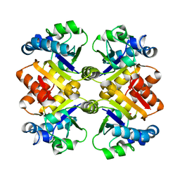
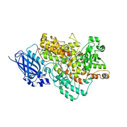
3EXT
 
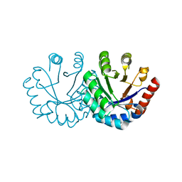 | | Crystal structure of KGPDC from Streptococcus mutans | | 分子名称: | MAGNESIUM ION, RmpD (Hexulose-6-phosphate synthase) | | 著者 | Liu, X, Li, G.L, Li, L.F, Su, X.D. | | 登録日 | 2008-10-17 | | 公開日 | 2009-08-25 | | 最終更新日 | 2023-11-01 | | 実験手法 | X-RAY DIFFRACTION (2 Å) | | 主引用文献 | Open-closed conformational change revealed by the crystal structures of 3-keto-L-gulonate 6-phosphate decarboxylase from Streptococcus mutans
Biochem.Biophys.Res.Commun., 381, 2009
|
|
5HP8
 | |
4KFW
 | |
4KFV
 | |
3S2S
 | |
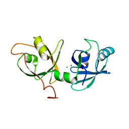
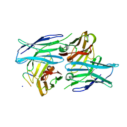
3S70
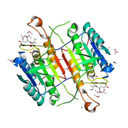 | | Crystal structure of active caspase-6 bound with Ac-VEID-CHO solved by As-SAD | | 分子名称: | ACETATE ION, CACODYLATE ION, Caspase-6, ... | | 著者 | Su, X.-D, Liu, X, Wang, X.-J. | | 登録日 | 2011-05-26 | | 公開日 | 2012-04-11 | | 最終更新日 | 2012-12-12 | | 実験手法 | X-RAY DIFFRACTION (1.625 Å) | | 主引用文献 | Get phases from arsenic anomalous scattering: de novo SAD phasing of two protein structures crystallized in cacodylate buffer
Plos One, 6, 2011
|
|
4NXE
 | | Crystal structure of iLOV-I486(2LT) at pH 6.5 | | 分子名称: | FLAVIN MONONUCLEOTIDE, Phototropin-2 | | 著者 | Wang, J, Liu, X, Li, J. | | 登録日 | 2013-12-09 | | 公開日 | 2014-09-24 | | 最終更新日 | 2023-11-08 | | 実験手法 | X-RAY DIFFRACTION (2.103 Å) | | 主引用文献 | Significant expansion of fluorescent protein sensing ability through the genetic incorporation of superior photo-induced electron-transfer quenchers.
J.Am.Chem.Soc., 136, 2014
|
|
4NXF
 | | Crystal structure of iLOV-I486(2LT) at pH 8.0 | | 分子名称: | FLAVIN MONONUCLEOTIDE, Phototropin-2 | | 著者 | Wang, J, Liu, X, Li, J. | | 登録日 | 2013-12-09 | | 公開日 | 2014-09-24 | | 最終更新日 | 2023-11-08 | | 実験手法 | X-RAY DIFFRACTION (1.766 Å) | | 主引用文献 | Significant expansion of fluorescent protein sensing ability through the genetic incorporation of superior photo-induced electron-transfer quenchers.
J.Am.Chem.Soc., 136, 2014
|
|
4QBA
 | |
4NXB
 | | Crystal structure of iLOV-I486(2LT) at pH 7.0 | | 分子名称: | FLAVIN MONONUCLEOTIDE, Phototropin-2 | | 著者 | Wang, J, Li, J, Liu, X. | | 登録日 | 2013-12-09 | | 公開日 | 2014-09-24 | | 最終更新日 | 2023-11-08 | | 実験手法 | X-RAY DIFFRACTION (2.561 Å) | | 主引用文献 | Significant expansion of fluorescent protein sensing ability through the genetic incorporation of superior photo-induced electron-transfer quenchers.
J.Am.Chem.Soc., 136, 2014
|
|
4NXG
 | | Crystal structure of iLOV-I486z(2LT) at pH 9.0 | | 分子名称: | FLAVIN MONONUCLEOTIDE, Phototropin-2 | | 著者 | Wang, J, Liu, X, Li, J. | | 登録日 | 2013-12-09 | | 公開日 | 2014-09-24 | | 最終更新日 | 2023-11-08 | | 実験手法 | X-RAY DIFFRACTION (2.09 Å) | | 主引用文献 | Significant expansion of fluorescent protein sensing ability through the genetic incorporation of superior photo-induced electron-transfer quenchers.
J.Am.Chem.Soc., 136, 2014
|
|
4GF6
 | | crystal structure of GFP with cuprum bound at the Incorporated metal Chelating Amino Acid PYZ151 | | 分子名称: | CALCIUM ION, COPPER (II) ION, green fluorescent protein | | 著者 | Dong, J, Liu, X, Li, J, Wang, J, Gong, W. | | 登録日 | 2012-08-03 | | 公開日 | 2012-08-29 | | 最終更新日 | 2023-11-15 | | 実験手法 | X-RAY DIFFRACTION (1.1 Å) | | 主引用文献 | Genetic incorporation of a metal-chelating amino Acid as a probe for protein electron transfer.
Angew.Chem.Int.Ed.Engl., 51, 2012
|
|
4GES
 | | crystal structure of GFP-TYR151PYZ with an unnatural amino acid incorporation | | 分子名称: | Green fluorescent protein | | 著者 | Dong, J, Liu, X, Li, J, Wang, J, Gong, W. | | 登録日 | 2012-08-02 | | 公開日 | 2012-08-29 | | 最終更新日 | 2023-11-15 | | 実験手法 | X-RAY DIFFRACTION (1.23 Å) | | 主引用文献 | Genetic incorporation of a metal-chelating amino Acid as a probe for protein electron transfer.
Angew.Chem.Int.Ed.Engl., 51, 2012
|
|
7JXG
 | | Structural model for Fe-containing human acireductone dioxygenase | | 分子名称: | 1,2-dihydroxy-3-keto-5-methylthiopentene dioxygenase, FE (II) ION | | 著者 | Pochapsky, T.C, Liu, X, Deshpande, A, Ringe, D, Garber, A, Ryan, J. | | 登録日 | 2020-08-27 | | 公開日 | 2020-11-18 | | 最終更新日 | 2024-05-01 | | 実験手法 | SOLUTION NMR | | 主引用文献 | A Model for the Solution Structure of Human Fe(II)-Bound Acireductone Dioxygenase and Interactions with the Regulatory Domain of Matrix Metalloproteinase I (MMP-I).
Biochemistry, 59, 2020
|
|
8I6Y
 | |
4JFG
 | | Crystal structure of sfGFP-66-HqAla | | 分子名称: | CESIUM ION, Green fluorescent protein, quinolin-8-ol | | 著者 | Wang, J, Liu, X, Li, J, Zhang, W, Hu, M, Zhou, J. | | 登録日 | 2013-02-28 | | 公開日 | 2013-10-02 | | 最終更新日 | 2023-11-15 | | 実験手法 | X-RAY DIFFRACTION (3.001 Å) | | 主引用文献 | Significant expansion of the fluorescent protein chromophore through the genetic incorporation of a metal-chelating unnatural amino acid.
Angew.Chem.Int.Ed.Engl., 52, 2013
|
|
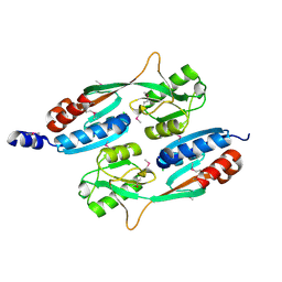
5XMZ
 | | Verticillium effector PevD1 | | 分子名称: | CALCIUM ION, CHLORIDE ION, Effector protein PevD1 | | 著者 | Liu, X, Zhou, R. | | 登録日 | 2017-05-17 | | 公開日 | 2017-07-05 | | 最終更新日 | 2017-09-13 | | 実験手法 | X-RAY DIFFRACTION (1.85 Å) | | 主引用文献 | The asparagine-rich protein NRP interacts with the Verticillium effector PevD1 and regulates the subcellular localization of cryptochrome 2
J. Exp. Bot., 68, 2017
|
|
7DF1
 | | Crystal structure of human CD98 heavy chain extracellular domain in complex with S1-F4 scFv | | 分子名称: | 4F2 cell-surface antigen heavy chain, IGL c2062_light_IGKV4-1_IGKJ5, S1-F4 VH | | 著者 | Liu, X, Ding, J, Sui, J, Tian, X. | | 登録日 | 2020-11-06 | | 公開日 | 2022-12-07 | | 最終更新日 | 2023-11-29 | | 実験手法 | X-RAY DIFFRACTION (2.806 Å) | | 主引用文献 | An anti-CD98 antibody displaying pH-dependent Fc-mediated tumour-specific activity against multiple cancers in CD98-humanized mice.
Nat Biomed Eng, 2022
|
|
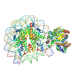
5X0Y
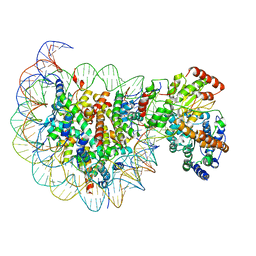 | | Complex of Snf2-Nucleosome complex with Snf2 bound to SHL2 of the nucleosome | | 分子名称: | DNA (167-MER), Histone H2A, Histone H2B 1.1, ... | | 著者 | Li, M, Liu, X, Xia, X, Chen, Z, Li, X. | | 登録日 | 2017-01-23 | | 公開日 | 2017-04-19 | | 最終更新日 | 2024-03-27 | | 実験手法 | ELECTRON MICROSCOPY (4.69 Å) | | 主引用文献 | Mechanism of chromatin remodelling revealed by the Snf2-nucleosome structure.
Nature, 544, 2017
|
|
5X0X
 | | Complex of Snf2-Nucleosome complex with Snf2 bound to position +6 of the nucleosome | | 分子名称: | DNA (167-MER), Histone H2A, Histone H2B 1.1, ... | | 著者 | Li, M, Liu, X, Xia, X, Chen, Z, Li, X. | | 登録日 | 2017-01-23 | | 公開日 | 2017-04-19 | | 最終更新日 | 2024-03-27 | | 実験手法 | ELECTRON MICROSCOPY (3.97 Å) | | 主引用文献 | Mechanism of chromatin remodelling revealed by the Snf2-nucleosome structure.
Nature, 544, 2017
|
|
5YAX
 | | Crystal structure of a human neutralizing antibody bound to a HBV preS1 peptide | | 分子名称: | Large envelope protein, SODIUM ION, scFv1 antibody | | 著者 | Liu, X, Zheng, S, Ye, K, Sui, J. | | 登録日 | 2017-09-02 | | 公開日 | 2017-10-11 | | 実験手法 | X-RAY DIFFRACTION (2.5 Å) | | 主引用文献 | A potent human neutralizing antibody Fc-dependently reduces established HBV infections
Elife, 6, 2017
|
|
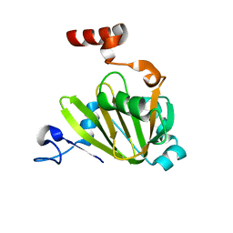
6IER
 | | Apo structure of a beta-glucosidase 1317 | | 分子名称: | 2-AMINO-2-HYDROXYMETHYL-PROPANE-1,3-DIOL, beta-glucosidase 1317 | | 著者 | Xie, W, Liu, X. | | 登録日 | 2018-09-16 | | 公開日 | 2019-07-24 | | 最終更新日 | 2023-11-22 | | 実験手法 | X-RAY DIFFRACTION (2.246 Å) | | 主引用文献 | Improving the cellobiose-hydrolysis activity and glucose-tolerance of a thermostable beta-glucosidase through rational design.
Int.J.Biol.Macromol., 136, 2019
|
|
7DZE
 | | Fabp ground state captured by XFELs | | 分子名称: | Fatty acid-binding protein, liver, PALMITIC ACID | | 著者 | Li, H, Yu, L.-J, Liu, X, Shen, J.-R, Wang, J. | | 登録日 | 2021-01-25 | | 公開日 | 2022-07-27 | | 最終更新日 | 2023-11-29 | | 実験手法 | X-RAY DIFFRACTION (1.55 Å) | | 主引用文献 | Excited-state intermediates in a designer protein encoding a phototrigger caught by an X-ray free-electron laser.
Nat.Chem., 14, 2022
|
|
7DZH
 | | intermediate of FABP with a delay time of 100 ns | | 分子名称: | Fatty acid-binding protein, liver, PALMITIC ACID | | 著者 | Li, H, Yu, L.-J, Liu, X, Shen, J.-R, Wang, J. | | 登録日 | 2021-01-25 | | 公開日 | 2022-07-27 | | 最終更新日 | 2023-11-29 | | 実験手法 | X-RAY DIFFRACTION (1.65 Å) | | 主引用文献 | Excited-state intermediates in a designer protein encoding a phototrigger caught by an X-ray free-electron laser.
Nat.Chem., 14, 2022
|
|
7DZF
 | | Intermediate of FABP with a delay time of 10 ns | | 分子名称: | Fatty acid-binding protein, liver, PALMITIC ACID | | 著者 | Li, H, Yu, L.-J, Liu, X, Shen, J.-R, Wang, J. | | 登録日 | 2021-01-25 | | 公開日 | 2022-07-27 | | 最終更新日 | 2023-11-29 | | 実験手法 | X-RAY DIFFRACTION (1.7 Å) | | 主引用文献 | Excited-state intermediates in a designer protein encoding a phototrigger caught by an X-ray free-electron laser.
Nat.Chem., 14, 2022
|
|


