5GXY
 
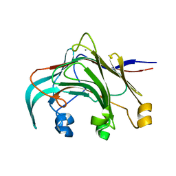
 | | Crystal structure of endoglucanase CelQ from Clostridium thermocellum complexed with cellobiose and Tris | | 分子名称: | 2-(2-(2-(2-(2-(2-ETHOXYETHOXY)ETHOXY)ETHOXY)ETHOXY)ETHOXY)ETHANOL, 2-AMINO-2-HYDROXYMETHYL-PROPANE-1,3-DIOL, BROMIDE ION, ... | | 著者 | Jeng, W.Y, Liu, C.I, Wang, A.H.J. | | 登録日 | 2016-09-21 | | 公開日 | 2017-09-27 | | 最終更新日 | 2023-11-08 | | 実験手法 | X-RAY DIFFRACTION (1.7 Å) | | 主引用文献 | Crystal Structures of the C-Terminally Truncated Endoglucanase Cel9Q from Clostridium thermocellum Complexed with Cellodextrins and Tris.
Chembiochem, 20, 2019
|
|
5GY0
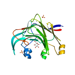
 | | Crystal structure of endoglucanase CelQ from Clostridium thermocellum complexed with cellotetraose | | 分子名称: | BROMIDE ION, CALCIUM ION, CHLORIDE ION, ... | | 著者 | Jeng, W.Y, Liu, C.I, Wang, A.H.J. | | 登録日 | 2016-09-21 | | 公開日 | 2017-09-27 | | 最終更新日 | 2023-11-08 | | 実験手法 | X-RAY DIFFRACTION (1.74 Å) | | 主引用文献 | Crystal Structures of the C-Terminally Truncated Endoglucanase Cel9Q from Clostridium thermocellum Complexed with Cellodextrins and Tris.
Chembiochem, 20, 2019
|
|
5GY1
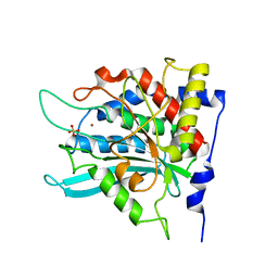
 | | Crystal structure of endoglucanase CelQ from Clostridium thermocellum complexed with cellotriose | | 分子名称: | BROMIDE ION, CALCIUM ION, CHLORIDE ION, ... | | 著者 | Jeng, W.Y, Liu, C.I, Wang, A.H.J. | | 登録日 | 2016-09-21 | | 公開日 | 2017-09-27 | | 最終更新日 | 2023-11-08 | | 実験手法 | X-RAY DIFFRACTION (1.99 Å) | | 主引用文献 | Crystal Structures of the C-Terminally Truncated Endoglucanase Cel9Q from Clostridium thermocellum Complexed with Cellodextrins and Tris.
Chembiochem, 20, 2019
|
|
5GXX
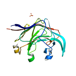
 | | Crystal structure of endoglucanase CelQ from Clostridium thermocellum complexed with Tris | | 分子名称: | 2-AMINO-2-HYDROXYMETHYL-PROPANE-1,3-DIOL, CALCIUM ION, CHLORIDE ION, ... | | 著者 | Jeng, W.Y, Liu, C.I, Wang, A.H.J. | | 登録日 | 2016-09-21 | | 公開日 | 2017-09-27 | | 最終更新日 | 2023-11-08 | | 実験手法 | X-RAY DIFFRACTION (1.5 Å) | | 主引用文献 | Crystal Structures of the C-Terminally Truncated Endoglucanase Cel9Q from Clostridium thermocellum Complexed with Cellodextrins and Tris.
Chembiochem, 20, 2019
|
|
5GXZ
 | | Crystal structure of endoglucanase CelQ from Clostridium thermocellum complexed with cellobiose and cellotriose | | 分子名称: | 2-(2-(2-(2-(2-(2-ETHOXYETHOXY)ETHOXY)ETHOXY)ETHOXY)ETHOXY)ETHANOL, BROMIDE ION, CALCIUM ION, ... | | 著者 | Jeng, W.Y, Liu, C.I, Wang, A.H.J. | | 登録日 | 2016-09-21 | | 公開日 | 2017-09-27 | | 最終更新日 | 2023-11-08 | | 実験手法 | X-RAY DIFFRACTION (2.05 Å) | | 主引用文献 | Crystal Structures of the C-Terminally Truncated Endoglucanase Cel9Q from Clostridium thermocellum Complexed with Cellodextrins and Tris.
Chembiochem, 20, 2019
|
|
2DF7
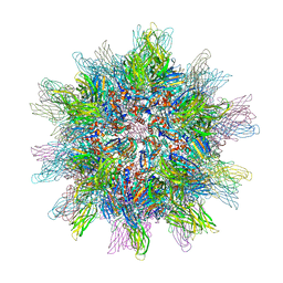 | | Crystal structure of infectious bursal disease virus VP2 subviral particle | | 分子名称: | CALCIUM ION, CHLORIDE ION, structural polyprotein VP2 | | 著者 | Ko, T.P, Lee, C.C, Wang, M.Y, Wang, A.H. | | 登録日 | 2006-02-27 | | 公開日 | 2006-06-27 | | 最終更新日 | 2023-10-25 | | 実験手法 | X-RAY DIFFRACTION (2.6 Å) | | 主引用文献 | Crystal structure of infectious bursal disease virus VP2 subviral particle at 2.6A resolution: Implications in virion assembly and immunogenicity.
J.Struct.Biol., 155, 2006
|
|
4MHZ
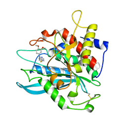 | | Crystal structure of apo-form glutaminyl cyclase from Ixodes scapularis in complex with PBD150 | | 分子名称: | 1-(3,4-dimethoxyphenyl)-3-[3-(1H-imidazol-1-yl)propyl]thiourea, Glutaminyl cyclase, putative | | 著者 | Huang, K.F, Hsu, H.L, Wang, A.H.J. | | 登録日 | 2013-08-30 | | 公開日 | 2014-03-12 | | 最終更新日 | 2023-11-08 | | 実験手法 | X-RAY DIFFRACTION (1.95 Å) | | 主引用文献 | Structural and functional analyses of a glutaminyl cyclase from Ixodes scapularis reveal metal-independent catalysis and inhibitor binding.
Acta Crystallogr.,Sect.D, 70, 2014
|
|
4MHN
 | | Crystal structure of a glutaminyl cyclase from Ixodes scapularis | | 分子名称: | Glutaminyl cyclase, putative, ZINC ION | | 著者 | Huang, K.F, Hsu, H.L, Wang, A.H.J. | | 登録日 | 2013-08-30 | | 公開日 | 2014-03-12 | | 最終更新日 | 2023-11-08 | | 実験手法 | X-RAY DIFFRACTION (1.15 Å) | | 主引用文献 | Structural and functional analyses of a glutaminyl cyclase from Ixodes scapularis reveal metal-independent catalysis and inhibitor binding.
Acta Crystallogr.,Sect.D, 70, 2014
|
|
4MHY
 | | Crystal structure of a glutaminyl cyclase from Ixodes scapularis in complex with PBD150 | | 分子名称: | 1-(3,4-dimethoxyphenyl)-3-[3-(1H-imidazol-1-yl)propyl]thiourea, Glutaminyl cyclase, putative, ... | | 著者 | Huang, K.F, Hsu, H.L, Wang, A.H.J. | | 登録日 | 2013-08-30 | | 公開日 | 2014-03-12 | | 最終更新日 | 2023-11-08 | | 実験手法 | X-RAY DIFFRACTION (1.38 Å) | | 主引用文献 | Structural and functional analyses of a glutaminyl cyclase from Ixodes scapularis reveal metal-independent catalysis and inhibitor binding.
Acta Crystallogr.,Sect.D, 70, 2014
|
|
4MHP
 | |
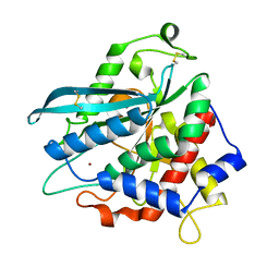
2AFX
 | | Crystal structure of human glutaminyl cyclase in complex with 1-benzylimidazole | | 分子名称: | 1-BENZYL-1H-IMIDAZOLE, Glutaminyl-peptide cyclotransferase, SULFATE ION, ... | | 著者 | Huang, K.F, Liu, Y.L, Cheng, W.J, Ko, T.P, Wang, A.H.J. | | 登録日 | 2005-07-26 | | 公開日 | 2005-08-23 | | 最終更新日 | 2024-03-13 | | 実験手法 | X-RAY DIFFRACTION (1.64 Å) | | 主引用文献 | Crystal structures of human glutaminyl cyclase, an enzyme responsible for protein N-terminal pyroglutamate formation
Proc.Natl.Acad.Sci.Usa, 102, 2005
|
|
2AFO
 | | Crystal structure of human glutaminyl cyclase at pH 8.0 | | 分子名称: | Glutaminyl-peptide cyclotransferase, SULFATE ION, ZINC ION | | 著者 | Huang, K.F, Liu, Y.L, Cheng, W.J, Ko, T.P, Wang, A.H.J. | | 登録日 | 2005-07-26 | | 公開日 | 2005-08-23 | | 最終更新日 | 2024-03-13 | | 実験手法 | X-RAY DIFFRACTION (2.35 Å) | | 主引用文献 | Crystal structures of human glutaminyl cyclase, an enzyme responsible for protein N-terminal pyroglutamate formation
Proc.Natl.Acad.Sci.Usa, 102, 2005
|
|
2AFW
 | | Crystal structure of human glutaminyl cyclase in complex with N-acetylhistamine | | 分子名称: | Glutaminyl-peptide cyclotransferase, N-[2-(1H-IMIDAZOL-4-YL)ETHYL]ACETAMIDE, SULFATE ION, ... | | 著者 | Huang, K.F, Liu, Y.L, Cheng, W.J, Ko, T.P, Wang, A.H.J. | | 登録日 | 2005-07-26 | | 公開日 | 2005-08-23 | | 最終更新日 | 2024-03-13 | | 実験手法 | X-RAY DIFFRACTION (1.56 Å) | | 主引用文献 | Crystal structures of human glutaminyl cyclase, an enzyme responsible for protein N-terminal pyroglutamate formation
Proc.Natl.Acad.Sci.Usa, 102, 2005
|
|
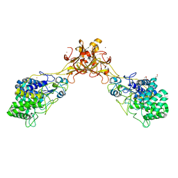
3B00
 | | Crystal structure of the laminarinase catalytic domain from Thermotoga maritima MSB8 in complex with cetyltrimethylammonium bromide | | 分子名称: | CALCIUM ION, CETYL-TRIMETHYL-AMMONIUM, Laminarinase | | 著者 | Jeng, W.Y, Wang, N.C, Wang, A.H.J. | | 登録日 | 2011-06-03 | | 公開日 | 2011-11-23 | | 最終更新日 | 2023-11-01 | | 実験手法 | X-RAY DIFFRACTION (1.74 Å) | | 主引用文献 | Crystal structures of the laminarinase catalytic domain from Thermotoga maritima MSB8 in complex with inhibitors: essential residues for beta-1,3 and beta-1,4 glucan selection.
J.Biol.Chem., 286, 2011
|
|
6JXG
 | | Crystasl Structure of Beta-glucosidase D2-BGL from Chaetomella Raphigera | | 分子名称: | 2-acetamido-2-deoxy-beta-D-glucopyranose, Beta-glucosidase, alpha-D-mannopyranose | | 著者 | Wang, A.H.-J, Lee, C.C, Kao, M.R, Ho, T.H.D. | | 登録日 | 2019-04-23 | | 公開日 | 2019-11-20 | | 最終更新日 | 2023-11-22 | | 実験手法 | X-RAY DIFFRACTION (1.9 Å) | | 主引用文献 | Chaetomella raphigerabeta-glucosidase D2-BGL has intriguing structural features and a high substrate affinity that renders it an efficient cellulase supplement for lignocellulosic biomass hydrolysis.
Biotechnol Biofuels, 12, 2019
|
|
2AFS
 | | Crystal structure of the genetic mutant R54W of human glutaminyl cyclase | | 分子名称: | Glutaminyl-peptide cyclotransferase, SULFATE ION, ZINC ION | | 著者 | Huang, K.F, Liu, Y.L, Cheng, W.J, Ko, T.P, Wang, A.H.J. | | 登録日 | 2005-07-26 | | 公開日 | 2005-08-23 | | 最終更新日 | 2024-05-29 | | 実験手法 | X-RAY DIFFRACTION (2.22 Å) | | 主引用文献 | Crystal structures of human glutaminyl cyclase, an enzyme responsible for protein N-terminal pyroglutamate formation
Proc.Natl.Acad.Sci.Usa, 102, 2005
|
|
2AFZ
 | | Crystal structure of human glutaminyl cyclase in complex with 1-vinylimidazole | | 分子名称: | 1-VINYLIMIDAZOLE, Glutaminyl-peptide cyclotransferase, SULFATE ION, ... | | 著者 | Huang, K.F, Liu, Y.L, Cheng, W.J, Ko, T.P, Wang, A.H.J. | | 登録日 | 2005-07-26 | | 公開日 | 2005-08-23 | | 最終更新日 | 2024-03-13 | | 実験手法 | X-RAY DIFFRACTION (1.68 Å) | | 主引用文献 | Crystal structures of human glutaminyl cyclase, an enzyme responsible for protein N-terminal pyroglutamate formation
Proc.Natl.Acad.Sci.Usa, 102, 2005
|
|
2AFU
 | | Crystal structure of human glutaminyl cyclase in complex with glutamine t-butyl ester | | 分子名称: | Glutaminyl-peptide cyclotransferase, TERT-BUTYL D-ALPHA-GLUTAMINATE, ZINC ION | | 著者 | Huang, K.F, Liu, Y.L, Cheng, W.J, Ko, T.P, Wang, A.H.J. | | 登録日 | 2005-07-26 | | 公開日 | 2005-08-23 | | 最終更新日 | 2024-05-29 | | 実験手法 | X-RAY DIFFRACTION (2.22 Å) | | 主引用文献 | Crystal structures of human glutaminyl cyclase, an enzyme responsible for protein N-terminal pyroglutamate formation
Proc.Natl.Acad.Sci.Usa, 102, 2005
|
|
3AZX
 | | Crystal structure of the laminarinase catalytic domain from Thermotoga maritima MSB8 | | 分子名称: | CALCIUM ION, Laminarinase | | 著者 | Jeng, W.Y, Wang, N.C, Wang, A.H.J. | | 登録日 | 2011-06-03 | | 公開日 | 2011-11-23 | | 最終更新日 | 2023-11-01 | | 実験手法 | X-RAY DIFFRACTION (1.8 Å) | | 主引用文献 | Crystal structures of the laminarinase catalytic domain from Thermotoga maritima MSB8 in complex with inhibitors: essential residues for beta-1,3 and beta-1,4 glucan selection.
J.Biol.Chem., 286, 2011
|
|
3AZZ
 | | Crystal structure of the laminarinase catalytic domain from Thermotoga maritima MSB8 in complex with gluconolactone | | 分子名称: | CALCIUM ION, D-glucono-1,5-lactone, Laminarinase, ... | | 著者 | Jeng, W.Y, Wang, N.C, Wang, A.H.J. | | 登録日 | 2011-06-03 | | 公開日 | 2011-11-23 | | 最終更新日 | 2023-11-01 | | 実験手法 | X-RAY DIFFRACTION (1.81 Å) | | 主引用文献 | Crystal structures of the laminarinase catalytic domain from Thermotoga maritima MSB8 in complex with inhibitors: essential residues for beta-1,3 and beta-1,4 glucan selection.
J.Biol.Chem., 286, 2011
|
|
2AFM
 | | Crystal structure of human glutaminyl cyclase at pH 6.5 | | 分子名称: | Glutaminyl-peptide cyclotransferase, SULFATE ION, ZINC ION | | 著者 | Huang, K.F, Liu, Y.L, Cheng, W.J, Ko, T.P, Wang, A.H.J. | | 登録日 | 2005-07-26 | | 公開日 | 2005-08-23 | | 最終更新日 | 2024-03-13 | | 実験手法 | X-RAY DIFFRACTION (1.66 Å) | | 主引用文献 | Crystal structures of human glutaminyl cyclase, an enzyme responsible for protein N-terminal pyroglutamate formation
Proc.Natl.Acad.Sci.Usa, 102, 2005
|
|
3B01
 | | Crystal structure of the laminarinase catalytic domain from Thermotoga maritima MSB8 | | 分子名称: | CALCIUM ION, CHLORIDE ION, Laminarinase, ... | | 著者 | Jeng, W.Y, Wang, N.C, Wang, A.H.J. | | 登録日 | 2011-06-03 | | 公開日 | 2011-11-23 | | 最終更新日 | 2023-11-01 | | 実験手法 | X-RAY DIFFRACTION (1.87 Å) | | 主引用文献 | Crystal structures of the laminarinase catalytic domain from Thermotoga maritima MSB8 in complex with inhibitors: essential residues for beta-1,3 and beta-1,4 glucan selection.
J.Biol.Chem., 286, 2011
|
|
3AZY
 | | Crystal structure of the laminarinase catalytic domain from Thermotoga maritima MSB8 | | 分子名称: | CALCIUM ION, Laminarinase, SULFATE ION | | 著者 | Jeng, W.Y, Wang, N.C, Wang, A.H.J. | | 登録日 | 2011-06-03 | | 公開日 | 2011-11-23 | | 最終更新日 | 2023-11-01 | | 実験手法 | X-RAY DIFFRACTION (1.65 Å) | | 主引用文献 | Crystal structures of the laminarinase catalytic domain from Thermotoga maritima MSB8 in complex with inhibitors: essential residues for beta-1,3 and beta-1,4 glucan selection.
J.Biol.Chem., 286, 2011
|
|
1EFM
 | |
5HXN
 | | Crystal Structure of Z,Z-Farnesyl Diphosphate Synthase (D71M and E75A mutants) from the Wild Tomato Solanum habrochaites | | 分子名称: | (2Z,6Z)-farnesyl diphosphate synthase, chloroplastic | | 著者 | Lee, C.C, Chan, Y.T, Wang, A.H.J. | | 登録日 | 2016-01-31 | | 公開日 | 2017-04-05 | | 最終更新日 | 2023-11-08 | | 実験手法 | X-RAY DIFFRACTION (2.05 Å) | | 主引用文献 | Crystal Structure and Potential Head-to-Middle Condensation Function of aZ,Z-Farnesyl Diphosphate Synthase.
Acs Omega, 2, 2017
|
|


