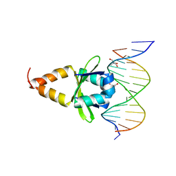
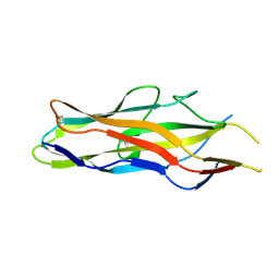
4ZM0
 
 | |
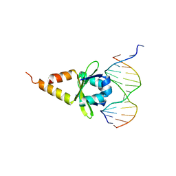
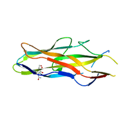
4ZM2
 | |
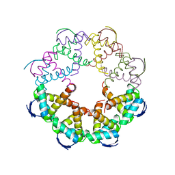
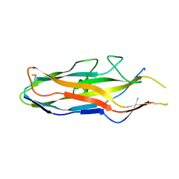
7B22
 | |
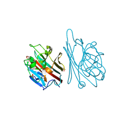
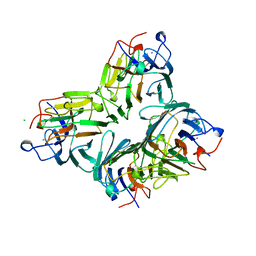
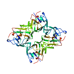
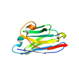
1FAT
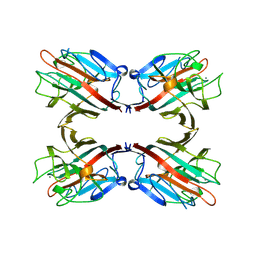 | | PHYTOHEMAGGLUTININ-L | | 分子名称: | 2-acetamido-2-deoxy-beta-D-glucopyranose, CALCIUM ION, MANGANESE (II) ION, ... | | 著者 | Hamelryck, T, Loris, R. | | 登録日 | 1996-06-12 | | 公開日 | 1996-12-23 | | 最終更新日 | 2020-07-29 | | 実験手法 | X-RAY DIFFRACTION (2.8 Å) | | 主引用文献 | The crystallographic structure of phytohemagglutinin-L.
J.Biol.Chem., 271, 1996
|
|
1FZU
 | | RNAse T1 V78A mutant | | 分子名称: | CALCIUM ION, GUANOSINE-2'-MONOPHOSPHATE, GUANYL-SPECIFIC RIBONUCLEASE T1 | | 著者 | De Vos, S, Loris, R, Steyaert, J. | | 登録日 | 2000-10-04 | | 公開日 | 2000-10-25 | | 最終更新日 | 2021-11-03 | | 実験手法 | X-RAY DIFFRACTION (1.8 Å) | | 主引用文献 | Hydrophobic core manipulations in ribonuclease T1.
Biochemistry, 40, 2001
|
|
1G02
 | | Ribonuclease T1 V16S mutant | | 分子名称: | CALCIUM ION, GUANOSINE-2'-MONOPHOSPHATE, GUANYL-SPECIFIC RIBONUCLEASE T1 | | 著者 | De Vos, S, Loris, R, Steyaert, J. | | 登録日 | 2000-10-05 | | 公開日 | 2000-10-25 | | 最終更新日 | 2011-07-13 | | 実験手法 | X-RAY DIFFRACTION (1.86 Å) | | 主引用文献 | Hydrophobic core manipulations in ribonuclease T1.
Biochemistry, 40, 2001
|
|
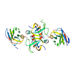
1G9F
 | | CRYSTAL STRUCTURE OF THE SOYBEAN AGGLUTININ IN A COMPLEX WITH A BIANTENNARY BLOOD GROUP ANTIGEN ANALOG | | 分子名称: | 2-acetamido-2-deoxy-beta-D-glucopyranose-(1-4)-2-acetamido-2-deoxy-beta-D-glucopyranose, CALCIUM ION, LECTIN, ... | | 著者 | Buts, L, Hamelryck, T.W, Dao-Thi, M.-H, Loris, R, Wyns, L, Etzler, M.E. | | 登録日 | 2000-11-23 | | 公開日 | 2001-06-13 | | 最終更新日 | 2024-04-03 | | 実験手法 | X-RAY DIFFRACTION (2.5 Å) | | 主引用文献 | Weak protein-protein interactions in lectins: the crystal structure of a vegetative lectin from the legume Dolichos biflorus.
J.Mol.Biol., 309, 2001
|
|
1FYS
 | | Ribonuclease T1 V16C mutant | | 分子名称: | CALCIUM ION, GUANOSINE-2'-MONOPHOSPHATE, GUANYL-SPECIFIC RIBONUCLEASE T1 | | 著者 | De Vos, S, Loris, R, Steyaert, J. | | 登録日 | 2000-10-03 | | 公開日 | 2000-10-25 | | 最終更新日 | 2021-11-03 | | 実験手法 | X-RAY DIFFRACTION (2 Å) | | 主引用文献 | Hydrophobic core manipulations in ribonuclease T1.
Biochemistry, 40, 2001
|
|
1GNZ
 | | LECTIN I-B4 FROM GRIFFONIA SIMPLICIFOLIA (GS I-B4)METAL FREE FORM | | 分子名称: | 2-acetamido-2-deoxy-beta-D-glucopyranose, GSI-B4 ISOLECTIN, PHOSPHATE ION | | 著者 | Lescar, J, Loris, R, Mitchell, E, Gautier, C, Imberty, A. | | 登録日 | 2001-10-11 | | 公開日 | 2001-11-29 | | 最終更新日 | 2023-12-13 | | 実験手法 | X-RAY DIFFRACTION (2.5 Å) | | 主引用文献 | Isolectins I-A and I-B of Griffonia (Bandeiraea) Simplicifolia. Crystal Structure of Metal-Free Gs I-B(4) and Molecular Basis for Metal Binding and Monosaccharide Specificity.
J.Biol.Chem., 277, 2002
|
|
1HYF
 | | RIBONUCLEASE T1 V16A MUTANT IN COMPLEX WITH SR2+ | | 分子名称: | GUANOSINE-2'-MONOPHOSPHATE, GUANYL-SPECIFIC RIBONUCLEASE T1, STRONTIUM ION | | 著者 | De Swarte, J, De Vos, S, Langhorst, U, Steyaert, J, Loris, R. | | 登録日 | 2001-01-19 | | 公開日 | 2001-02-14 | | 最終更新日 | 2021-11-10 | | 実験手法 | X-RAY DIFFRACTION (1.7 Å) | | 主引用文献 | The contribution of metal ions to the conformational stability of ribonuclease T1: crystal versus solution.
Eur.J.Biochem., 268, 2001
|
|
1HZ1
 | | RIBONUCLEASE T1 V16A MUTANT IN COMPLEX WITH MG2+ | | 分子名称: | GUANOSINE-2'-MONOPHOSPHATE, MAGNESIUM ION, RIBONUCLEASE T1 | | 著者 | De Swarte, J, De Vos, S, Langhorst, U, Steyaert, J, Loris, R. | | 登録日 | 2001-01-23 | | 公開日 | 2001-01-31 | | 最終更新日 | 2021-10-27 | | 実験手法 | X-RAY DIFFRACTION (1.8 Å) | | 主引用文献 | The contribution of metal ions to the conformational stability of ribonuclease T1: crystal versus solution.
Eur.J.Biochem., 268, 2001
|
|
9G2A
 | | Staphylococcus aureus MazF in complex with nanobody 4 | | 分子名称: | 3[N-MORPHOLINO]PROPANE SULFONIC ACID, 4-(2-HYDROXYETHYL)-1-PIPERAZINE ETHANESULFONIC ACID, Endoribonuclease MazF, ... | | 著者 | Prolic-Kalinsek, M, Zorzini, V, Haesaerts, S, Loris, R. | | 登録日 | 2024-07-10 | | 公開日 | 2024-07-17 | | 実験手法 | X-RAY DIFFRACTION (2.046594 Å) | | 主引用文献 | Staphylococcus aureus MazF in complex with nanobody 4
To Be Published
|
|
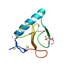
1MMC
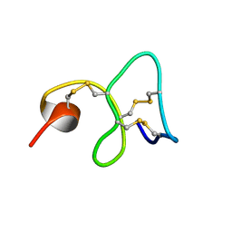 | | 1H NMR STUDY OF THE SOLUTION STRUCTURE OF AC-AMP2 | | 分子名称: | ANTIMICROBIAL PEPTIDE 2 | | 著者 | Martins, J.C, Maes, D, Loris, R, Pepermans, H.A.M, Wyns, L, Willem, R, Verheyden, P. | | 登録日 | 1995-10-25 | | 公開日 | 1996-03-08 | | 最終更新日 | 2017-11-29 | | 実験手法 | SOLUTION NMR | | 主引用文献 | H NMR study of the solution structure of Ac-AMP2, a sugar binding antimicrobial protein isolated from Amaranthus caudatus.
J.Mol.Biol., 258, 1996
|
|
1O9W
 | | F17-aG lectin domain from Escherichia coli in complex with N-acetyl-glucosamine | | 分子名称: | 2-acetamido-2-deoxy-beta-D-glucopyranose, F17A-G FIMBRIAL ADHESIN | | 著者 | Buts, L, De Genst, E, Loris, R, Oscarson, S, Lahmann, M, Messens, J, Brosens, E, Wyns, L, Bouckaert, J, De Greve, H. | | 登録日 | 2002-12-20 | | 公開日 | 2003-05-29 | | 最終更新日 | 2023-12-13 | | 実験手法 | X-RAY DIFFRACTION (1.65 Å) | | 主引用文献 | The Fimbrial Adhesin F17-G of Enterotoxigenic Escherichia Coli Has an Immunoglobulin-Like Lectin Domain that Binds N-Acetylglucosamine
Mol.Microbiol., 49, 2003
|
|
1O9V
 | | F17-aG lectin domain from Escherichia coli in complex with a selenium carbohydrate derivative | | 分子名称: | F17-AG LECTIN DOMAIN, methyl 2-acetamido-2-deoxy-1-seleno-beta-D-glucopyranoside | | 著者 | Buts, L, De Genst, E, Loris, R, Oscarson, S, Lahmann, M, Messens, J, Brosens, E, Wyns, L, Bouckaert, J, De Greve, H. | | 登録日 | 2002-12-20 | | 公開日 | 2003-05-29 | | 最終更新日 | 2020-07-29 | | 実験手法 | X-RAY DIFFRACTION (1.75 Å) | | 主引用文献 | The Fimbrial Adhesin F17-G of Enterotoxigenic Escherichia Coli Has an Immunoglobulin-Like Lectin Domain that Binds N-Acetylglucosamine
Mol.Microbiol., 49, 2003
|
|
1O9Z
 | | F17-aG lectin domain from Escherichia coli (ligand free) | | 分子名称: | F17-AG LECTIN DOMAIN | | 著者 | Buts, L, De Genst, E, Loris, R, Oscarson, S, Lahmann, M, Messens, J, Brosens, E, Wyns, L, Bouckaert, J, De Greve, H. | | 登録日 | 2002-12-23 | | 公開日 | 2003-05-29 | | 最終更新日 | 2023-12-13 | | 実験手法 | X-RAY DIFFRACTION (1.75 Å) | | 主引用文献 | The Fimbrial Adhesin F17-G of Enterotoxigenic Escherichia Coli Has an Immunoglobulin-Like Lectin Domain that Binds N-Acetylglucosamine
Mol.Microbiol., 49, 2003
|
|
1QDC
 | | MAN(APLHA1-6)MAN(ALPHA1-O)METHYL CONCANAVALIN A COMPLEX | | 分子名称: | CALCIUM ION, CHLORIDE ION, MANGANESE (II) ION, ... | | 著者 | Bouckaert, J, Loris, R, Wyns, L. | | 登録日 | 1998-07-14 | | 公開日 | 1999-10-14 | | 最終更新日 | 2023-08-16 | | 実験手法 | X-RAY DIFFRACTION (2 Å) | | 主引用文献 | The crystal structures of Man(alpha1-3)Man(alpha1-O)Me and Man(alpha1-6)Man(alpha1-O)Me in complex with concanavalin A.
J.Biol.Chem., 274, 1999
|
|
1QDO
 | | MAN(APLHA1-3)MAN(ALPHA1-O)METHYL CONCANAVALIN A COMPLEX | | 分子名称: | CALCIUM ION, MANGANESE (II) ION, PROTEIN (CONCANAVALIN A), ... | | 著者 | Bouckaert, J, Loris, R, Wyns, L. | | 登録日 | 1999-05-24 | | 公開日 | 1999-10-14 | | 最終更新日 | 2023-08-16 | | 実験手法 | X-RAY DIFFRACTION (2.8 Å) | | 主引用文献 | The crystal structures of Man(alpha1-3)Man(alpha1-O)Me and Man(alpha1-6)Man(alpha1-O)Me in complex with concanavalin A.
J.Biol.Chem., 274, 1999
|
|
1BIR
 | | RIBONUCLEASE T1, PHE 100 TO ALA MUTANT COMPLEXED WITH 2' GMP | | 分子名称: | CALCIUM ION, GUANOSINE-2'-MONOPHOSPHATE, RIBONUCLEASE T1 | | 著者 | Doumen, J, Gonciarz, M, Zegers, I, Loris, R, Wyns, L, Steyaert, J. | | 登録日 | 1996-01-04 | | 公開日 | 1996-08-17 | | 最終更新日 | 2021-11-03 | | 実験手法 | X-RAY DIFFRACTION (1.8 Å) | | 主引用文献 | A catalytic function for the structurally conserved residue Phe 100 of ribonuclease T1.
Protein Sci., 5, 1996
|
|
3DWT
 | |
3G7Z
 | | CcdB dimer in complex with two C-terminal CcdA domains | | 分子名称: | Cytotoxic protein ccdB, Protein ccdA | | 著者 | De Jonge, N, Loris, R, Garcia-Pino, A, Buts, L. | | 登録日 | 2009-02-11 | | 公開日 | 2009-08-11 | | 最終更新日 | 2023-09-06 | | 実験手法 | X-RAY DIFFRACTION (2.351 Å) | | 主引用文献 | Rejuvenation of CcdB-Poisoned Gyrase by an Intrinsically Disordered Protein Domain.
Mol.Cell, 35, 2009
|
|
3DIE
 | |
3ENR
 | | ZINC-CALCIUM CONCANAVALIN A AT PH 6.15 | | 分子名称: | CALCIUM ION, Concanavalin-A, ZINC ION | | 著者 | Bouckaert, J, Loris, R, Wyns, L. | | 登録日 | 1999-02-11 | | 公開日 | 2000-09-18 | | 最終更新日 | 2023-09-06 | | 実験手法 | X-RAY DIFFRACTION (2.4 Å) | | 主引用文献 | Zinc/calcium- and cadmium/cadmium-substituted concanavalin A: interplay of metal binding, pH and molecular packing.
Acta Crystallogr.,Sect.D, 56, 2000
|
|
3EAK
 | | NbBCII10 humanized (FGLA mutant) | | 分子名称: | NbBCII10-FGLA, SULFATE ION | | 著者 | Vincke, C, Loris, R, Saerens, D, Martinez-Rodriguez, S, Muyldermans, S, Conrath, K. | | 登録日 | 2008-08-26 | | 公開日 | 2008-12-02 | | 最終更新日 | 2023-11-01 | | 実験手法 | X-RAY DIFFRACTION (1.95 Å) | | 主引用文献 | General strategy to humanize a camelid single-domain antibody and identification of a universal humanized nanobody scaffold.
J.Biol.Chem., 2008
|
|
3DD9
 | | Structure of DocH66Y dimer | | 分子名称: | Death on curing protein | | 著者 | Garcia-Pino, A, Loris, R. | | 登録日 | 2008-06-05 | | 公開日 | 2009-06-09 | | 最終更新日 | 2023-11-01 | | 実験手法 | X-RAY DIFFRACTION (2.45 Å) | | 主引用文献 | The intrinsically disordered domain of the antitoxin Phd chaperones the toxin Doc against irreversible inactivation and misfolding
J. Biol. Chem., 289, 2014
|
|


