7ALL
 
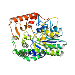 | | A single sulfatase is required for metabolism of colonic mucin O-glycans and intestinal colonization by a symbiotic human gut bacterium (BT4683-S1_4) | | 分子名称: | Arylsulfatase, CALCIUM ION, IODIDE ION, ... | | 著者 | Sofia de Jesus Vaz Luis, A, Martens, E.C, Basle, A, Cartmell, A. | | 登録日 | 2020-10-06 | | 公開日 | 2021-10-13 | | 最終更新日 | 2024-01-31 | | 実験手法 | X-RAY DIFFRACTION (1.63 Å) | | 主引用文献 | A single sulfatase is required to access colonic mucin by a gut bacterium.
Nature, 598, 2021
|
|
7AN1
 
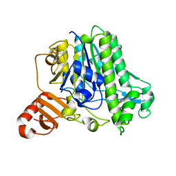 | | A single sulfatase is required for metabolism of colonic mucin O-glycans and intestinal colonization by a symbiotic human gut bacterium (BT1636-S1_20) | | 分子名称: | Arylsulfatase, CALCIUM ION, beta-D-galactopyranose-(1-4)-2-acetamido-2-deoxy-beta-D-glucopyranose | | 著者 | Sofia de Jesus Vaz Luis, A, Basle, A, Martens, E.C, Cartmell, A. | | 登録日 | 2020-10-10 | | 公開日 | 2021-10-27 | | 最終更新日 | 2024-01-31 | | 実験手法 | X-RAY DIFFRACTION (1.5 Å) | | 主引用文献 | A single sulfatase is required to access colonic mucin by a gut bacterium.
Nature, 598, 2021
|
|
7ANB
 
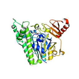 | | A single sulfatase is required for metabolism of colonic mucin O-glycans and intestinal colonization by a symbiotic human gut bacterium (BT1622-S1_20) | | 分子名称: | 1,2-ETHANEDIOL, CALCIUM ION, N-acetylgalactosamine-6-sulfatase, ... | | 著者 | Sofia de Jesus Vaz Luis, A, Basle, A, Martens, E.C, Cartmell, A. | | 登録日 | 2020-10-11 | | 公開日 | 2021-10-27 | | 最終更新日 | 2024-05-01 | | 実験手法 | X-RAY DIFFRACTION (2.6 Å) | | 主引用文献 | A single sulfatase is required to access colonic mucin by a gut bacterium.
Nature, 598, 2021
|
|
7ANA
 
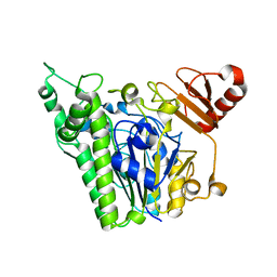 | | A single sulfatase is required for metabolism of colonic mucin O-glycans and intestinal colonization by a symbiotic human gut bacterium (BT1622-S1_20) | | 分子名称: | 1,2-ETHANEDIOL, 2-acetamido-2-deoxy-alpha-D-galactopyranose, 2-acetamido-2-deoxy-beta-D-galactopyranose, ... | | 著者 | Sofia de Jesus Vaz Luis, A, Basle, A, Martens, E.C, Cartmell, A. | | 登録日 | 2020-10-11 | | 公開日 | 2021-11-10 | | 最終更新日 | 2024-05-01 | | 実験手法 | X-RAY DIFFRACTION (2.3 Å) | | 主引用文献 | A single sulfatase is required to access colonic mucin by a gut bacterium.
Nature, 598, 2021
|
|
4EBY
 
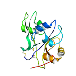 | | Crystal structure of the ectodomain of a receptor like kinase | | 分子名称: | 2-acetamido-2-deoxy-beta-D-glucopyranose, 2-acetamido-2-deoxy-beta-D-glucopyranose-(1-4)-2-acetamido-2-deoxy-beta-D-glucopyranose, Chitin elicitor receptor kinase 1, ... | | 著者 | Chai, J, Liu, T, Han, Z, She, J, Wang, J. | | 登録日 | 2012-03-25 | | 公開日 | 2012-06-27 | | 最終更新日 | 2024-11-06 | | 実験手法 | X-RAY DIFFRACTION (1.65 Å) | | 主引用文献 | Chitin-induced dimerization activates a plant immune receptor.
Science, 336, 2012
|
|
4EBZ
 
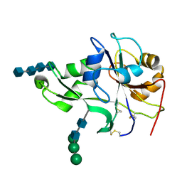 | | Crystal structure of the ectodomain of a receptor like kinase | | 分子名称: | 2-acetamido-2-deoxy-beta-D-glucopyranose-(1-4)-2-acetamido-2-deoxy-beta-D-glucopyranose, 2-acetamido-2-deoxy-beta-D-glucopyranose-(1-4)-2-acetamido-2-deoxy-beta-D-glucopyranose-(1-4)-2-acetamido-2-deoxy-beta-D-glucopyranose-(1-4)-2-acetamido-2-deoxy-beta-D-glucopyranose, Chitin elicitor receptor kinase 1, ... | | 著者 | Chai, J, Liu, T, Han, Z, She, J, Wang, J. | | 登録日 | 2012-03-26 | | 公開日 | 2012-06-27 | | 最終更新日 | 2024-10-16 | | 実験手法 | X-RAY DIFFRACTION (1.792 Å) | | 主引用文献 | Chitin-induced dimerization activates a plant immune receptor.
Science, 336, 2012
|
|
9JNN
 
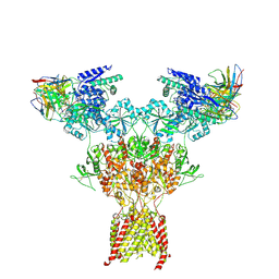 | | Structure of native di-heteromeric GluN1-GluN2B NMDA receptor in rat cortex and hippocampus | | 分子名称: | (2R)-4-(3-phosphonopropyl)piperazine-2-carboxylic acid, 2-acetamido-2-deoxy-beta-D-glucopyranose, 2-acetamido-2-deoxy-beta-D-glucopyranose-(1-4)-2-acetamido-2-deoxy-beta-D-glucopyranose, ... | | 著者 | Zhang, M, Feng, J, Li, Y, Zhu, S. | | 登録日 | 2024-09-23 | | 公開日 | 2025-02-05 | | 最終更新日 | 2025-03-19 | | 実験手法 | ELECTRON MICROSCOPY (5.4 Å) | | 主引用文献 | Assembly and architecture of endogenous NMDA receptors in adult cerebral cortex and hippocampus.
Cell, 188, 2025
|
|
9DNT
 
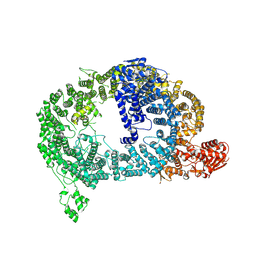 | | Cryo-EM structure of Tom1 (S. cerevisiae) | | 分子名称: | E3 ubiquitin-protein ligase TOM1 | | 著者 | Warner, K.M, Hunkeler, M, Baek, K, Roy Burman, S.S, Fischer, E.S. | | 登録日 | 2024-09-18 | | 公開日 | 2025-05-28 | | 実験手法 | ELECTRON MICROSCOPY (3 Å) | | 主引用文献 | Structural ubiquitin contributes to K48 linkage specificity of the HECT ligase Tom1.
Cell Rep, 44, 2025
|
|
9DNS
 
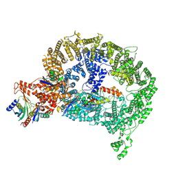 | | Cryo-EM structure of Tom1-UBE2D2-ubiquitin complex | | 分子名称: | E3 ubiquitin-protein ligase TOM1, Ubiquitin, Ubiquitin-conjugating enzyme E2 D2 | | 著者 | Warner, K.M, Hunkeler, M, Baek, K, Roy Burman, S.S, Fischer, E.S. | | 登録日 | 2024-09-18 | | 公開日 | 2025-05-28 | | 実験手法 | ELECTRON MICROSCOPY (2.8 Å) | | 主引用文献 | Structural ubiquitin contributes to K48 linkage specificity of the HECT ligase Tom1.
Cell Rep, 44, 2025
|
|
8HN6
 
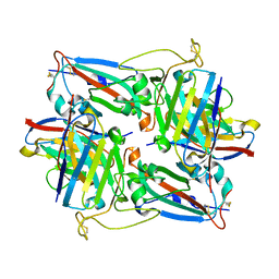 | | Crystal structure of monoclonal antibody complexed with SARS-CoV-2 RBD | | 分子名称: | Heavy chain of monoclonal antibody 3G10, Light chain of monoclonal antibody 3G10, Spike protein S1 | | 著者 | Qi, J, Chen, Y. | | 登録日 | 2022-12-07 | | 公開日 | 2023-05-17 | | 最終更新日 | 2024-10-23 | | 実験手法 | X-RAY DIFFRACTION (2.07 Å) | | 主引用文献 | Characterization of RBD-specific cross-neutralizing antibodies responses against SARS-CoV-2 variants from COVID-19 convalescents.
Front Immunol, 14, 2023
|
|
8HN7
 
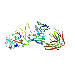 | | Crystal structure of monoclonal antibody complexed with SARS-CoV-2 RBD | | 分子名称: | 2-acetamido-2-deoxy-beta-D-glucopyranose, Heavy chain of monoclonal antibody 3C11, Light chain of monoclonal antibody 3C11, ... | | 著者 | Qi, J, Chen, Y. | | 登録日 | 2022-12-07 | | 公開日 | 2023-05-17 | | 最終更新日 | 2024-11-13 | | 実験手法 | X-RAY DIFFRACTION (3 Å) | | 主引用文献 | Characterization of RBD-specific cross-neutralizing antibodies responses against SARS-CoV-2 variants from COVID-19 convalescents.
Front Immunol, 14, 2023
|
|
4BMA
 
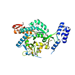 | | structural of Aspergillus fumigatus UDP-N-acetylglucosamine pyrophosphorylase | | 分子名称: | GLYCEROL, UDP-N-ACETYLGLUCOSAMINE PYROPHOSPHORYLASE | | 著者 | Fang, W, Raimi, O.G, HurtadoGuerrero, R, vanAalten, D.M.F. | | 登録日 | 2013-05-07 | | 公開日 | 2013-05-15 | | 最終更新日 | 2023-12-20 | | 実験手法 | X-RAY DIFFRACTION (2.08 Å) | | 主引用文献 | Genetic and Structural Validation of Aspergillus Fumigatus Udp-N-Acetylglucosamine Pyrophosphorylase as an Antifungal Target.
Mol.Microbiol., 89, 2013
|
|
4BJU
 
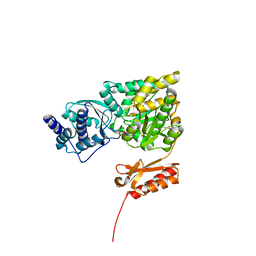 | | Genetic and structural validation of Aspergillus fumigatus N- acetylphosphoglucosamine mutase as an antifungal target | | 分子名称: | MAGNESIUM ION, N-ACETYLGLUCOSAMINE-PHOSPHATE MUTASE | | 著者 | Fang, W, Raimi, O.G, Hurtado Guerrero, R, van Aalten, D.M.F. | | 登録日 | 2013-04-19 | | 公開日 | 2013-05-01 | | 最終更新日 | 2024-11-13 | | 実験手法 | X-RAY DIFFRACTION (2.35 Å) | | 主引用文献 | Genetic and Structural Validation of Aspergillus Fumigatus N-Acetylphosphoglucosamine Mutase as an Antifungal Target.
Biosci.Rep, 33, 2013
|
|
8H2D
 
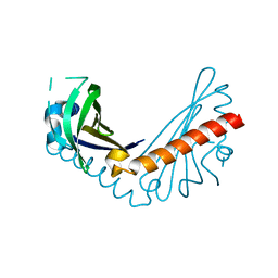 | |
8H0H
 
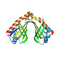 | |
7P26
 
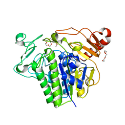 | |
7P24
 
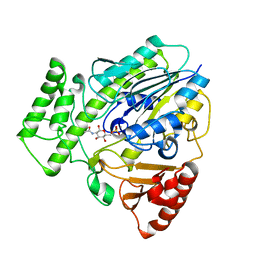 | |
4DRA
 
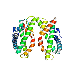 | | Crystal structure of MHF complex | | 分子名称: | Centromere protein S, Centromere protein X | | 著者 | Tao, Y, Niu, L, Teng, M. | | 登録日 | 2012-02-17 | | 公開日 | 2012-05-16 | | 最終更新日 | 2024-03-20 | | 実験手法 | X-RAY DIFFRACTION (2.414 Å) | | 主引用文献 | The structure of the FANCM-MHF complex reveals physical features for functional assembly
Nat Commun, 3, 2012
|
|
4DRB
 
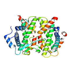 | |
3IWM
 
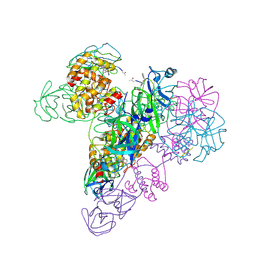 | | The octameric SARS-CoV main protease | | 分子名称: | 3C-like proteinase, N-[(5-METHYLISOXAZOL-3-YL)CARBONYL]ALANYL-L-VALYL-N~1~-((1R,2Z)-4-(BENZYLOXY)-4-OXO-1-{[(3R)-2-OXOPYRROLIDIN-3-YL]METHYL}BUT-2-ENYL)-L-LEUCINAMIDE | | 著者 | Zhong, N, Zhang, S, Xue, F, Lou, Z, Rao, Z, Xia, B. | | 登録日 | 2009-09-02 | | 公開日 | 2010-07-21 | | 最終更新日 | 2024-11-06 | | 実験手法 | X-RAY DIFFRACTION (3.2 Å) | | 主引用文献 | Three-dimensional domain swapping as a mechanism to lock the active conformation in a super-active octamer of SARS-CoV main protease
Protein Cell, 1, 2010
|
|
1Z7P
 
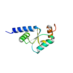 | | Solution structure of reduced glutaredoxin C1 from Populus tremula x tremuloides | | 分子名称: | glutaredoxin | | 著者 | Feng, Y, Zhong, N, Rouhier, N, Jacquot, J.P, Xia, B. | | 登録日 | 2005-03-26 | | 公開日 | 2006-03-28 | | 最終更新日 | 2024-05-29 | | 実験手法 | SOLUTION NMR | | 主引用文献 | Structural Insight into Poplar Glutaredoxin C1 with a Bridging Iron-Sulfur Cluster at the Active Site
Biochemistry, 45, 2006
|
|
1Z7R
 
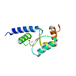 | | Solution Structure of reduced glutaredoxin C1 from Populus tremula x tremuloides | | 分子名称: | glutaredoxin | | 著者 | Feng, Y, Zhong, N, Rouhier, N, Jacquot, J.P, Xia, B. | | 登録日 | 2005-03-26 | | 公開日 | 2006-03-28 | | 最終更新日 | 2024-05-01 | | 実験手法 | SOLUTION NMR | | 主引用文献 | Structural Insight into Poplar Glutaredoxin C1 with a Bridging Iron-Sulfur Cluster at the Active Site
Biochemistry, 45, 2006
|
|
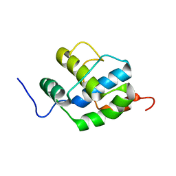
2K7X
 
 | |
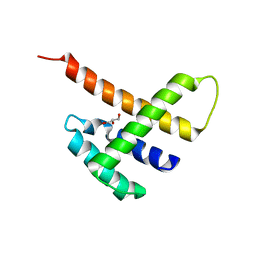
5JDK
 
 | |
2I39
 
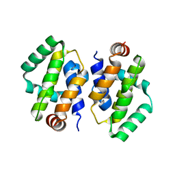 | | Crystal structure of Vaccinia virus N1L protein | | 分子名称: | (4S)-2-METHYL-2,4-PENTANEDIOL, Protein N1 | | 著者 | Aoyagi, M, Aleshin, A.E, Stec, B, Liddington, R.C. | | 登録日 | 2006-08-17 | | 公開日 | 2006-11-21 | | 最終更新日 | 2024-02-21 | | 実験手法 | X-RAY DIFFRACTION (2.2 Å) | | 主引用文献 | Vaccinia virus N1L protein resembles a B cell lymphoma-2 (Bcl-2) family protein.
Protein Sci., 16, 2007
|
|
