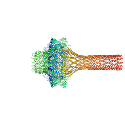
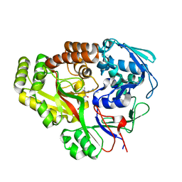
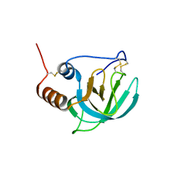
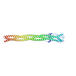
9BGM
 
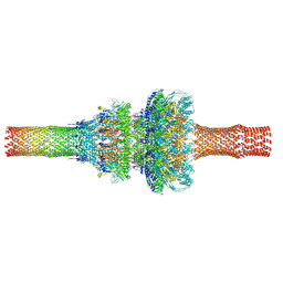 | | Pseudomonas phage DEV neck and tail (portal, head-to-tail and tail tube proteins) | | 分子名称: | gp75 tail tube, gp80 portal protein, gp83 head-to-tail | | 著者 | Iglesias, S.M, Hou, C.-F.D, Li, F, Cingolani, G. | | 登録日 | 2024-04-19 | | 公開日 | 2024-09-04 | | 実験手法 | ELECTRON MICROSCOPY (3.1 Å) | | 主引用文献 | Integrative structural analysis of Pseudomonas phage DEV reveals a genome ejection motor
To Be Published
|
|
3LJ5
 | |
3LWW
 | |
1ADR
 | |
1AOY
 | |
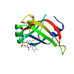
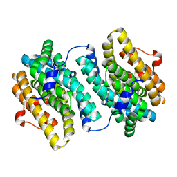
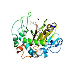
4BFR
 | | Discovery and Optimization of Pyrimidone Indoline Amide PI3Kbeta Inhibitors for the Treatment of Phosphatase and TENsin homologue (PTEN)-Deficient Cancers | | 分子名称: | 2-[2-(2-METHYL-2,3-DIHYDRO-INDOL-1-YL)-2-OXO-ETHYL]-6-MORPHOLIN-4-YL-3H-PYRIMIDIN-4-ONE, PHOSPHATIDYLINOSITOL 4,5-BISPHOSPHATE 3-KINASE CATALYTIC S SUBUNIT BETA ISOFORM | | 著者 | Certal, V, Carry, J.C, Halley, F, Virone-Oddos, A, Thompson, F, Filoche-Romme, B, El-Ahmad, Y, Karlsson, A, Charrier, V, Delorme, C, Rak, A, Abecassis, P.Y, Amara, C, Vincent, L, Bonnevaux, H, Nicolas, J.P, Mathieu, M, Bertrand, T, Marquette, J.P, Michot, N, Benard, T, Perrin, M.A, Perron, S, Monget, S, Gruss-Leleu, F, Doerflinger, G, Guizani, H, Brollo, M, Delbarre, L, Bertin, L, Richepin, P, Loyau, V, Garcia-Echeverria, C, Lengauer, C, Schio, L. | | 登録日 | 2013-03-22 | | 公開日 | 2014-01-15 | | 最終更新日 | 2023-12-20 | | 実験手法 | X-RAY DIFFRACTION (2.8 Å) | | 主引用文献 | Discovery and Optimization of Pyrimidone Indoline Amide Pi3Kbeta Inhibitors for the Treatment of Phosphatase and Tensin Homologue (Pten)-Deficient Cancers.
J.Med.Chem., 57, 2014
|
|
1AXJ
 | |
4DKW
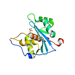 | |
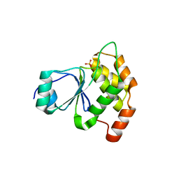
2YAK
 | | Structure of death-associated protein Kinase 1 (dapk1) in complex with a ruthenium octasporine ligand (OSV) | | 分子名称: | DEATH-ASSOCIATED PROTEIN KINASE 1, RUTHENIUM OCTASPORINE 4 | | 著者 | Feng, L, Geisselbrecht, Y, Blanck, S, Wilbuer, A, Atilla-Gokcumen, G.E, Filippakopoulos, P, Kraeling, K, Celik, M.A, Harms, K, Maksimoska, J, Marmorstein, R, Frenking, G, Knapp, S, Essen, L.-O, Meggers, E. | | 登録日 | 2011-02-23 | | 公開日 | 2011-04-27 | | 最終更新日 | 2024-05-01 | | 実験手法 | X-RAY DIFFRACTION (2.2 Å) | | 主引用文献 | Structurally Sophisticated Octahedral Metal Complexes as Highly Selective Protein Kinase Inhibitors.
J.Am.Chem.Soc., 133, 2011
|
|
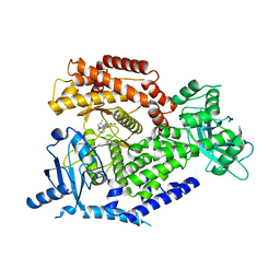
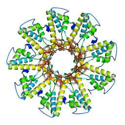
3MJO
 | | Small subunit (R2F) of native ribonucleotide reductase from Corynebacterium ammoniagenes | | 分子名称: | MANGANESE (III) ION, Ribonucleotide reductase subunit R2F | | 著者 | Ogata, H, Stolle, P, Stehr, M, Auling, G, Lubitz, W. | | 登録日 | 2010-04-13 | | 公開日 | 2010-08-25 | | 最終更新日 | 2023-09-06 | | 実験手法 | X-RAY DIFFRACTION (1.36 Å) | | 主引用文献 | A Tyrosyl-Dimanganese Coupled Spin System is the Native Metalloradical Cofactor of the R2F Subunit of the Ribonucleotide Reductase of Corynebacterium ammoniagenes.
J.Am.Chem.Soc., 132, 2010
|
|
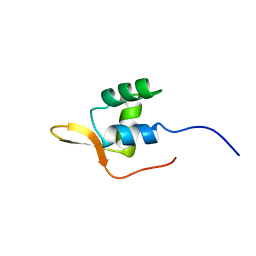
3ZOB
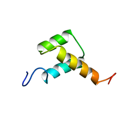 | | Solution structure of chicken Engrailed 2 homeodomain | | 分子名称: | HOMEOBOX PROTEIN ENGRAILED-2 | | 著者 | Carlier, L, Balayssac, S, Cantrelle, F.X, Khemtemourian, L, Chassaing, G, Joliot, A, Lequin, O. | | 登録日 | 2013-02-21 | | 公開日 | 2013-08-28 | | 最終更新日 | 2024-05-15 | | 実験手法 | SOLUTION NMR | | 主引用文献 | Investigation of Homeodomain Membrane Translocation Properties: Insights from the Structure Determination of Engrailed-2 Homeodomain in Aqueous and Membrane-Mimetic Environments.
Biophys.J., 105, 2013
|
|
3P9A
 | |
6E3B
 | |
6E4D
 | | Atomic structure of Mycobacterium tuberculosis DppA | | 分子名称: | Periplasmic dipeptide-binding lipoprotein DPPA, VAL-VAL-VAL-ALA | | 著者 | Ko, Y, Mitra, A, Niederweis, M, Cingolani, G. | | 登録日 | 2018-07-17 | | 公開日 | 2019-09-11 | | 最終更新日 | 2023-10-11 | | 実験手法 | X-RAY DIFFRACTION (1.252 Å) | | 主引用文献 | Heme and hemoglobin utilization by Mycobacterium tuberculosis.
Nat Commun, 10, 2019
|
|
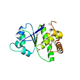
8K62
 | | Crystal structure of ALKBH1 and 13h complex. | | 分子名称: | 1-[5-[[3-(trifluoromethyloxy)phenyl]methoxy]pyrimidin-2-yl]pyrazole-4-carboxylic acid, MANGANESE (II) ION, Nucleic acid dioxygenase ALKBH1 | | 著者 | Liang, X, Yinping, G, Feng, L, Jiang, Z, Ke, X, Shengyong, Y. | | 登録日 | 2023-07-24 | | 公開日 | 2024-07-31 | | 実験手法 | X-RAY DIFFRACTION (1.991 Å) | | 主引用文献 | Crystal structure of ALKBH1 and 13h complex
To Be Published
|
|
4R30
 | |
4EI2
 | |
4ZKP
 | |
1UAP
 | | NMR structure of the NTR domain from human PCOLCE1 | | 分子名称: | Procollagen C-proteinase enhancer protein | | 著者 | Liepinsh, E, Banyai, L, Pintacuda, G, Trexler, M, Patthy, L, Otting, G. | | 登録日 | 2003-03-14 | | 公開日 | 2003-07-15 | | 最終更新日 | 2023-12-27 | | 実験手法 | SOLUTION NMR | | 主引用文献 | NMR Structure of the Netrin-like Domain (NTR) of Human Type I Procollagen C-Proteinase Enhancer Defines Structural Consensus of NTR Domains and Assesses Potential Proteinase Inhibitory Activity and Ligand Binding.
J.Biol.Chem., 278, 2003
|
|
4HRF
 | |
5BU5
 | |
5BVZ
 | |
4ZXQ
 | |
1T3W
 | | Crystal Structure of the E.coli DnaG C-terminal domain (residues 434 to 581) | | 分子名称: | ACETIC ACID, DNA primase | | 著者 | Oakley, A.J, Loscha, K.V, Schaeffer, P.M, Liepinsh, E, Wilce, M.C.J, Otting, G, Dixon, N.E. | | 登録日 | 2004-04-28 | | 公開日 | 2004-11-02 | | 最終更新日 | 2016-09-28 | | 実験手法 | X-RAY DIFFRACTION (2.8 Å) | | 主引用文献 | Crystal and solution structures of the helicase-binding domain of Escherichia coli primase
J.Biol.Chem., 280, 2005
|
|
5BU8
 | |


