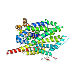
4XP6
 
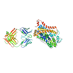 | | X-ray structure of Drosophila dopamine transporter bound to psychostimulant methamphetamine | | 分子名称: | (2S)-N-methyl-1-phenylpropan-2-amine, 2-acetamido-2-deoxy-beta-D-glucopyranose, Antibody fragment heavy chain-protein, ... | | 著者 | Aravind, P, Wang, K, Gouaux, E. | | 登録日 | 2015-01-16 | | 公開日 | 2015-05-13 | | 最終更新日 | 2023-09-27 | | 実験手法 | X-RAY DIFFRACTION (3.1 Å) | | 主引用文献 | Neurotransmitter and psychostimulant recognition by the dopamine transporter.
Nature, 521, 2015
|
|
4XPH
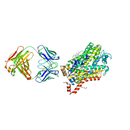 | | X-ray structure of Drosophila dopamine transporter with subsiteB mutations (D121G/S426M) bound to 3,4dichlorophenethylamine | | 分子名称: | 1-ETHOXY-2-(2-ETHOXYETHOXY)ETHANE, 2-(3,4-dichlorophenyl)ethanamine, Antibody fragment heavy chain, ... | | 著者 | Penmatsa, A, Wang, K, Gouaux, E. | | 登録日 | 2015-01-17 | | 公開日 | 2015-05-06 | | 最終更新日 | 2020-07-29 | | 実験手法 | X-RAY DIFFRACTION (2.9 Å) | | 主引用文献 | Neurotransmitter and psychostimulant recognition by the dopamine transporter.
Nature, 521, 2015
|
|
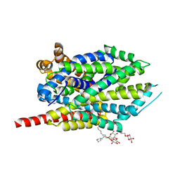
4XP9
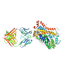 | | X-ray structure of Drosophila dopamine transporter bound to psychostimulant D-amphetamine | | 分子名称: | (2S)-1-phenylpropan-2-amine, 1-ETHOXY-2-(2-ETHOXYETHOXY)ETHANE, 2-acetamido-2-deoxy-beta-D-glucopyranose, ... | | 著者 | Aravind, P, Wang, K, Gouaux, E. | | 登録日 | 2015-01-16 | | 公開日 | 2015-05-06 | | 最終更新日 | 2023-09-27 | | 実験手法 | X-RAY DIFFRACTION (2.8 Å) | | 主引用文献 | Neurotransmitter and psychostimulant recognition by the dopamine transporter.
Nature, 521, 2015
|
|
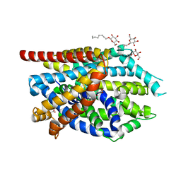
4XPA
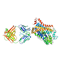 | | X-ray structure of Drosophila dopamine transporter bound to 3,4dichlorophenethylamine | | 分子名称: | 2-(3,4-dichlorophenyl)ethanamine, Antibody fragment heavy chain-protein, 9D5-heavy chain, ... | | 著者 | Aravind, P, Wang, K, Gouaux, E. | | 登録日 | 2015-01-16 | | 公開日 | 2015-05-20 | | 最終更新日 | 2023-09-27 | | 実験手法 | X-RAY DIFFRACTION (2.95 Å) | | 主引用文献 | Neurotransmitter and psychostimulant recognition by the dopamine transporter.
Nature, 521, 2015
|
|
3QS6
 | |
3QS4
 | |
3QS5
 | |
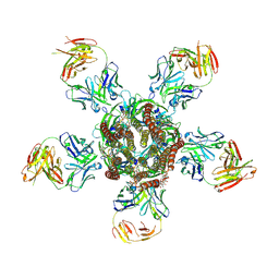
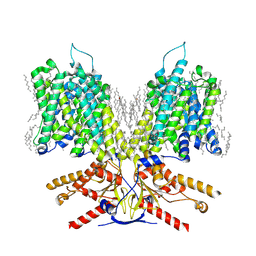
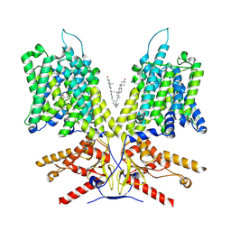
3RHW
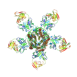 | | C. elegans glutamate-gated chloride channel (GluCl) in complex with Fab and ivermectin | | 分子名称: | (2aE,4E,5'S,6S,6'R,7S,8E,11R,13R,15S,17aR,20R,20aR,20bS)-6'-[(2S)-butan-2-yl]-20,20b-dihydroxy-5',6,8,19-tetramethyl-17 -oxo-3',4',5',6,6',10,11,14,15,17,17a,20,20a,20b-tetradecahydro-2H,7H-spiro[11,15-methanofuro[4,3,2-pq][2,6]benzodioxacy clooctadecine-13,2'-pyran]-7-yl 2,6-dideoxy-4-O-(2,6-dideoxy-3-O-methyl-alpha-L-arabino-hexopyranosyl)-3-O-methyl-alpha-L-arabino-hexopyranoside, 2-acetamido-2-deoxy-beta-D-glucopyranose, Avermectin-sensitive glutamate-gated chloride channel GluCl alpha, ... | | 著者 | Hibbs, R.E, Gouaux, E. | | 登録日 | 2011-04-12 | | 公開日 | 2011-05-25 | | 最終更新日 | 2023-09-13 | | 実験手法 | X-RAY DIFFRACTION (3.26 Å) | | 主引用文献 | Principles of activation and permeation in an anion-selective Cys-loop receptor.
Nature, 474, 2011
|
|
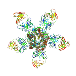
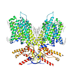
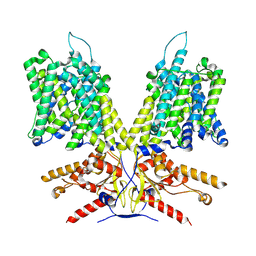
3RIF
 | | C. elegans glutamate-gated chloride channel (GluCl) in complex with Fab, ivermectin and glutamate. | | 分子名称: | (2aE,4E,5'S,6S,6'R,7S,8E,11R,13R,15S,17aR,20R,20aR,20bS)-6'-[(2S)-butan-2-yl]-20,20b-dihydroxy-5',6,8,19-tetramethyl-17 -oxo-3',4',5',6,6',10,11,14,15,17,17a,20,20a,20b-tetradecahydro-2H,7H-spiro[11,15-methanofuro[4,3,2-pq][2,6]benzodioxacy clooctadecine-13,2'-pyran]-7-yl 2,6-dideoxy-4-O-(2,6-dideoxy-3-O-methyl-alpha-L-arabino-hexopyranosyl)-3-O-methyl-alpha-L-arabino-hexopyranoside, 2-acetamido-2-deoxy-beta-D-glucopyranose, Avermectin-sensitive glutamate-gated chloride channel GluCl alpha, ... | | 著者 | Hibbs, R.E, Gouaux, E. | | 登録日 | 2011-04-13 | | 公開日 | 2011-05-25 | | 最終更新日 | 2023-09-13 | | 実験手法 | X-RAY DIFFRACTION (3.345 Å) | | 主引用文献 | Principles of activation and permeation in an anion-selective Cys-loop receptor.
Nature, 474, 2011
|
|
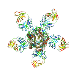
3RIA
 | | C. elegans glutamate-gated chloride channel (GluCl) in complex with Fab, ivermectin and iodide. | | 分子名称: | (2aE,4E,5'S,6S,6'R,7S,8E,11R,13R,15S,17aR,20R,20aR,20bS)-6'-[(2S)-butan-2-yl]-20,20b-dihydroxy-5',6,8,19-tetramethyl-17 -oxo-3',4',5',6,6',10,11,14,15,17,17a,20,20a,20b-tetradecahydro-2H,7H-spiro[11,15-methanofuro[4,3,2-pq][2,6]benzodioxacy clooctadecine-13,2'-pyran]-7-yl 2,6-dideoxy-4-O-(2,6-dideoxy-3-O-methyl-alpha-L-arabino-hexopyranosyl)-3-O-methyl-alpha-L-arabino-hexopyranoside, Avermectin-sensitive glutamate-gated chloride channel GluCl alpha, CHLORIDE ION, ... | | 著者 | Hibbs, R.E, Gouaux, E. | | 登録日 | 2011-04-13 | | 公開日 | 2011-05-25 | | 最終更新日 | 2023-09-13 | | 実験手法 | X-RAY DIFFRACTION (3.8 Å) | | 主引用文献 | Principles of activation and permeation in an anion-selective Cys-loop receptor.
Nature, 474, 2011
|
|
3RI5
 | | C. elegans glutamate-gated chloride channel (GluCl) in complex with Fab, ivermectin and picrotoxin | | 分子名称: | (1aR,2aR,3S,6R,6aS,8aS,8bR,9R)-2a-hydroxy-8b-methyl-9-(prop-1-en-2-yl)hexahydro-3,6-methano-1,5,7-trioxacyclopenta[ij]c yclopropa[a]azulene-4,8(3H)-dione, (2aE,4E,5'S,6S,6'R,7S,8E,11R,13R,15S,17aR,20R,20aR,20bS)-6'-[(2S)-butan-2-yl]-20,20b-dihydroxy-5',6,8,19-tetramethyl-17 -oxo-3',4',5',6,6',10,11,14,15,17,17a,20,20a,20b-tetradecahydro-2H,7H-spiro[11,15-methanofuro[4,3,2-pq][2,6]benzodioxacy clooctadecine-13,2'-pyran]-7-yl 2,6-dideoxy-4-O-(2,6-dideoxy-3-O-methyl-alpha-L-arabino-hexopyranosyl)-3-O-methyl-alpha-L-arabino-hexopyranoside, 2-acetamido-2-deoxy-beta-D-glucopyranose, ... | | 著者 | Hibbs, R.E, Gouaux, E. | | 登録日 | 2011-04-12 | | 公開日 | 2011-05-25 | | 最終更新日 | 2023-09-13 | | 実験手法 | X-RAY DIFFRACTION (3.4 Å) | | 主引用文献 | Principles of activation and permeation in an anion-selective Cys-loop receptor.
Nature, 474, 2011
|
|
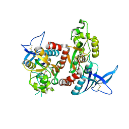
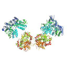
2A5S
 | | Crystal Structure Of The NR2A Ligand Binding Core In Complex With Glutamate | | 分子名称: | GLUTAMIC ACID, N-methyl-D-aspartate receptor NMDAR2A subunit | | 著者 | Furukawa, H, Singh, S.K, Mancusso, R, Gouaux, E. | | 登録日 | 2005-06-30 | | 公開日 | 2005-11-15 | | 最終更新日 | 2017-07-26 | | 実験手法 | X-RAY DIFFRACTION (1.7 Å) | | 主引用文献 | Subunit arrangement and function in NMDA receptors
Nature, 438, 2005
|
|
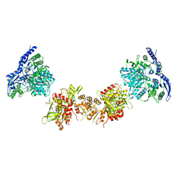
2A5T
 | | Crystal Structure Of The NR1/NR2A ligand-binding cores complex | | 分子名称: | GLUTAMIC ACID, GLYCINE, N-methyl-D-aspartate receptor NMDAR1-4a subunit, ... | | 著者 | Furukawa, H, Singh, S.K, Mancusso, R, Gouaux, E. | | 登録日 | 2005-06-30 | | 公開日 | 2005-11-15 | | 最終更新日 | 2017-07-26 | | 実験手法 | X-RAY DIFFRACTION (2 Å) | | 主引用文献 | Subunit arrangement and function in NMDA receptors
Nature, 438, 2005
|
|
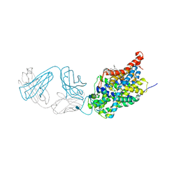
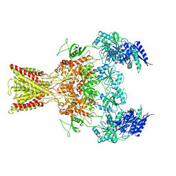
2A65
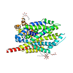 | | Crystal structure of LEUTAA, a bacterial homolog of Na+/Cl--dependent neurotransmitter transporters | | 分子名称: | CHLORIDE ION, LEUCINE, Na(+):neurotransmitter symporter (Snf family), ... | | 著者 | Yamashita, A, Singh, S.K, Kawate, T, Jin, Y, Gouaux, E. | | 登録日 | 2005-07-01 | | 公開日 | 2005-08-02 | | 最終更新日 | 2024-02-14 | | 実験手法 | X-RAY DIFFRACTION (1.65 Å) | | 主引用文献 | Crystal structure of a bacterial homologue of Na(+)/Cl(-)-dependent neurotransmitter transporters.
Nature, 437, 2005
|
|
7LGU
 | |
7LGW
 | |
7LH2
 | |
7LH3
 | |
5I74
 | | X-ray structure of the ts3 human serotonin transporter complexed with Br-citalopram at the central site | | 分子名称: | (1S)-1-(4-bromophenyl)-1-[3-(dimethylamino)propyl]-1,3-dihydro-2-benzofuran-5-carbonitrile, 2-acetamido-2-deoxy-beta-D-glucopyranose, 8B6 antibody, ... | | 著者 | Coleman, J.A, Green, E.M, Gouaux, E. | | 登録日 | 2016-02-16 | | 公開日 | 2016-04-13 | | 最終更新日 | 2020-07-29 | | 実験手法 | X-RAY DIFFRACTION (3.395 Å) | | 主引用文献 | X-ray structures and mechanism of the human serotonin transporter.
Nature, 532, 2016
|
|
5I71
 | | X-ray structure of the ts3 human serotonin transporter complexed with s-citalopram at the central site | | 分子名称: | (1S)-1-[3-(dimethylamino)propyl]-1-(4-fluorophenyl)-1,3-dihydro-2-benzofuran-5-carbonitrile, 2-acetamido-2-deoxy-beta-D-glucopyranose, 8B6 antibody, ... | | 著者 | Coleman, J.A, Green, E.M, Gouaux, E. | | 登録日 | 2016-02-16 | | 公開日 | 2016-04-13 | | 最終更新日 | 2020-07-29 | | 実験手法 | X-RAY DIFFRACTION (3.15 Å) | | 主引用文献 | X-ray structures and mechanism of the human serotonin transporter.
Nature, 532, 2016
|
|
5IPT
 | | Cryo-EM structure of GluN1/GluN2B NMDA receptor in the DCKA/D-APV-bound conformation, state 5 | | 分子名称: | Ionotropic glutamate receptor subunit NR2B, N-methyl-D-aspartate receptor subunit NR1-8a | | 著者 | Zhu, S, Stein, A.R, Yoshioka, C, Lee, C.H, Goehring, A, Mchaourab, S.H, Gouaux, E. | | 登録日 | 2016-03-09 | | 公開日 | 2016-04-20 | | 最終更新日 | 2024-03-06 | | 実験手法 | ELECTRON MICROSCOPY (14.1 Å) | | 主引用文献 | Mechanism of NMDA Receptor Inhibition and Activation.
Cell, 165, 2016
|
|
5IOU
 | | Cryo-EM structure of GluN1/GluN2B NMDA receptor in the glutamate/glycine-bound conformation | | 分子名称: | GLUTAMIC ACID, GLYCINE, Ionotropic glutamate receptor subunit NR2B, ... | | 著者 | Zhu, S, Stein, A.R, Yoshioka, C, Lee, C.H, Goehring, A, Mchaourab, S.H, Gouaux, E. | | 登録日 | 2016-03-09 | | 公開日 | 2016-04-20 | | 最終更新日 | 2024-03-06 | | 実験手法 | ELECTRON MICROSCOPY (7 Å) | | 主引用文献 | Mechanism of NMDA Receptor Inhibition and Activation.
Cell, 165, 2016
|
|
5IPU
 | | Cryo-EM structure of GluN1/GluN2B NMDA receptor in the DCKA/D-APV-bound conformation, state 6 | | 分子名称: | Ionotropic glutamate receptor subunit NR2B, N-methyl-D-aspartate receptor subunit NR1-8a | | 著者 | Zhu, S, Stein, A.R, Yoshioka, C, Lee, C.H, Goehring, A, Mchaourab, S.H, Gouaux, E. | | 登録日 | 2016-03-09 | | 公開日 | 2016-04-20 | | 最終更新日 | 2024-03-06 | | 実験手法 | ELECTRON MICROSCOPY (15.4 Å) | | 主引用文献 | Mechanism of NMDA Receptor Inhibition and Activation.
Cell, 165, 2016
|
|
5I6X
 | | X-ray structure of the ts3 human serotonin transporter complexed with paroxetine at the central site | | 分子名称: | 2-acetamido-2-deoxy-beta-D-glucopyranose, 8B6 antibody, heavy chain, ... | | 著者 | Coleman, J.A, Green, E.M, Gouaux, E. | | 登録日 | 2016-02-16 | | 公開日 | 2016-04-13 | | 最終更新日 | 2020-07-29 | | 実験手法 | X-RAY DIFFRACTION (3.14 Å) | | 主引用文献 | X-ray structures and mechanism of the human serotonin transporter.
Nature, 532, 2016
|
|
5I66
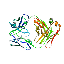 | | X-ray structure of the Fab fragment of 8B6, a murine monoclonal antibody specific for the human serotonin transporter | | 分子名称: | 8B6 antibody, heavy chain, light chain | | 著者 | Coleman, J.A, Green, E.M, Gouaux, E. | | 登録日 | 2016-02-16 | | 公開日 | 2016-04-13 | | 最終更新日 | 2023-09-27 | | 実験手法 | X-RAY DIFFRACTION (1.624 Å) | | 主引用文献 | X-ray structures and mechanism of the human serotonin transporter.
Nature, 532, 2016
|
|


