1HHA
 
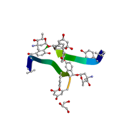 | | Decaplanin first P6122-Form | | 分子名称: | 4-epi-vancosamine, DECAPLANIN, GLYCEROL, ... | | 著者 | Lehmann, C, Vertessy, L, Sheldrick, G.M, Dauter, Z, Dauter, M. | | 登録日 | 2000-12-22 | | 公開日 | 2005-07-11 | | 最終更新日 | 2025-04-09 | | 実験手法 | X-RAY DIFFRACTION (1.9 Å) | | 主引用文献 | Structures of Four Crystal Forms of Decaplanin
Helv.Chim.Acta, 86, 2003
|
|
1HHC
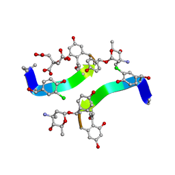 | | Crystal structure of Decaplanin - space group P21, second form | | 分子名称: | 4-epi-vancosamine, CITRIC ACID, DECAPLANIN, ... | | 著者 | Lehmann, C, Vertessy, L, Sheldrick, G.M, Dauter, Z, Dauter, M. | | 登録日 | 2000-12-22 | | 公開日 | 2005-07-11 | | 最終更新日 | 2023-12-13 | | 実験手法 | X-RAY DIFFRACTION (1.13 Å) | | 主引用文献 | Structures of Four Crystal Forms of Decaplanin
Helv.Chim.Acta, 86, 2003
|
|
1HHF
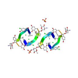 | | Decaplanin second P6122-Form | | 分子名称: | 4-epi-vancosamine, CHLORIDE ION, DECAPLANIN, ... | | 著者 | Lehmann, C, Vertessy, L, Sheldrick, G.M, Dauter, Z, Dauter, M. | | 登録日 | 2000-12-22 | | 公開日 | 2005-07-11 | | 最終更新日 | 2025-04-09 | | 実験手法 | X-RAY DIFFRACTION (1.47 Å) | | 主引用文献 | Structures of Four Crystal Forms of Decaplanin
Helv.Chim.Acta, 86, 2003
|
|
1HH3
 | | Decaplanin first P21-Form | | 分子名称: | 4-epi-vancosamine, DECAPLANIN, GLYCEROL, ... | | 著者 | Lehmann, C, Vertessy, L, Sheldrick, G.M, Dauter, Z, Dauter, M. | | 登録日 | 2000-12-19 | | 公開日 | 2005-07-11 | | 最終更新日 | 2025-04-09 | | 実験手法 | X-RAY DIFFRACTION (1 Å) | | 主引用文献 | Structures of Four Crystal Forms of Decaplanin
Helv.Chim.Acta, 86, 2003
|
|
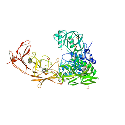
6VBK
 | | Crystal structure of N-terminal domain of Mycobacterium tuberculosis complex Lon protease | | 分子名称: | GLYCEROL, Lon211 | | 著者 | Bi, F.K, Chen, C, Chen, X.Y, Guo, C.Y, Lin, D.H. | | 登録日 | 2019-12-19 | | 公開日 | 2020-12-23 | | 最終更新日 | 2023-10-11 | | 実験手法 | X-RAY DIFFRACTION (2 Å) | | 主引用文献 | Crystal structure of the N domain of Lon protease from Mycobacterium avium complex.
Protein Sci., 28, 2019
|
|
6UTO
 | | Native E. coli Glyceraldehyde 3-phosphate dehydrogenase | | 分子名称: | ACETATE ION, Glyceraldehyde-3-phosphate dehydrogenase, SN-GLYCEROL-3-PHOSPHATE, ... | | 著者 | Rodriguez-Hernandez, A, Romo-Arevalo, E, Rodriguez-Romero, A. | | 登録日 | 2019-10-29 | | 公開日 | 2019-12-11 | | 最終更新日 | 2024-10-09 | | 実験手法 | X-RAY DIFFRACTION (1.64 Å) | | 主引用文献 | A Novel Substrate-Binding Site in the X-Ray Structure of an Oxidized E. coli Glyceraldehyde 3-Phosphate Dehydrogenase Elucidated by Single-Wavelength Anomalous Dispersion
Crystals, 9, 2019
|
|
6UP0
 | |
6PHN
 | |
6PHL
 | |
6PHQ
 | |
6PHI
 | |
6PHM
 | |
7UPH
 | | Structure of a ribosome with tethered subunits | | 分子名称: | 30S ribosomal protein S10, 30S ribosomal protein S11, 30S ribosomal protein S12, ... | | 著者 | Kim, D.S, Watkins, A, Bidstrup, E, Lee, J, Topkar, V.V, Kofman, C, Schwarz, K.J, Liu, Y, Pintilie, G, Roney, E, Das, R, Jewett, M.C. | | 登録日 | 2022-04-15 | | 公開日 | 2022-08-17 | | 最終更新日 | 2025-03-19 | | 実験手法 | ELECTRON MICROSCOPY (4.18 Å) | | 主引用文献 | Three-dimensional structure-guided evolution of a ribosome with tethered subunits.
Nat.Chem.Biol., 18, 2022
|
|
7BJ3
 | | ScpA from Streptococcus pyogenes, S512A active site mutant | | 分子名称: | 4-(2-HYDROXYETHYL)-1-PIPERAZINE ETHANESULFONIC ACID, C5a peptidase, CALCIUM ION, ... | | 著者 | Kagawa, T.F, O'Connell, M.R, Cooney, J.C. | | 登録日 | 2021-01-13 | | 公開日 | 2021-05-12 | | 最終更新日 | 2024-01-31 | | 実験手法 | X-RAY DIFFRACTION (2.6 Å) | | 主引用文献 | Enzyme kinetic and binding studies identify determinants of specificity for the immunomodulatory enzyme ScpA, a C5a inactivating bacterial protease.
Comput Struct Biotechnol J, 19, 2021
|
|
4YE7
 | |
6PQ7
 | |
5MJL
 | | Single-shot pink beam serial crystallography: Proteinase K | | 分子名称: | 2-[N-CYCLOHEXYLAMINO]ETHANE SULFONIC ACID, 4-(2-HYDROXYETHYL)-1-PIPERAZINE ETHANESULFONIC ACID, CALCIUM ION, ... | | 著者 | Meents, A, Oberthuer, D, Lieske, J, Srajer, V. | | 登録日 | 2016-12-01 | | 公開日 | 2017-11-15 | | 最終更新日 | 2024-11-13 | | 実験手法 | X-RAY DIFFRACTION (2.21013784 Å) | | 主引用文献 | Pink-beam serial crystallography.
Nat Commun, 8, 2017
|
|
5H31
 | |
5MJG
 | | Single-shot pink beam serial crystallography: Thaumatin | | 分子名称: | S,R MESO-TARTARIC ACID, SODIUM ION, Thaumatin-1 | | 著者 | Meents, A, Oberthuer, D, Lieske, J, Srajer, V. | | 登録日 | 2016-12-01 | | 公開日 | 2017-12-20 | | 最終更新日 | 2024-10-23 | | 実験手法 | X-RAY DIFFRACTION (2.1 Å) | | 主引用文献 | Single-shot pink beam serial crystallography: Thaumatin
To Be Published
|
|
8G9B
 | | Human IMPDH2 mutant - L245P, treated with GTP, ATP, IMP, and NAD+; compressed filament segment reconstruction | | 分子名称: | ADENOSINE-5'-TRIPHOSPHATE, GUANOSINE-5'-TRIPHOSPHATE, INOSINIC ACID, ... | | 著者 | O'Neill, A.G, Kollman, J.M. | | 登録日 | 2023-02-21 | | 公開日 | 2023-04-19 | | 最終更新日 | 2024-11-13 | | 実験手法 | ELECTRON MICROSCOPY (3 Å) | | 主引用文献 | Neurodevelopmental disorder mutations in the purine biosynthetic enzyme IMPDH2 disrupt its allosteric regulation.
J.Biol.Chem., 299, 2023
|
|
8G8F
 | | Human IMPDH2 mutant - L245P, treated with ATP, IMP, and NAD+; extended filament segment reconstruction | | 分子名称: | ADENOSINE-5'-TRIPHOSPHATE, INOSINIC ACID, Inosine-5'-monophosphate dehydrogenase 2, ... | | 著者 | O'Neill, A.G, Kollman, J.M. | | 登録日 | 2023-02-17 | | 公開日 | 2023-04-19 | | 最終更新日 | 2024-11-06 | | 実験手法 | ELECTRON MICROSCOPY (2.6 Å) | | 主引用文献 | Neurodevelopmental disorder mutations in the purine biosynthetic enzyme IMPDH2 disrupt its allosteric regulation.
J.Biol.Chem., 299, 2023
|
|
5ZYR
 | | Crystal structure of the reductase (C1) component of p-hydroxyphenylacetate 3-hydroxylase (HPAH) from Acinetobacter baumannii | | 分子名称: | ACETATE ION, FLAVIN MONONUCLEOTIDE, p-hydroxyphenylacetate 3-hydroxylase, ... | | 著者 | Oonanant, W, Phongsak, T, Sucharitakul, J, Chaiyen, P, Yuvaniyama, J. | | 登録日 | 2018-05-28 | | 公開日 | 2019-06-05 | | 最終更新日 | 2024-03-27 | | 実験手法 | X-RAY DIFFRACTION (2.20001316 Å) | | 主引用文献 | Crystal structure of the reductase (C1) component of p-hydroxyphenylacetate 3-hydroxylase (HPAH) from Acinetobacter baumannii
To Be Published
|
|
8FSD
 | | P130R mutant of soybean SHMT8 in complex with PLP-glycine and formylTHF | | 分子名称: | 1,2-ETHANEDIOL, N-GLYCINE-[3-HYDROXY-2-METHYL-5-PHOSPHONOOXYMETHYL-PYRIDIN-4-YL-METHANE], N-[4-({[(6S)-2-amino-5-formyl-4-oxo-3,4,5,6,7,8-hexahydropteridin-6-yl]methyl}amino)benzoyl]-L-glutamic acid, ... | | 著者 | Beamer, L.J, Korasick, D.A. | | 登録日 | 2023-01-09 | | 公開日 | 2023-10-18 | | 最終更新日 | 2024-01-31 | | 実験手法 | X-RAY DIFFRACTION (1.49 Å) | | 主引用文献 | Structural and functional analysis of two SHMT8 variants associated with soybean cyst nematode resistance.
Febs J., 291, 2024
|
|
8ERX
 | | Structure of chimeric HLA-A*11:01-A*02:01 bound to HIV-1 RT peptide | | 分子名称: | Beta-2-microglobulin, HIV-1 RT, HLA-A*02:01 | | 著者 | Florio, T.J, Ani, O, Young, M.C, Mallik, L, Sgourakis, N.G. | | 登録日 | 2022-10-13 | | 公開日 | 2023-01-25 | | 最終更新日 | 2024-11-06 | | 実験手法 | X-RAY DIFFRACTION (2.07 Å) | | 主引用文献 | Decoupling peptide binding from T cell receptor recognition with engineered chimeric MHC-I molecules.
Front Immunol, 14, 2023
|
|
8EGK
 | | Re-refinement of Crystal Structure of NosGet3d, the All4481 protein from Nostoc sp. PCC 7120 | | 分子名称: | AZIDE ION, All4481 protein | | 著者 | Barlow, A.N, Manu, M.S, Ramasamy, S, Clemons Jr, W.M. | | 登録日 | 2022-09-12 | | 公開日 | 2023-05-24 | | 最終更新日 | 2024-11-13 | | 実験手法 | X-RAY DIFFRACTION (1.98 Å) | | 主引用文献 | Structures of Get3d reveal a distinct architecture associated with the emergence of photosynthesis.
J.Biol.Chem., 299, 2023
|
|


