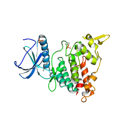
7A53
 
 | |
7A4R
 
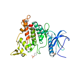 | | Structure of DYRK1A in complex with compound 1 | | 分子名称: | 2-amino-3H,4H,7H-pyrrolo[2,3-d]pyrimidin-4-one, DIMETHYL SULFOXIDE, Dual specificity tyrosine-phosphorylation-regulated kinase 1A | | 著者 | Dokurno, P, Surgenor, A.E, Hubbard, R.E. | | 登録日 | 2020-08-20 | | 公開日 | 2021-06-30 | | 最終更新日 | 2024-10-23 | | 実験手法 | X-RAY DIFFRACTION (1.8 Å) | | 主引用文献 | Fragment-Derived Selective Inhibitors of Dual-Specificity Kinases DYRK1A and DYRK1B.
J.Med.Chem., 64, 2021
|
|
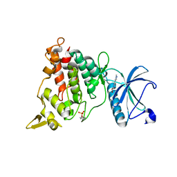
7A55
 
 | |
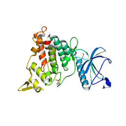
7A4W
 
 | |
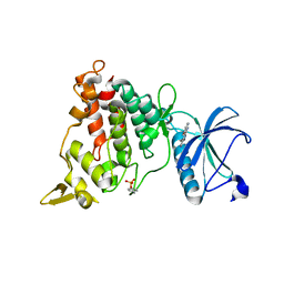
7A5B
 
 | |
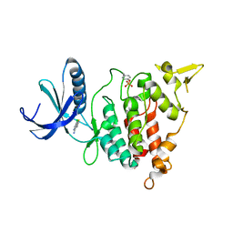
7A4S
 
 | |
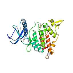
7A4Z
 
 | |
7A5D
 
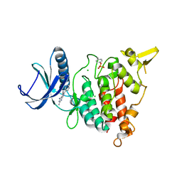 | | Structure of DYRK1A in complex with compound 16 | | 分子名称: | 4-azanyl-2-methyl-6-pyridin-3-yl-7~{H}-pyrrolo[2,3-d]pyrimidine-5-carbonitrile, CHLORIDE ION, Dual specificity tyrosine-phosphorylation-regulated kinase 1A | | 著者 | Dokurno, P, Surgenor, A.E, Hubbard, R.E. | | 登録日 | 2020-08-21 | | 公開日 | 2021-06-30 | | 最終更新日 | 2024-01-31 | | 実験手法 | X-RAY DIFFRACTION (1.8 Å) | | 主引用文献 | Fragment-Derived Selective Inhibitors of Dual-Specificity Kinases DYRK1A and DYRK1B.
J.Med.Chem., 64, 2021
|
|
7A51
 
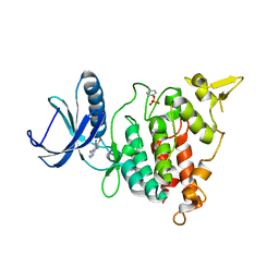 | |
7A52
 
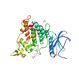 | | Structure of DYRK1A in complex with compound 6 | | 分子名称: | 3-phenyl-1H,4H,5H-pyrazolo[3,4-d]pyrimidin-4-one, DIMETHYL SULFOXIDE, Dual specificity tyrosine-phosphorylation-regulated kinase 1A, ... | | 著者 | Dokurno, P, Surgenor, A.E, Hubbard, R.E. | | 登録日 | 2020-08-20 | | 公開日 | 2021-06-30 | | 最終更新日 | 2024-01-31 | | 実験手法 | X-RAY DIFFRACTION (2.1 Å) | | 主引用文献 | Fragment-Derived Selective Inhibitors of Dual-Specificity Kinases DYRK1A and DYRK1B.
J.Med.Chem., 64, 2021
|
|
7A5N
 
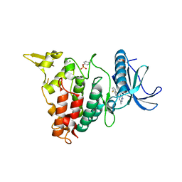 | | Structure of DYRK1A in complex with compound 34 | | 分子名称: | 5-(2-azanylpyridin-4-yl)-~{N}-[[2,6-bis(fluoranyl)phenyl]methyl]-2-methyl-7~{H}-pyrrolo[2,3-d]pyrimidin-4-amine, CHLORIDE ION, Dual specificity tyrosine-phosphorylation-regulated kinase 1A | | 著者 | Dokurno, P, Surgenor, A.E, Hubbard, R.E. | | 登録日 | 2020-08-21 | | 公開日 | 2021-06-30 | | 最終更新日 | 2024-01-31 | | 実験手法 | X-RAY DIFFRACTION (2.3 Å) | | 主引用文献 | Fragment-Derived Selective Inhibitors of Dual-Specificity Kinases DYRK1A and DYRK1B.
J.Med.Chem., 64, 2021
|
|
7A5L
 
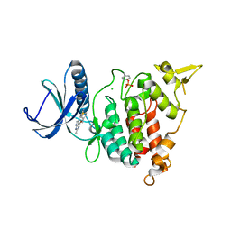 | | tructure of DYRK1A in complex with compound 24 | | 分子名称: | 4-[2-methyl-4-(thiophen-3-ylmethoxy)-7~{H}-pyrrolo[2,3-d]pyrimidin-5-yl]pyridin-2-amine, CHLORIDE ION, Dual specificity tyrosine-phosphorylation-regulated kinase 1A | | 著者 | Dokurno, P, Surgenor, A.E, Hubbard, R.E. | | 登録日 | 2020-08-21 | | 公開日 | 2021-06-30 | | 最終更新日 | 2024-10-16 | | 実験手法 | X-RAY DIFFRACTION (2.1 Å) | | 主引用文献 | Fragment-Derived Selective Inhibitors of Dual-Specificity Kinases DYRK1A and DYRK1B.
J.Med.Chem., 64, 2021
|
|
1QJZ
 
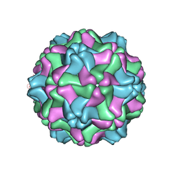 | | Three Dimensional Structure of Physalis Mottle Virus : Implications for the Viral Assembly | | 分子名称: | COAT PROTEIN | | 著者 | Krishna, S.S, Hiremath, C.N, Munshi, S.K, Prahadeeswaran, D, Sastri, M, Savithri, H.S, Murthy, M.R.N. | | 登録日 | 1999-07-07 | | 公開日 | 1999-07-08 | | 最終更新日 | 2023-12-13 | | 実験手法 | X-RAY DIFFRACTION (3.8 Å) | | 主引用文献 | Three Dimensional Structure of Physalis Movirus: Implications for the Viral Assembly
J.Mol.Biol., 289, 1999
|
|
4UVH
 
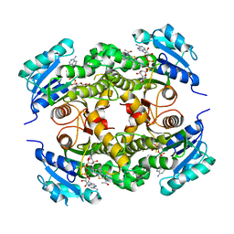 | | Discovery of pyrimidine isoxazoles InhA in complex with compound 10 | | 分子名称: | ACETATE ION, ENOYL-[ACYL-CARRIER-PROTEIN] REDUCTASE [NADH], N-(1,3-BENZOTHIAZOL-2-YL)ACETAMIDE, ... | | 著者 | Read, J.A, Gingell, H, Madhavapeddi, P, Ghorpade, S, Cowan, S. | | 登録日 | 2014-08-05 | | 公開日 | 2015-09-30 | | 最終更新日 | 2024-05-08 | | 実験手法 | X-RAY DIFFRACTION (1.89 Å) | | 主引用文献 | Hitting the Target in More Than One Way: Novel, Direct Inhibitors of Mycobacterium Tuberculosis Enoyl Acp Reductase
To be Published
|
|
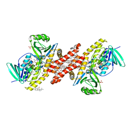
6M1W
 
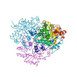 | | Structure of the 2-Aminoisobutyric acid Monooxygenase Hydroxylase | | 分子名称: | Amidohydrolase, CHLORIDE ION, FE (III) ION, ... | | 著者 | Hibi, M, Mikami, B, Ogawa, J. | | 登録日 | 2020-02-26 | | 公開日 | 2021-01-06 | | 最終更新日 | 2021-01-27 | | 実験手法 | X-RAY DIFFRACTION (2.75 Å) | | 主引用文献 | A three-component monooxygenase from Rhodococcus wratislaviensis may expand industrial applications of bacterial enzymes.
Commun Biol, 4, 2021
|
|
6M2I
 
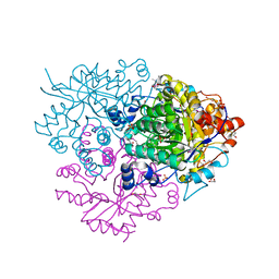 | | Structure of the 2-Aminoisobutyric acid Monooxygenase Hydroxylase | | 分子名称: | 1,2-ETHANEDIOL, Amidohydrolase, FE (III) ION, ... | | 著者 | Hibi, M, Mikami, B, Ogawa, J. | | 登録日 | 2020-02-27 | | 公開日 | 2021-01-06 | | 最終更新日 | 2023-11-29 | | 実験手法 | X-RAY DIFFRACTION (2.45 Å) | | 主引用文献 | A three-component monooxygenase from Rhodococcus wratislaviensis may expand industrial applications of bacterial enzymes.
Commun Biol, 4, 2021
|
|
4UVI
 
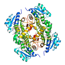 | | Discovery of pyrimidine isoxazoles InhA in complex with compound 23 | | 分子名称: | 5-{[(4,6-dimethylpyrimidin-2-yl)sulfanyl]methyl}-N-[(2-methylpyridin-4-yl)methyl]-1,2-oxazole-3-carboxamide, ENOYL-[ACYL-CARRIER-PROTEIN] REDUCTASE [NADH], NICOTINAMIDE-ADENINE-DINUCLEOTIDE | | 著者 | Read, J.A, Gingell, H, Madhavapeddi, P, Ghorpade, S, Cowan, S. | | 登録日 | 2014-08-05 | | 公開日 | 2015-09-30 | | 最終更新日 | 2024-05-08 | | 実験手法 | X-RAY DIFFRACTION (1.73 Å) | | 主引用文献 | Hitting the Target in More Than One Way: Novel, Direct Inhibitors of Mycobacterium Tuberculosis Enoyl Acp Reductase
To be Published
|
|
4UVD
 
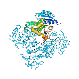 | | Discovery of pyrimidine isoxazoles InhA in complex with compound 6 | | 分子名称: | 2-[(4,6-dimethylpyrimidin-2-yl)sulfanyl]-N-[(2Z)-5-[3-(trifluoromethyl)benzyl]-1,3-thiazol-2(3H)-ylidene]acetamide, ENOYL-[ACYL-CARRIER-PROTEIN] REDUCTASE [NADH], MAGNESIUM ION, ... | | 著者 | Read, J.A, Gingell, H, Madhavapeddi, P, Ghorpade, S, Cowan, S. | | 登録日 | 2014-08-05 | | 公開日 | 2015-09-30 | | 最終更新日 | 2024-05-08 | | 実験手法 | X-RAY DIFFRACTION (1.82 Å) | | 主引用文献 | Hitting the Target in More Than One Way: Novel, Direct Inhibitors of Mycobacterium Tuberculosis Enoyl Acp Reductase
To be Published
|
|
4MS8
 
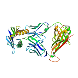 | | 42F3 TCR pCPB9/H-2Ld Complex | | 分子名称: | 42F3 alpha, 42F3 beta, H-2 class I histocompatibility antigen, ... | | 著者 | Birnbaum, M.E, Adams, J.J, Garcia, K.C. | | 登録日 | 2013-09-18 | | 公開日 | 2014-09-24 | | 最終更新日 | 2024-10-09 | | 実験手法 | X-RAY DIFFRACTION (1.922 Å) | | 主引用文献 | Structural interplay between germline interactions and adaptive recognition determines the bandwidth of TCR-peptide-MHC cross-reactivity.
Nat. Immunol., 17, 2016
|
|
6Z46
 
 | |
7S3E
 
 | |
1BA2
 
 | |
1GCA
 
 | |
1URP
 
 | |
6C6N
 
 | |
