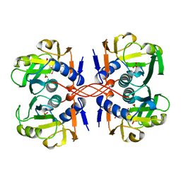
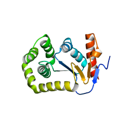
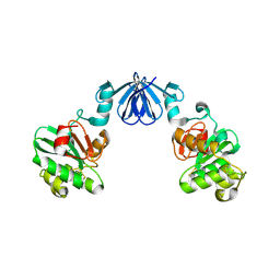
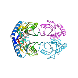
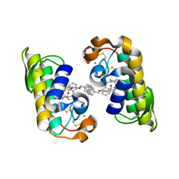
2AN3
 
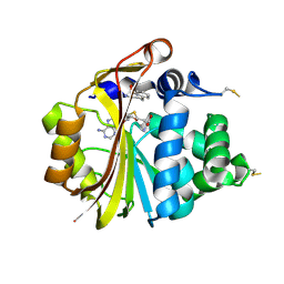 | | Structure of PNMT with S-adenosyl-L-homocysteine and the semi-rigid analogue acceptor substrate cis-(1R,2S)-2-amino-1-tetralol. | | 分子名称: | CIS-(1R,2S)-2-AMINO-1,2,3,4-TETRAHYDRONAPHTHALEN-1-OL, Phenylethanolamine N-methyltransferase, S-ADENOSYL-L-HOMOCYSTEINE | | 著者 | Gee, C.L, Tyndall, J.D.A, Grunewald, G.L, Wu, Q, McLeish, M.J, Martin, J.L. | | 登録日 | 2005-08-11 | | 公開日 | 2006-03-14 | | 最終更新日 | 2024-10-30 | | 実験手法 | X-RAY DIFFRACTION (2.2 Å) | | 主引用文献 | Mode of binding of methyl acceptor substrates to the adrenaline-synthesizing enzyme phenylethanolamine N-methyltransferase: implications for catalysis
Biochemistry, 44, 2005
|
|
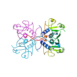
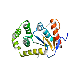
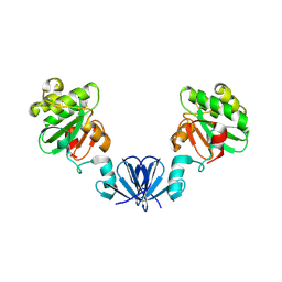
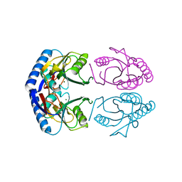
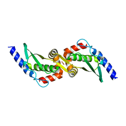
2AN4
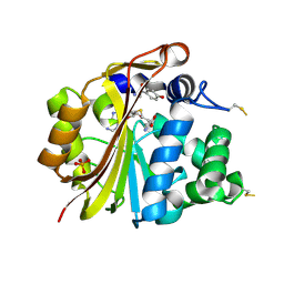 | | Structure of PNMT complexed with S-adenosyl-L-homocysteine and the acceptor substrate octopamine | | 分子名称: | 4-(2R-AMINO-1-HYDROXYETHYL)PHENOL, PHOSPHATE ION, Phenylethanolamine N-methyltransferase, ... | | 著者 | Gee, C.L, Tyndall, J.D.A, Grunewald, G.L, Wu, Q, McLeish, M.J, Martin, J.L. | | 登録日 | 2005-08-11 | | 公開日 | 2006-03-14 | | 最終更新日 | 2024-11-20 | | 実験手法 | X-RAY DIFFRACTION (2.2 Å) | | 主引用文献 | Mode of binding of methyl acceptor substrates to the adrenaline-synthesizing enzyme phenylethanolamine N-methyltransferase: implications for catalysis
Biochemistry, 44, 2005
|
|
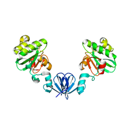
1A95
 | | XPRTASE FROM E. COLI COMPLEXED WITH MG:CPRPP AND GUANINE | | 分子名称: | 1-ALPHA-PYROPHOSPHORYL-2-ALPHA,3-ALPHA-DIHYDROXY-4-BETA-CYCLOPENTANE-METHANOL-5-PHOSPHATE, BORIC ACID, GUANINE, ... | | 著者 | Vos, S, Parry, R.J, Burns, M.R, De Jersey, J, Martin, J.L. | | 登録日 | 1998-04-16 | | 公開日 | 1998-11-11 | | 最終更新日 | 2024-05-22 | | 実験手法 | X-RAY DIFFRACTION (2 Å) | | 主引用文献 | Structures of free and complexed forms of Escherichia coli xanthine-guanine phosphoribosyltransferase.
J.Mol.Biol., 282, 1998
|
|
1A0M
 | | 1.1 ANGSTROM CRYSTAL STRUCTURE OF A-CONOTOXIN [TYR15]-EPI | | 分子名称: | ALPHA-CONOTOXIN [TYR15]-EPI | | 著者 | Hu, S.-H, Loughnan, M, Miller, R, Weeks, C.M, Blessing, R.H, Alewood, P.F, Lewis, R.J, Martin, J.L. | | 登録日 | 1997-12-03 | | 公開日 | 1999-01-13 | | 最終更新日 | 2024-10-30 | | 実験手法 | X-RAY DIFFRACTION (1.1 Å) | | 主引用文献 | The 1.1 A resolution crystal structure of [Tyr15]EpI, a novel alpha-conotoxin from Conus episcopatus, solved by direct methods.
Biochemistry, 37, 1998
|
|
3PUJ
 | | Crystal structure of the MUNC18-1 and SYNTAXIN4 N-Peptide complex | | 分子名称: | Syntaxin-4 N-terminal peptide, Syntaxin-binding protein 1 | | 著者 | Hu, S.-H, Christie, M.P, Saez, N.J, Latham, C.F, Jarrott, R, Lua, L.H.L, Collins, B.M, Martin, J.L. | | 登録日 | 2010-12-05 | | 公開日 | 2011-01-19 | | 最終更新日 | 2023-11-01 | | 実験手法 | X-RAY DIFFRACTION (3.313 Å) | | 主引用文献 | Possible roles for Munc18-1 domain 3a and Syntaxin1 N-peptide and C-terminal anchor in SNARE complex formation
Proc.Natl.Acad.Sci.USA, 108, 2011
|
|
1A97
 | | XPRTASE FROM E. COLI COMPLEXED WITH GMP | | 分子名称: | BORIC ACID, GUANOSINE-5'-MONOPHOSPHATE, XANTHINE-GUANINE PHOSPHORIBOSYLTRANSFERASE | | 著者 | Vos, S, Parry, R.J, Burns, M.R, De Jersey, J, Martin, J.L. | | 登録日 | 1998-04-16 | | 公開日 | 1998-11-11 | | 最終更新日 | 2024-05-22 | | 実験手法 | X-RAY DIFFRACTION (2.6 Å) | | 主引用文献 | Structures of free and complexed forms of Escherichia coli xanthine-guanine phosphoribosyltransferase.
J.Mol.Biol., 282, 1998
|
|
1AKG
 | |
1A98
 | | XPRTASE FROM E. COLI COMPLEXED WITH GMP | | 分子名称: | XANTHINE-GUANINE PHOSPHORIBOSYLTRANSFERASE | | 著者 | Vos, S, Parry, R.J, Burns, M.R, De Jersey, J, Martin, J.L. | | 登録日 | 1998-04-16 | | 公開日 | 1998-06-17 | | 最終更新日 | 2024-05-22 | | 実験手法 | X-RAY DIFFRACTION (2.25 Å) | | 主引用文献 | Structures of free and complexed forms of Escherichia coli xanthine-guanine phosphoribosyltransferase.
J.Mol.Biol., 282, 1998
|
|
1A96
 | | XPRTASE FROM E. COLI WITH BOUND CPRPP AND XANTHINE | | 分子名称: | 1-ALPHA-PYROPHOSPHORYL-2-ALPHA,3-ALPHA-DIHYDROXY-4-BETA-CYCLOPENTANE-METHANOL-5-PHOSPHATE, BORIC ACID, MAGNESIUM ION, ... | | 著者 | Vos, S, Parry, R.J, Burns, M.R, De Jersey, J, Martin, J.L. | | 登録日 | 1998-04-16 | | 公開日 | 1998-11-11 | | 最終更新日 | 2024-05-22 | | 実験手法 | X-RAY DIFFRACTION (2 Å) | | 主引用文献 | Structures of free and complexed forms of Escherichia coli xanthine-guanine phosphoribosyltransferase.
J.Mol.Biol., 282, 1998
|
|
4MCU
 | |
1PEN
 | | ALPHA-CONOTOXIN PNI1 | | 分子名称: | ALPHA-CONOTOXIN PNIA | | 著者 | Hu, S.-H, Gehrmann, J, Guddat, L.W, Alewood, P.F, Craik, D.J, Martin, J.L. | | 登録日 | 1996-01-29 | | 公開日 | 1997-04-21 | | 最終更新日 | 2024-10-16 | | 実験手法 | X-RAY DIFFRACTION (1.1 Å) | | 主引用文献 | The 1.1 A crystal structure of the neuronal acetylcholine receptor antagonist, alpha-conotoxin PnIA from Conus pennaceus.
Structure, 4, 1996
|
|
4MLY
 | | Disulfide isomerase from multidrug resistance IncA/C related integrative and conjugative elements in oxidized state (P21 space group) | | 分子名称: | 1,3-BUTANEDIOL, DsbP | | 著者 | Premkumar, L, Kurth, F, Neyer, S, Martin, J.L. | | 登録日 | 2013-09-06 | | 公開日 | 2013-12-11 | | 最終更新日 | 2024-10-16 | | 実験手法 | X-RAY DIFFRACTION (2.207 Å) | | 主引用文献 | The Multidrug Resistance IncA/C Transferable Plasmid Encodes a Novel Domain-swapped Dimeric Protein-disulfide Isomerase.
J.Biol.Chem., 289, 2014
|
|
4OCF
 | |
4ML6
 | |
4ML1
 | |
4OCE
 | |
4OD7
 | |
1G9T
 | | CRYSTAL STRUCTURE OF E.COLI HPRT-GMP COMPLEX | | 分子名称: | ANY 5'-MONOPHOSPHATE NUCLEOTIDE, GUANOSINE-5'-MONOPHOSPHATE, HYPOXANTHINE PHOSPHORIBOSYLTRANSFERASE | | 著者 | Guddat, L.W, Vos, S, Martin, J.L, keough, D.T, de Jersey, J. | | 登録日 | 2000-11-28 | | 公開日 | 2002-08-28 | | 最終更新日 | 2024-04-03 | | 実験手法 | X-RAY DIFFRACTION (2.8 Å) | | 主引用文献 | Crystal structures of free, IMP-, and GMP-bound Escherichia coli hypoxanthine phosphoribosyltransferase.
Protein Sci., 11, 2002
|
|
1GRV
 | | Hypoxanthine Phosphoribosyltransferase from E. coli | | 分子名称: | HYPOXANTHINE PHOSPHORIBOSYLTRANSFERASE, MAGNESIUM ION | | 著者 | Guddat, L.W, Vos, S, Martin, J.L, Keough, D.T, De Jersey, J. | | 登録日 | 2001-12-17 | | 公開日 | 2002-12-13 | | 最終更新日 | 2023-12-13 | | 実験手法 | X-RAY DIFFRACTION (2.9 Å) | | 主引用文献 | Crystal Structures of Free, Imp-, and Gmp- Bound Escherichia Coli Hypoxanthine Phosphoribosyltransferase
Protein Sci., 11, 2002
|
|
1G9S
 | | CRYSTAL STRUCTURE OF A COMPLEX BETWEEN E.COLI HPRT AND IMP | | 分子名称: | ANY 5'-MONOPHOSPHATE NUCLEOTIDE, HYPOXANTHINE PHOSPHORIBOSYLTRANSFERASE, INOSINIC ACID | | 著者 | Guddat, L.W, Vos, S, Martin, J.L, Keough, D.T, de Jersey, J. | | 登録日 | 2000-11-27 | | 公開日 | 2002-08-28 | | 最終更新日 | 2024-04-03 | | 実験手法 | X-RAY DIFFRACTION (2.8 Å) | | 主引用文献 | Crystal structures of free, IMP-, and GMP-bound Escherichia coli hypoxanthine phosphoribosyltransferase.
Protein Sci., 11, 2002
|
|
3UX2
 | |
1J1A
 | | PANCREATIC SECRETORY PHOSPHOLIPASE A2 (IIa) WITH ANTI-INFLAMMATORY ACTIVITY | | 分子名称: | (S)-5-(4-BENZYLOXY-PHENYL)-4-(7-PHENYL-HEPTANOYLAMINO)-PENTANOIC ACID, CALCIUM ION, Phospholipase A2 | | 著者 | Hansford, K.A, Reid, R.C, Clark, C.I, Tyndall, J.D.A, Whitehouse, M.W, Guthrie, T, McGeary, R.P, Schafer, K, Martin, J.L, Fairlie, D.P. | | 登録日 | 2002-12-03 | | 公開日 | 2003-03-18 | | 最終更新日 | 2024-11-13 | | 実験手法 | X-RAY DIFFRACTION (2.2 Å) | | 主引用文献 | D-Tyrosine as a Chiral Precusor to Potent Inhibitors of Human Nonpancreatic Secretory Phospholipase A2 (IIa) with Antiinflammatory Activity
Chembiochem, 4, 2003
|
|
3UX3
 | | Crystal Structure of Domain-Swapped Fam96a minor dimer | | 分子名称: | ACETATE ION, MIP18 family protein FAM96A, ZINC ION | | 著者 | Chen, K.-E, Kobe, B, Martin, J.L. | | 登録日 | 2011-12-03 | | 公開日 | 2012-05-30 | | 最終更新日 | 2023-11-08 | | 実験手法 | X-RAY DIFFRACTION (1.8 Å) | | 主引用文献 | The mammalian DUF59 protein Fam96a forms two distinct types of domain-swapped dimer.
Acta Crystallogr.,Sect.D, 68, 2012
|
|
1BED
 | |
1CJM
 | | HUMAN SULT1A3 WITH SULFATE BOUND | | 分子名称: | PROTEIN (ARYL SULFOTRANSFERASE), SULFATE ION | | 著者 | Bidwell, L.M, Mcmanus, M.E, Gaedigk, A, Kakuta, Y, Negishi, M, Pedersen, L, Martin, J.L. | | 登録日 | 1999-04-18 | | 公開日 | 1999-11-10 | | 最終更新日 | 2023-08-09 | | 実験手法 | X-RAY DIFFRACTION (2.4 Å) | | 主引用文献 | Crystal structure of human catecholamine sulfotransferase.
J.Mol.Biol., 293, 1999
|
|


