6E80
 
 | |
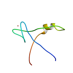
6E84
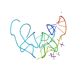
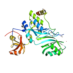
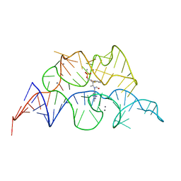
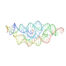
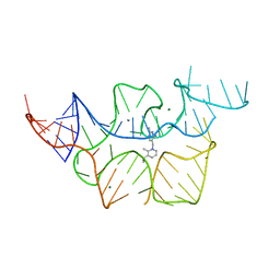
 | | Crystal structure of the Corn aptamer in complex with TO | | 分子名称: | 1-methyl-4-[(Z)-(3-methyl-1,3-benzothiazol-2(3H)-ylidene)methyl]quinolin-1-ium, POTASSIUM ION, RNA (36-MER) | | 著者 | Sjekloca, L, Ferre-D'Amare, A.R. | | 登録日 | 2018-07-27 | | 公開日 | 2019-07-31 | | 最終更新日 | 2023-10-11 | | 実験手法 | X-RAY DIFFRACTION (2.901 Å) | | 主引用文献 | Binding between G Quadruplexes at the Homodimer Interface of the Corn RNA Aptamer Strongly Activates Thioflavin T Fluorescence.
Cell Chem Biol, 26, 2019
|
|
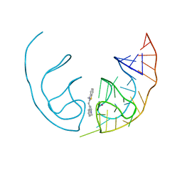
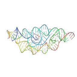
6E81
 | |
1ZL3
 | | Coupling of active site motions and RNA binding | | 分子名称: | 5'-R(*GP*GP*CP*AP*AP*CP*GP*GP*UP*(FLO) UP*CP*GP*AP*UP*CP*CP*CP*GP*UP*UP*GP*C)-3', SULFATE ION, tRNA pseudouridine synthase B | | 著者 | Hoang, C, Hamilton, C.S, Mueller, E.G, Ferre-D'Amare, A.R. | | 登録日 | 2005-05-05 | | 公開日 | 2005-08-09 | | 最終更新日 | 2023-08-23 | | 実験手法 | X-RAY DIFFRACTION (2.8 Å) | | 主引用文献 | Precursor complex structure of pseudouridine synthase TruB suggests coupling of active site perturbations to an RNA-sequestering peripheral protein domain
Protein Sci., 14, 2005
|
|
2APO
 | | Crystal Structure of the Methanococcus jannaschii Cbf5 Nop10 Complex | | 分子名称: | POTASSIUM ION, Probable tRNA pseudouridine synthase B, Ribosome biogenesis protein Nop10, ... | | 著者 | Hamma, T, Reichow, S.L, Varani, G, Ferre-D'Amare, A.R. | | 登録日 | 2005-08-16 | | 公開日 | 2005-11-15 | | 最終更新日 | 2011-07-13 | | 実験手法 | X-RAY DIFFRACTION (1.95 Å) | | 主引用文献 | The Cbf5-Nop10 complex is a molecular bracket that organizes box H/ACA RNPs.
Nat.Struct.Mol.Biol., 12, 2005
|
|
2AQA
 | |
2GCS
 | |
2GCV
 | |
2AQC
 | | NMR Structural analysis of archaeal Nop10 | | 分子名称: | Ribosome biogenesis protein Nop10, ZINC ION | | 著者 | Hamma, T, Reichow, S.L, Varani, G, Ferre-D'Amare, A.R. | | 登録日 | 2005-08-17 | | 公開日 | 2005-11-15 | | 最終更新日 | 2024-05-22 | | 実験手法 | SOLUTION NMR | | 主引用文献 | The Cbf5-Nop10 complex is a molecular bracket that organizes box H/ACA RNPs.
Nat.Struct.Mol.Biol., 12, 2005
|
|
2H0Z
 | |
2HO7
 | |
2HOK
 | |
2HOL
 | |
2HOO
 | |
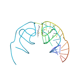
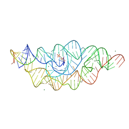
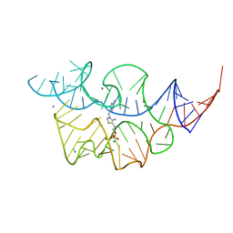
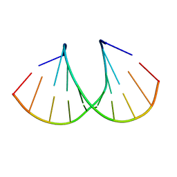
5V3F
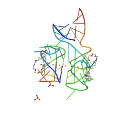 | | Co-crystal structure of the fluorogenic RNA Mango | | 分子名称: | 4-{[(2S)-3-{2,16-dioxo-20-[(3aS,4S,6aR)-2-oxohexahydro-1H-thieno[3,4-d]imidazol-4-yl]-6,9,12-trioxa-3,15-diazaicosan-1-yl}-2,3-dihydro-1,3-benzothiazol-2-yl]methyl}-1-methylquinolin-1-ium, PHOSPHATE ION, POTASSIUM ION, ... | | 著者 | Trachman, R.J, Ferre-D'Amare, A.R. | | 登録日 | 2017-03-07 | | 公開日 | 2017-05-24 | | 最終更新日 | 2024-03-06 | | 実験手法 | X-RAY DIFFRACTION (1.7 Å) | | 主引用文献 | Structural basis for high-affinity fluorophore binding and activation by RNA Mango.
Nat. Chem. Biol., 13, 2017
|
|
2H0X
 | |
2H0S
 | |
2H0W
 | |
2HO6
 | |
2HOM
 | |
2HOJ
 | |
2HOP
 | |
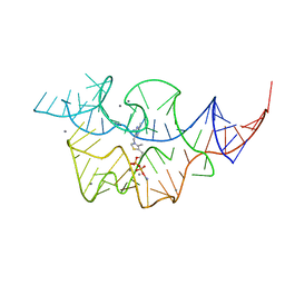
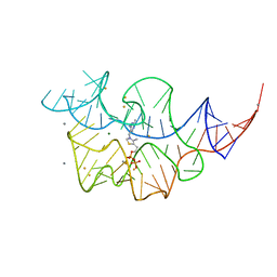
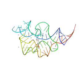
6OD9
 | | Co-crystal structure of the Fusobacterium ulcerans ZTP riboswitch using an X-ray free-electron laser | | 分子名称: | AMINOIMIDAZOLE 4-CARBOXAMIDE RIBONUCLEOTIDE, CESIUM ION, MAGNESIUM ION, ... | | 著者 | Jones, C.P, Tran, B, Ferre-D'Amare, A.R. | | 登録日 | 2019-03-26 | | 公開日 | 2019-07-17 | | 最終更新日 | 2023-10-11 | | 実験手法 | X-RAY DIFFRACTION (4.102 Å) | | 主引用文献 | Co-crystal structure of the Fusobacterium ulcerans ZTP riboswitch using an X-ray free-electron laser.
Acta Crystallogr.,Sect.F, 75, 2019
|
|
3OT0
 | |
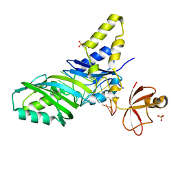
1AM9
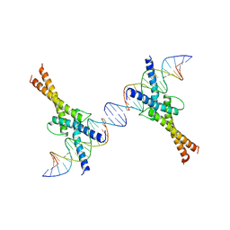 | | HUMAN SREBP-1A BOUND TO LDL RECEPTOR PROMOTER | | 分子名称: | DNA (5'-D(*CP*AP*TP*GP*AP*GP*AP*TP*CP*AP*CP*CP*CP*CP*AP*CP*T P*GP*CP*AP*A)-3'), DNA (5'-D(*TP*TP*GP*CP*AP*GP*TP*GP*GP*GP*GP*TP*GP*AP*TP*CP*T )-3'), MAGNESIUM ION, ... | | 著者 | Parraga, A, Burley, S.K. | | 登録日 | 1997-06-25 | | 公開日 | 1998-07-10 | | 最終更新日 | 2024-04-03 | | 実験手法 | X-RAY DIFFRACTION (2.3 Å) | | 主引用文献 | Co-crystal structure of sterol regulatory element binding protein 1a at 2.3 A resolution.
Structure, 6, 1998
|
|


