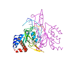
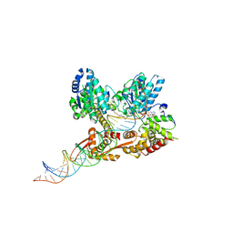
1JL2
 
 | |
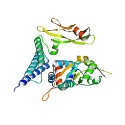
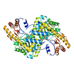
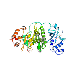
1MU5
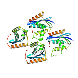
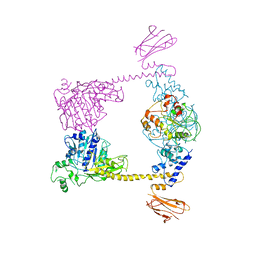
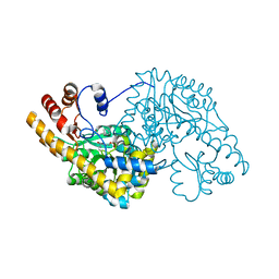
 | | Structure of topoisomerase subunit | | 分子名称: | CALCIUM ION, Type II DNA topoisomerase VI Subunit B | | 著者 | Corbett, K.D, Berger, J.M. | | 登録日 | 2002-09-23 | | 公開日 | 2003-01-07 | | 最終更新日 | 2024-02-14 | | 実験手法 | X-RAY DIFFRACTION (2 Å) | | 主引用文献 | Structure of the topoisomerase VI-B subunit: implications for type II topoisomerase mechanism and evolution
Embo J., 22, 2003
|
|
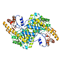
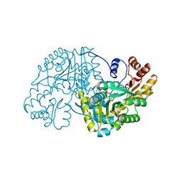
1MX0
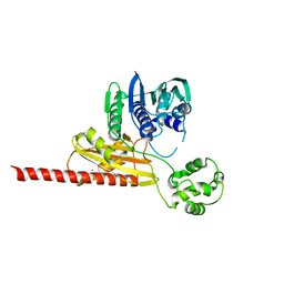
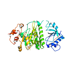
 | | Structure of topoisomerase subunit | | 分子名称: | MAGNESIUM ION, PHOSPHOAMINOPHOSPHONIC ACID-ADENYLATE ESTER, SODIUM ION, ... | | 著者 | Corbett, K.D, Berger, J.M. | | 登録日 | 2002-10-01 | | 公開日 | 2003-01-07 | | 最終更新日 | 2011-07-13 | | 実験手法 | X-RAY DIFFRACTION (2.3 Å) | | 主引用文献 | Structure of the topoisomerase VI-B subunit: implications for type II topoisomerase mechanism and evolution
Embo J., 22, 2003
|
|
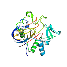
1L6Z
 | | CRYSTAL STRUCTURE OF MURINE CEACAM1A[1,4]: A CORONAVIRUS RECEPTOR AND CELL ADHESION MOLECULE IN THE CEA FAMILY | | 分子名称: | 2-acetamido-2-deoxy-beta-D-glucopyranose, beta-D-mannopyranose-(1-4)-2-acetamido-2-deoxy-beta-D-glucopyranose-(1-4)-2-acetamido-2-deoxy-beta-D-glucopyranose, biliary glycoprotein C | | 著者 | Tan, K, Zelus, B.D, Meijers, R, Liu, J.-H, Bergelson, J.M, Duke, N, Zhang, R, Joachimiak, A, Holmes, K.V, Wang, J.-H. | | 登録日 | 2002-03-14 | | 公開日 | 2002-09-14 | | 最終更新日 | 2023-08-16 | | 実験手法 | X-RAY DIFFRACTION (3.32 Å) | | 主引用文献 | CRYSTAL STRUCTURE OF MURINE sCEACAM1a[1,4]: A CORONAVIRUS RECEPTOR IN THE CEA FAMILY
Embo J., 21, 2002
|
|
3HVD
 | | The Protective Antigen Component of Anthrax Toxin Forms Functional Octameric Complexes | | 分子名称: | CALCIUM ION, Protective antigen | | 著者 | Kintzer, A.F, Dong, K.C, Berger, J.M, Krantz, B.A. | | 登録日 | 2009-06-15 | | 公開日 | 2009-08-25 | | 最終更新日 | 2024-03-13 | | 実験手法 | X-RAY DIFFRACTION (3.209 Å) | | 主引用文献 | The protective antigen component of anthrax toxin forms functional octameric complexes.
J.Mol.Biol., 392, 2009
|
|
3ICE
 | |
437D
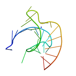 | | CRYSTAL STRUCTURE OF AN RNA PSEUDOKNOT FROM BEET WESTERN YELLOW VIRUS INVOLVED IN RIBOSOMAL FRAMESHIFTING | | 分子名称: | MAGNESIUM ION, RNA PSEUDOKNOT, SODIUM ION | | 著者 | Su, L, Chen, L, Egli, M, Berger, J.M, Rich, A. | | 登録日 | 1998-11-24 | | 公開日 | 1998-12-03 | | 最終更新日 | 2024-02-28 | | 実験手法 | X-RAY DIFFRACTION (1.6 Å) | | 主引用文献 | Minor groove RNA triplex in the crystal structure of a ribosomal frameshifting viral pseudoknot.
Nat.Struct.Biol., 6, 1999
|
|
3IBP
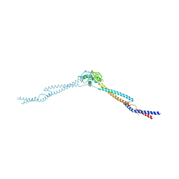 | | The Crystal Structure of the Dimerization Domain of Escherichia coli Structural Maintenance of Chromosomes Protein MukB | | 分子名称: | AMMONIUM ION, Chromosome partition protein mukB | | 著者 | Li, Y, Schoeffler, A.J, Berger, J.M, Oakley, M.G. | | 登録日 | 2009-07-16 | | 公開日 | 2010-01-26 | | 最終更新日 | 2017-11-01 | | 実験手法 | X-RAY DIFFRACTION (3.099 Å) | | 主引用文献 | The crystal structure of the hinge domain of the Escherichia coli structural maintenance of chromosomes protein MukB.
J.Mol.Biol., 395, 2010
|
|
3ILW
 | | Structure of DNA gyrase subunit A N-terminal domain | | 分子名称: | DNA gyrase subunit A, GLYCEROL | | 著者 | Tretter, E.M, Schoeffler, A.J, Weisfield, S.R, Berger, J.M, TB Structural Genomics Consortium (TBSGC) | | 登録日 | 2009-08-07 | | 公開日 | 2009-10-06 | | 最終更新日 | 2023-09-06 | | 実験手法 | X-RAY DIFFRACTION (1.603 Å) | | 主引用文献 | Crystal structure of the DNA gyrase GyrA N-terminal domain from Mycobacterium tuberculosis.
Proteins, 78, 2010
|
|
1TOE
 | | Unliganded structure of Hexamutant + A293D mutant of E. coli aspartate aminotransferase | | 分子名称: | Aspartate aminotransferase, SULFATE ION | | 著者 | Chow, M.A, McElroy, K.E, Corbett, K.D, Berger, J.M, Kirsch, J.F. | | 登録日 | 2004-06-14 | | 公開日 | 2004-10-05 | | 最終更新日 | 2023-11-15 | | 実験手法 | X-RAY DIFFRACTION (2 Å) | | 主引用文献 | Narrowing substrate specificity in a directly evolved enzyme: the A293D mutant of aspartate aminotransferase
Biochemistry, 43, 2004
|
|
2OXV
 | |
1TUE
 | |
1TOG
 | | Hydrocinnamic acid-bound structure of SRHEPT + A293D mutant of E. coli aspartate aminotransferase | | 分子名称: | Aspartate aminotransferase, HYDROCINNAMIC ACID | | 著者 | Chow, M.A, McElroy, K.E, Corbett, K.D, Berger, J.M, Kirsch, J.F. | | 登録日 | 2004-06-14 | | 公開日 | 2004-10-05 | | 最終更新日 | 2023-11-15 | | 実験手法 | X-RAY DIFFRACTION (2.31 Å) | | 主引用文献 | Narrowing substrate specificity in a directly evolved enzyme: the A293D mutant of aspartate aminotransferase
Biochemistry, 43, 2004
|
|
1VS0
 | | Crystal Structure of the Ligase Domain from M. tuberculosis LigD at 2.4A | | 分子名称: | CHLORIDE ION, MAGNESIUM ION, Putative DNA ligase-like protein Rv0938/MT0965, ... | | 著者 | Akey, D, Martins, A, Aniukwu, J, Glickman, M.S, Shuman, S, Berger, J.M, TB Structural Genomics Consortium (TBSGC) | | 登録日 | 2006-01-27 | | 公開日 | 2006-02-28 | | 最終更新日 | 2011-07-13 | | 実験手法 | X-RAY DIFFRACTION (2.4 Å) | | 主引用文献 | Crystal Structure and Nonhomologous End-joining Function of the Ligase Component of Mycobacterium DNA Ligase D.
J.Biol.Chem., 281, 2006
|
|
2Q17
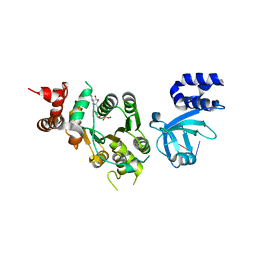
 | | Formylglycine Generating Enzyme from Streptomyces coelicolor | | 分子名称: | CALCIUM ION, formylglycine generating enzyme | | 著者 | Carlson, B.L, Ballister, E.R, Skordalakes, E, King, D.S, Breidenbach, M.A, Gilmore, S.A, Berger, J.M, Bertozzi, C.R. | | 登録日 | 2007-05-23 | | 公開日 | 2008-04-01 | | 最終更新日 | 2023-08-30 | | 実験手法 | X-RAY DIFFRACTION (2.1 Å) | | 主引用文献 | Function and structure of a prokaryotic formylglycine-generating enzyme.
J.Biol.Chem., 283, 2008
|
|
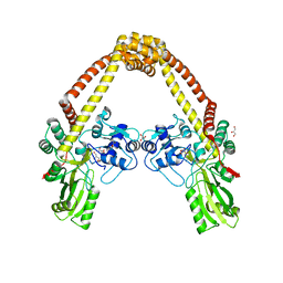
2QBY
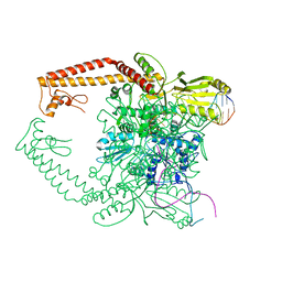
 | | Crystal structure of a heterodimer of Cdc6/Orc1 initiators bound to origin DNA (from S. solfataricus) | | 分子名称: | ADENOSINE-5'-DIPHOSPHATE, Cell division control protein 6 homolog 1, Cell division control protein 6 homolog 3, ... | | 著者 | Cunningham Dueber, E.L, Corn, J.E, Bell, S.D, Berger, J.M. | | 登録日 | 2007-06-18 | | 公開日 | 2007-09-11 | | 最終更新日 | 2023-08-30 | | 実験手法 | X-RAY DIFFRACTION (3.35 Å) | | 主引用文献 | Replication origin recognition and deformation by a heterodimeric archaeal Orc1 complex.
Science, 317, 2007
|
|
1TOK
 | | Maleic acid-bound structure of SRHEPT mutant of E. coli aspartate aminotransferase | | 分子名称: | Aspartate aminotransferase, MALEIC ACID | | 著者 | Chow, M.A, McElroy, K.E, Corbett, K.D, Berger, J.M, Kirsch, J.F. | | 登録日 | 2004-06-14 | | 公開日 | 2004-10-05 | | 最終更新日 | 2023-11-15 | | 実験手法 | X-RAY DIFFRACTION (1.85 Å) | | 主引用文献 | Narrowing substrate specificity in a directly evolved enzyme: the A293D mutant of aspartate aminotransferase
Biochemistry, 43, 2004
|
|
2Q2E
 | |
1TOJ
 | | Hydrocinnamic acid-bound structure of SRHEPT mutant of E. coli aspartate aminotransferase | | 分子名称: | Aspartate aminotransferase, HYDROCINNAMIC ACID | | 著者 | Chow, M.A, McElroy, K.E, Corbett, K.D, Berger, J.M, Kirsch, J.F. | | 登録日 | 2004-06-14 | | 公開日 | 2004-10-05 | | 最終更新日 | 2023-11-15 | | 実験手法 | X-RAY DIFFRACTION (1.9 Å) | | 主引用文献 | Narrowing substrate specificity in a directly evolved enzyme: the A293D mutant of aspartate aminotransferase
Biochemistry, 43, 2004
|
|
1TOI
 | | Hydrocinnamic acid-bound structure of Hexamutant + A293D mutant of E. coli aspartate aminotransferase | | 分子名称: | Aspartate aminotransferase, HYDROCINNAMIC ACID | | 著者 | Chow, M.A, McElroy, K.E, Corbett, K.D, Berger, J.M, Kirsch, J.F. | | 登録日 | 2004-06-14 | | 公開日 | 2004-10-05 | | 最終更新日 | 2023-11-15 | | 実験手法 | X-RAY DIFFRACTION (1.9 Å) | | 主引用文献 | Narrowing substrate specificity in a directly evolved enzyme: the A293D mutant of aspartate aminotransferase
Biochemistry, 43, 2004
|
|
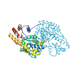
1XPR
 | | Structural mechanism of inhibition of the Rho transcription termination factor by the antibiotic 5a-formylbicyclomycin (FB) | | 分子名称: | 5'-R(*CP*UP*CP*UP*CP*UP*CP*U)-3', 5A-FORMYLBICYCLOMYCIN, MAGNESIUM ION, ... | | 著者 | Skordalakes, E, Brogan, A.P, Park, B.S, Kohn, H, Berger, J.M. | | 登録日 | 2004-10-09 | | 公開日 | 2004-11-02 | | 最終更新日 | 2024-02-14 | | 実験手法 | X-RAY DIFFRACTION (3.15 Å) | | 主引用文献 | Structural mechanism of inhibition of the rho transcription termination factor by the antibiotic bicyclomycin
Structure, 13, 2005
|
|
2RGR
 | |
1XPU
 | | Structural mechanism of inhibition of the Rho transcription termination factor by the antibiotic 5a-(3-formylphenylsulfanyl)-dihydrobicyclomycin (FPDB) | | 分子名称: | 5'-R(*CP*UP*CP*UP*CP*UP*CP*U)-3', 5A-(3-FORMYLPHENYLSULFANYL)-DIHYDROBICYCLOMYCIN, MAGNESIUM ION, ... | | 著者 | Skordalakes, E, Brogan, A.P, Park, B.S, Kohn, H, Berger, J.M. | | 登録日 | 2004-10-09 | | 公開日 | 2004-11-02 | | 最終更新日 | 2011-07-13 | | 実験手法 | X-RAY DIFFRACTION (3.05 Å) | | 主引用文献 | Structural mechanism of inhibition of the rho transcription termination factor by the antibiotic bicyclomycin
Structure, 13, 2005
|
|
1XPO
 | | Structural mechanism of inhibition of the Rho transcription termination factor by the antibiotic bicyclomycin | | 分子名称: | 5'-R(*CP*UP*CP*UP*CP*UP*CP*U)-3', BICYCLOMYCIN, MAGNESIUM ION, ... | | 著者 | Skordalakes, E, Brogan, A.P, Park, B.S, Kohn, H, Berger, J.M. | | 登録日 | 2004-10-09 | | 公開日 | 2005-02-08 | | 最終更新日 | 2018-01-31 | | 実験手法 | X-RAY DIFFRACTION (3.15 Å) | | 主引用文献 | Structural mechanism of inhibition of the rho transcription termination factor by the antibiotic bicyclomycin
Structure, 13, 2005
|
|
1QC9
 | |


