7NGI
 
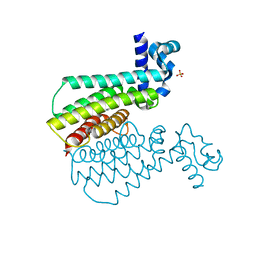 | |
7NGY
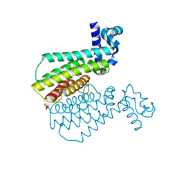 | |
7NGW
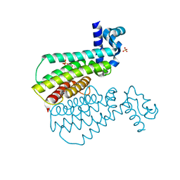 | |
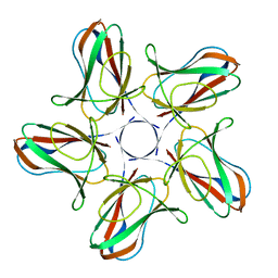
6L6O
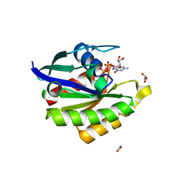 | | Crystal structure of stabilized Rab5a GTPase domain from Leishmania donovani | | 分子名称: | ACETATE ION, GLYCEROL, GUANOSINE-5'-DIPHOSPHATE, ... | | 著者 | Arora, A, Zohib, M, Biswal, B.K, Maheshwari, D, Pal, R.K. | | 登録日 | 2019-10-29 | | 公開日 | 2020-11-11 | | 最終更新日 | 2023-11-22 | | 実験手法 | X-RAY DIFFRACTION (1.8 Å) | | 主引用文献 | Crystal structure of the GDP-bound GTPase domain of Rab5a from Leishmania donovani.
Acta Crystallogr.,Sect.F, 76, 2020
|
|
6I5O
 | |
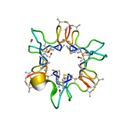
6I56
 | | Crystal structure of PBSX exported protein XepA | | 分子名称: | GLYCEROL, Phage-like element PBSX protein XepA | | 著者 | Hakansson, M, Svensson, L.A, Welin, M, Al-Karadaghi, S. | | 登録日 | 2018-11-13 | | 公開日 | 2019-11-20 | | 最終更新日 | 2024-05-15 | | 実験手法 | X-RAY DIFFRACTION (2.12 Å) | | 主引用文献 | Crystal structures of the Bacillus subtilis prophage lytic cassette proteins XepA and YomS.
Acta Crystallogr D Struct Biol, 75, 2019
|
|
1CED
 | | THE STRUCTURE OF CYTOCHROME C6 FROM MONORAPHIDIUM BRAUNII, NMR, MINIMIZED AVERAGE STRUCTURE | | 分子名称: | CYTOCHROME C6, PROTOPORPHYRIN IX CONTAINING FE | | 著者 | Banci, L, Bertini, I, Quacquarini, G, Walter, O, Diaz, A, Hervas, M, De La Rosa, M.A. | | 登録日 | 1996-03-06 | | 公開日 | 1996-08-17 | | 最終更新日 | 2022-02-16 | | 実験手法 | SOLUTION NMR | | 主引用文献 | The solution structure of cytochrome c6 from the green alga Monoraphidium braunii
J.Biol.Inorg.Chem., 1, 1996
|
|
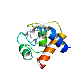
7OXW
 | | CrabP2 mutant R30DK31D | | 分子名称: | ACETATE ION, Cellular retinoic acid-binding protein 2, SULFATE ION | | 著者 | Pastok, M.W, Basle, A, Endicott, J.A. | | 登録日 | 2021-06-23 | | 公開日 | 2022-07-13 | | 最終更新日 | 2024-01-31 | | 実験手法 | X-RAY DIFFRACTION (1.16 Å) | | 主引用文献 | Structural requirements for the specific binding of CRABP2 to cyclin D3
To Be Published
|
|
1IBJ
 | | Crystal structure of cystathionine beta-lyase from Arabidopsis thaliana | | 分子名称: | CARBONATE ION, CYSTATHIONINE BETA-LYASE, PYRIDOXAL-5'-PHOSPHATE, ... | | 著者 | Breitinger, U, Clausen, T, Messerschmidt, A. | | 登録日 | 2001-03-28 | | 公開日 | 2001-04-04 | | 最終更新日 | 2023-08-09 | | 実験手法 | X-RAY DIFFRACTION (2.3 Å) | | 主引用文献 | The three-dimensional structure of cystathionine beta-lyase from Arabidopsis and its substrate specificity
Plant Physiol., 126, 2001
|
|
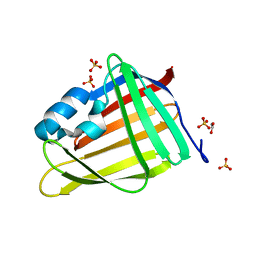
1QOW
 | |
1CTJ
 | | CRYSTAL STRUCTURE OF CYTOCHROME C6 | | 分子名称: | CYTOCHROME C6, PROTOPORPHYRIN IX CONTAINING FE | | 著者 | Sheldrick, G.M. | | 登録日 | 1995-08-08 | | 公開日 | 1996-06-10 | | 最終更新日 | 2011-07-13 | | 実験手法 | X-RAY DIFFRACTION (1.1 Å) | | 主引用文献 | Ab initio determination of the crystal structure of cytochrome c6 and comparison with plastocyanin.
Structure, 3, 1995
|
|
1OAE
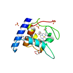 | | Crystal structure of the reduced form of cytochrome c" from Methylophilus methylotrophus | | 分子名称: | CYTOCHROME C", GLYCEROL, HEME C, ... | | 著者 | Enguita, F.J, Grenha, R, Santos, H, Carrondo, M.A. | | 登録日 | 2003-01-09 | | 公開日 | 2004-03-26 | | 最終更新日 | 2024-05-01 | | 実験手法 | X-RAY DIFFRACTION (1.95 Å) | | 主引用文献 | Structural Evidence for a Proton Transfer Pathway Coupled with Haem Reduction of Cytochrome C" from Methylophilus Methylotrophus.
J.Biol.Inorg.Chem., 11, 2006
|
|
1SOC
 | |
2SOC
 | |
1KMH
 | |
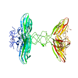
7ZMZ
 | | Engineered Interleukin 2 bound to CD25 receptor | | 分子名称: | Interleukin-2, Interleukin-2 receptor subunit alpha | | 著者 | Fyfe, P.K, Moraga, I, Gaggero, S, Mitra, S. | | 登録日 | 2022-04-20 | | 公開日 | 2022-12-14 | | 最終更新日 | 2024-01-31 | | 実験手法 | X-RAY DIFFRACTION (3.2 Å) | | 主引用文献 | IL-2 is inactivated by the acidic pH environment of tumors enabling engineering of a pH-selective mutein.
Sci Immunol, 7, 2022
|
|


