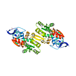
1Y6A
 
 | | Crystal structure of VEGFR2 in complex with a 2-anilino-5-aryl-oxazole inhibitor | | 分子名称: | N-[5-(ETHYLSULFONYL)-2-METHOXYPHENYL]-5-[3-(2-PYRIDINYL)PHENYL]-1,3-OXAZOL-2-AMINE, Vascular endothelial growth factor receptor 2 | | 著者 | Harris, P.A, Cheung, M, Hunter, R.N, Brown, M.L, Veal, J.M, Nolte, R.T, Wang, L, Liu, W, Crosby, R.M, Johnson, J.H, Epperly, A.H, Kumar, R, Luttrell, D.K, Stafford, J.A. | | 登録日 | 2004-12-05 | | 公開日 | 2005-06-07 | | 最終更新日 | 2024-02-14 | | 実験手法 | X-RAY DIFFRACTION (2.1 Å) | | 主引用文献 | Discovery and evaluation of 2-anilino-5-aryloxazoles as a novel class of VEGFR2 kinase inhibitors.
J.Med.Chem., 48, 2005
|
|
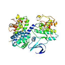
5YLO
 | | Structural of Pseudomonas aeruginosa PA4980 | | 分子名称: | GLYCEROL, Probable enoyl-CoA hydratase/isomerase | | 著者 | Liu, L, Li, T, Peng, C.T, Li, C.C, Xiao, Q.J, He, L.H, Wang, N.Y, Bao, R. | | 登録日 | 2017-10-18 | | 公開日 | 2018-08-22 | | 最終更新日 | 2024-03-27 | | 実験手法 | X-RAY DIFFRACTION (2.39 Å) | | 主引用文献 | Structural characterization of a Delta3, Delta2-enoyl-CoA isomerase from Pseudomonas aeruginosa: implications for its involvement in unsaturated fatty acid metabolism.
J.Biomol.Struct.Dyn., 37, 2019
|
|
1Y89
 | | Crystal Structure of devB protein | | 分子名称: | 3,6,9,12,15,18,21,24-OCTAOXAHEXACOSAN-1-OL, DI(HYDROXYETHYL)ETHER, NONAETHYLENE GLYCOL, ... | | 著者 | Lazarski, K, Cymborowski, M, Chruszcz, M, Zheng, H, Zhang, R, Lezondra, L, Joachimiak, A, Minor, W, Midwest Center for Structural Genomics (MCSG) | | 登録日 | 2004-12-10 | | 公開日 | 2005-01-25 | | 最終更新日 | 2022-04-13 | | 実験手法 | X-RAY DIFFRACTION (2 Å) | | 主引用文献 | Crystal Structure of devB protein
To be Published
|
|
5YL9
 | |
5MIU
 | | G307E variant of Murine Apoptosis Inducing Factor (oxidized state) | | 分子名称: | Apoptosis-inducing factor 1, mitochondrial, FLAVIN-ADENINE DINUCLEOTIDE | | 著者 | Sorrentino, L, Cossu, F, Milani, M, Aliverti, A, Mastrangelo, E. | | 登録日 | 2016-11-29 | | 公開日 | 2017-07-12 | | 最終更新日 | 2024-01-17 | | 実験手法 | X-RAY DIFFRACTION (3.5 Å) | | 主引用文献 | Structural bases of the altered catalytic properties of a pathogenic variant of apoptosis inducing factor.
Biochem. Biophys. Res. Commun., 490, 2017
|
|
5YNB
 | | Crystal structure of MERS-CoV nsp16/nsp10 complex bound to Sinefungin | | 分子名称: | SINEFUNGIN, ZINC ION, nsp10 protein, ... | | 著者 | Wei, S.M, Yang, L, Ke, Z.H, Chen, Y, Guo, D.Y, Fan, C.P. | | 登録日 | 2017-10-24 | | 公開日 | 2018-12-05 | | 最終更新日 | 2023-11-22 | | 実験手法 | X-RAY DIFFRACTION (1.98 Å) | | 主引用文献 | Structural insights into the molecular mechanism of MERS Coronavirus RNA ribose 2'-O-methylation by nsp16/nsp10 protein complex
To Be Published
|
|
1YD6
 | | Crystal structure of the GIY-YIG N-terminal endonuclease domain of UvrC from Bacillus caldotenax | | 分子名称: | CHLORIDE ION, SULFATE ION, UvrC | | 著者 | Truglio, J.J, Rhau, B, Croteau, D.L, Wang, L, Skorvaga, M, Karakas, E, DellaVecchia, M.J, Wang, H, Van Houten, B, Kisker, C. | | 登録日 | 2004-12-23 | | 公開日 | 2005-03-01 | | 最終更新日 | 2024-03-13 | | 実験手法 | X-RAY DIFFRACTION (2 Å) | | 主引用文献 | Structural insights into the first incision reaction during nucleotide excision repair
Embo J., 24, 2005
|
|
5YNM
 | | Crystal structure of MERS-CoV nsp16/nsp10 complex bound to SAM and m7GpppA | | 分子名称: | P1-7-METHYLGUANOSINE-P3-ADENOSINE-5',5'-TRIPHOSPHATE, S-ADENOSYLMETHIONINE, ZINC ION, ... | | 著者 | Wei, S.M, Yang, L, Ke, Z.H, Guo, D.Y, Fan, C.P. | | 登録日 | 2017-10-24 | | 公開日 | 2018-12-05 | | 最終更新日 | 2023-11-22 | | 実験手法 | X-RAY DIFFRACTION (1.68 Å) | | 主引用文献 | Structural insights into the molecular mechanism of MERS Coronavirus RNA ribose 2'-O-methylation by nsp16/nsp10 protein complex
To Be Published
|
|
1NZR
 | | CRYSTAL STRUCTURE OF THE AZURIN MUTANT NICKEL-TRP48MET FROM PSEUDOMONAS AERUGINOSA AT 2.2 ANGSTROMS RESOLUTION | | 分子名称: | AZURIN, NICKEL (II) ION, NITRATE ION | | 著者 | Tsai, L.-C, Sjolin, L, Langer, V, Bonander, N, Karlsson, B.G, Vanngard, T, Hammann, C, Nar, H. | | 登録日 | 1994-12-09 | | 公開日 | 1995-02-27 | | 最終更新日 | 2024-06-05 | | 実験手法 | X-RAY DIFFRACTION (2.2 Å) | | 主引用文献 | Structure of the azurin mutant nickel-Trp48Met from Pseudomonas aeruginosa at 2.2 A resolution.
Acta Crystallogr.,Sect.D, 51, 1995
|
|
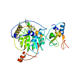
5C7L
 | | Structure of human testis-specific glyceraldehyde-3-phosphate dehydrogenase apo form | | 分子名称: | Glyceraldehyde-3-phosphate dehydrogenase, testis-specific | | 著者 | Betts, L, Machius, M, Danshina, P, O'Brien, D. | | 登録日 | 2015-06-24 | | 公開日 | 2016-03-09 | | 最終更新日 | 2019-12-11 | | 実験手法 | X-RAY DIFFRACTION (1.862 Å) | | 主引用文献 | Structural analyses to identify selective inhibitors of glyceraldehyde 3-phosphate dehydrogenase-S, a sperm-specific glycolytic enzyme.
Mol. Hum. Reprod., 22, 2016
|
|
1YB5
 | | Crystal structure of human Zeta-Crystallin with bound NADP | | 分子名称: | ACETATE ION, CHLORIDE ION, GLYCEROL, ... | | 著者 | Debreczeni, J, Berridge, G, Kavanagh, K, Colbrook, S, Bray, J, Williams, L, Oppermann, U, Sundstrom, M, Arrowsmith, C, Edwards, A, Gileadi, O, von Delft, F, Structural Genomics Consortium (SGC) | | 登録日 | 2004-12-20 | | 公開日 | 2004-12-28 | | 最終更新日 | 2024-02-14 | | 実験手法 | X-RAY DIFFRACTION (1.85 Å) | | 主引用文献 | Crystal Structure of human Zeta-Crystallin at 1.85A
To be Published
|
|
1OJ6
 | | Human brain neuroglobin three-dimensional structure | | 分子名称: | Neuroglobin, PROTOPORPHYRIN IX CONTAINING FE, SULFATE ION | | 著者 | Pesce, A, Dewilde, S, Nardini, M, Moens, L, Ascenzi, P, Hankeln, T, Burmester, T, Bolognesi, M. | | 登録日 | 2003-07-03 | | 公開日 | 2003-09-11 | | 最終更新日 | 2024-05-08 | | 実験手法 | X-RAY DIFFRACTION (1.95 Å) | | 主引用文献 | Human Brain Neuroglobin Structure Reveals a Distinct Mode of Controlling Oxygen Affinity
Structure, 11, 2003
|
|
1YJX
 | | Crystal structure of human B type phosphoglycerate mutase | | 分子名称: | CHLORIDE ION, CITRIC ACID, Phosphoglycerate mutase 1 | | 著者 | Wang, Y, Wei, Z, Liu, L, Gong, W. | | 登録日 | 2005-01-16 | | 公開日 | 2005-05-17 | | 最終更新日 | 2023-10-25 | | 実験手法 | X-RAY DIFFRACTION (2.8 Å) | | 主引用文献 | Crystal structure of human B-type phosphoglycerate mutase bound with citrate.
Biochem.Biophys.Res.Commun., 331, 2005
|
|
1YJU
 | | Solution structure of the apo form of the sixth soluble domain of Menkes protein | | 分子名称: | Copper-transporting ATPase 1 | | 著者 | Banci, L, Bertini, I, Cantini, F, Migliardi, M, Rosato, A, Wang, S, Structural Proteomics in Europe (SPINE) | | 登録日 | 2005-01-15 | | 公開日 | 2006-01-03 | | 最終更新日 | 2024-05-29 | | 実験手法 | SOLUTION NMR | | 主引用文献 | An atomic-level investigation of the disease-causing A629P mutant of the Menkes protein, ATP7A
J.Mol.Biol., 352, 2005
|
|
1YJT
 | | Solution structure of the Cu(I) form of the sixth soluble domain A69P mutant of Menkes protein | | 分子名称: | COPPER (I) ION, Copper-transporting ATPase 1 | | 著者 | Banci, L, Bertini, I, Cantini, F, Migliardi, M, Rosato, A, Wang, S. | | 登録日 | 2005-01-15 | | 公開日 | 2006-01-03 | | 最終更新日 | 2024-05-29 | | 実験手法 | SOLUTION NMR | | 主引用文献 | An atomic-level investigation of the disease-causing A629P mutant of the Menkes protein, ATP7A
J.Mol.Biol., 352, 2005
|
|
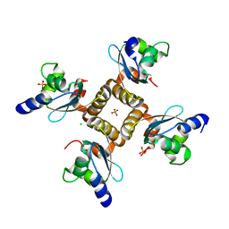
5MOA
 | | ABA RECEPTOR FROM TOMATO, SlPYL1 | | 分子名称: | SlPYL1 | | 著者 | Moreno-Alvero, M, Yunta, C, Gonzalez-Guzman, M, Arbona, V, Granell, A, Martinez-Ripoll, M, Infantes, L, Rodriguez, P.L, Albert, A. | | 登録日 | 2016-12-14 | | 公開日 | 2017-08-02 | | 最終更新日 | 2024-05-08 | | 実験手法 | X-RAY DIFFRACTION (1.65 Å) | | 主引用文献 | Structure of Ligand-Bound Intermediates of Crop ABA Receptors Highlights PP2C as Necessary ABA Co-receptor.
Mol Plant, 10, 2017
|
|
1YJR
 | | Solution structure of the apo form of the sixth soluble domain A69P mutant of Menkes protein | | 分子名称: | Copper-transporting ATPase 1 | | 著者 | Banci, L, Bertini, I, Cantini, F, Migliardi, M, Rosato, A, Wang, S, Structural Proteomics in Europe (SPINE) | | 登録日 | 2005-01-15 | | 公開日 | 2006-01-03 | | 最終更新日 | 2024-05-29 | | 実験手法 | SOLUTION NMR | | 主引用文献 | An atomic-level investigation of the disease-causing A629P mutant of the Menkes protein, ATP7A
J.Mol.Biol., 352, 2005
|
|
1OGS
 | | human acid-beta-glucosidase | | 分子名称: | 2-acetamido-2-deoxy-beta-D-glucopyranose, 2-acetamido-2-deoxy-beta-D-glucopyranose-(1-4)-2-acetamido-2-deoxy-beta-D-glucopyranose, Glucosylceramidase, ... | | 著者 | Dvir, H, Harel, M, McCarthy, A.A, Toker, L, Silman, I, Futerman, A.H, Sussman, J.L. | | 登録日 | 2003-05-13 | | 公開日 | 2003-07-03 | | 最終更新日 | 2020-07-29 | | 実験手法 | X-RAY DIFFRACTION (2 Å) | | 主引用文献 | X-Ray Structure of Human Acid-Beta-Glucosidase, the Defective Enzyme in Gaucher Disease
Embo Rep., 4, 2003
|
|
5YP7
 | | p62/SQSTM1 ZZ domain | | 分子名称: | Sequestosome-1, ZINC ION | | 著者 | Kwon, D.H, Kim, L, Song, H.K. | | 登録日 | 2017-11-01 | | 公開日 | 2018-08-29 | | 最終更新日 | 2024-03-27 | | 実験手法 | X-RAY DIFFRACTION (1.424 Å) | | 主引用文献 | Insights into degradation mechanism of N-end rule substrates by p62/SQSTM1 autophagy adapter.
Nat Commun, 9, 2018
|
|
7QSX
 | | Non-obligately L8S8-complex forming RubisCO derived from ancestral sequence reconstruction and rational engineering in L8 complex | | 分子名称: | 2-CARBOXYARABINITOL-1,5-DIPHOSPHATE, MAGNESIUM ION, RubisCO large subunit | | 著者 | Zarzycki, J, Schulz, L, Erb, T.J, Hochberg, G.K.A. | | 登録日 | 2022-01-14 | | 公開日 | 2022-10-12 | | 最終更新日 | 2024-01-31 | | 実験手法 | X-RAY DIFFRACTION (2.7 Å) | | 主引用文献 | Evolution of increased complexity and specificity at the dawn of form I Rubiscos.
Science, 378, 2022
|
|
1YJV
 | | Solution structure of the Cu(I) form of the sixth soluble domain of Menkes protein | | 分子名称: | COPPER (I) ION, Copper-transporting ATPase 1 | | 著者 | Banci, L, Bertini, I, Cantini, F, Migliardi, M, Rosato, A, Wang, S. | | 登録日 | 2005-01-15 | | 公開日 | 2006-01-03 | | 最終更新日 | 2024-05-29 | | 実験手法 | SOLUTION NMR | | 主引用文献 | An atomic-level investigation of the disease-causing A629P mutant of the Menkes protein, ATP7A
J.Mol.Biol., 352, 2005
|
|
1Y8Z
 | | alpha-glucosyltransferase in complex with UDP and a 13-mer DNA containing a HMU base at 1.9 A resolution | | 分子名称: | 5'-D(*CP*TP*AP*TP*CP*TP*GP*AP*G)-3', 5'-D(*GP*AP*TP*AP*CP*TP*(5HU)P*AP*GP*AP*TP*AP*G)-3', CHLORIDE ION, ... | | 著者 | Lariviere, L, Sommer, N, Morera, S. | | 登録日 | 2004-12-14 | | 公開日 | 2005-08-30 | | 最終更新日 | 2023-10-25 | | 実験手法 | X-RAY DIFFRACTION (1.9 Å) | | 主引用文献 | Structural evidence of a passive base-flipping mechanism for AGT, an unusual GT-B glycosyltransferase.
J.Mol.Biol., 352, 2005
|
|
5YSJ
 | | SdeA mART-C domain WT apo | | 分子名称: | Ubiquitinating/deubiquitinating enzyme SdeA | | 著者 | Kim, L, Kwon, D.H, Song, H.K. | | 登録日 | 2017-11-14 | | 公開日 | 2018-08-29 | | 最終更新日 | 2024-03-27 | | 実験手法 | X-RAY DIFFRACTION (2.059 Å) | | 主引用文献 | Structural and Biochemical Study of the Mono-ADP-Ribosyltransferase Domain of SdeA, a Ubiquitylating/Deubiquitylating Enzyme from Legionella pneumophila
J. Mol. Biol., 430, 2018
|
|
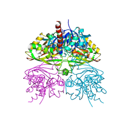
6JQN
 | | Structure of PaaZ, a bifunctional enzyme in complex with NADP+ and OCoA | | 分子名称: | Bifunctional protein PaaZ, NADP NICOTINAMIDE-ADENINE-DINUCLEOTIDE PHOSPHATE, OCTANOYL-COENZYME A | | 著者 | Gakher, L, Vinothkumar, K.R, Katagihallimath, N, Sowdhamini, R, Sathyanarayanan, N, Cannone, G. | | 登録日 | 2019-03-31 | | 公開日 | 2019-09-11 | | 最終更新日 | 2024-03-27 | | 実験手法 | ELECTRON MICROSCOPY (3.1 Å) | | 主引用文献 | Molecular basis for metabolite channeling in a ring opening enzyme of the phenylacetate degradation pathway.
Nat Commun, 10, 2019
|
|
1OKV
 | | Cyclin A binding groove inhibitor H-Arg-Arg-Leu-Ile-Phe-NH2 | | 分子名称: | CELL DIVISION PROTEIN KINASE 2, CYCLIN A2, H-ARG-ARG-LEU-ILE-PHE-NH2 | | 著者 | Kontopidis, G, Andrews, M, McInnes, C, Cowan, A, Powers, H, Innes, L, Plater, A, Griffiths, G, Paterson, D, Zheleva, D, Lane, D, Green, S, Walkinshaw, M, Fischer, P. | | 登録日 | 2003-07-30 | | 公開日 | 2003-12-11 | | 最終更新日 | 2023-12-13 | | 実験手法 | X-RAY DIFFRACTION (2.4 Å) | | 主引用文献 | Insights Into Cyclin Groove Recognition. Complex Crystal Structures and Inhibitor Design Through Ligand Exchange
Structure, 11, 2003
|
|


