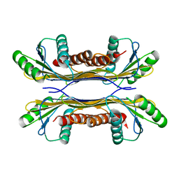
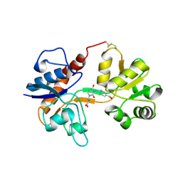
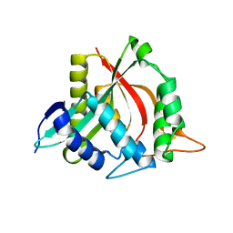
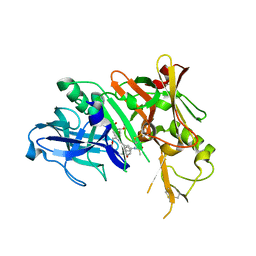
3H78
 
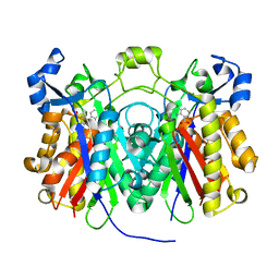 | | Crystal structure of Pseudomonas aeruginosa PqsD C112A mutant in complex with anthranilic acid | | 分子名称: | 2-AMINOBENZOIC ACID, PQS biosynthetic enzyme | | 著者 | Bera, A.K, Atanasova, V, Parsons, J.F. | | 登録日 | 2009-04-24 | | 公開日 | 2009-09-15 | | 最終更新日 | 2023-11-15 | | 実験手法 | X-RAY DIFFRACTION (1.7 Å) | | 主引用文献 | Structure of PqsD, a Pseudomonas quinolone signal biosynthetic enzyme, in complex with anthranilate.
Biochemistry, 48, 2009
|
|
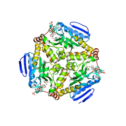
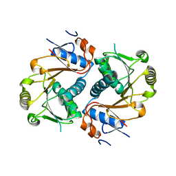
3HGU
 | |
3HGV
 | |
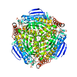
5T10
 | |
5T11
 | |
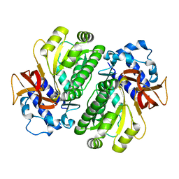
5T0Z
 | | PelC from Geobacter metallireducens | | 分子名称: | Lipoprotein, putative | | 著者 | Marmont, L.S, Howell, P.L. | | 登録日 | 2016-08-17 | | 公開日 | 2017-03-15 | | 最終更新日 | 2024-03-06 | | 実験手法 | X-RAY DIFFRACTION (2.255 Å) | | 主引用文献 | Oligomeric lipoprotein PelC guides Pel polysaccharide export across the outer membrane of Pseudomonas aeruginosa.
Proc. Natl. Acad. Sci. U.S.A., 114, 2017
|
|
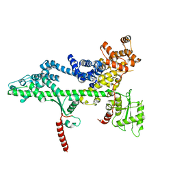
2XHS
 | |
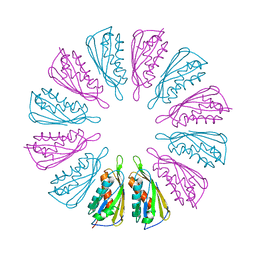
7M72
 | | MHC-like protein complex structure | | 分子名称: | (3R)-N-[(2S,3R)-1-(alpha-D-galactopyranosyloxy)-3-hydroxy-15-methylhexadecan-2-yl]-3-hydroxyheptadecanamide, 2-acetamido-2-deoxy-beta-D-glucopyranose-(1-4)-2-acetamido-2-deoxy-beta-D-glucopyranose, Antigen-presenting glycoprotein CD1d1, ... | | 著者 | Thirunavukkarasu, P, Le Nours, J, Rossjohn, J. | | 登録日 | 2021-03-26 | | 公開日 | 2021-11-10 | | 最終更新日 | 2024-10-16 | | 実験手法 | X-RAY DIFFRACTION (2.4 Å) | | 主引用文献 | Host immunomodulatory lipids created by symbionts from dietary amino acids.
Nature, 600, 2021
|
|
3H03
 | |
3H06
 | |
4I52
 | | scMenB im complex with 1-hydroxy-2-naphthoyl-CoA | | 分子名称: | 1-hydroxy-2-naphthoyl-CoA, CHLORIDE ION, Naphthoate synthase | | 著者 | Song, H.G, Sun, Y.R, Li, J, Li, Y, Jiang, M, Zhou, J.H, Guo, Z.H. | | 登録日 | 2012-11-28 | | 公開日 | 2013-05-08 | | 最終更新日 | 2023-11-08 | | 実験手法 | X-RAY DIFFRACTION (2.35 Å) | | 主引用文献 | Structural basis of the induced-fit mechanism of 1,4-dihydroxy-2-naphthoyl coenzyme a synthase from the crotonase fold superfamily
Plos One, 8, 2013
|
|
4I4Z
 | | Synechocystis sp. PCC 6803 1,4-dihydroxy-2-naphthoyl-coenzyme A synthase (MenB) in complex with salicylyl-CoA | | 分子名称: | BICARBONATE ION, MALONATE ION, Naphthoate synthase, ... | | 著者 | Song, H.G, Sun, Y.R, Li, J, Li, Y, Jiang, M, Zhou, J.H, Guo, Z.H. | | 登録日 | 2012-11-28 | | 公開日 | 2013-05-08 | | 最終更新日 | 2023-11-08 | | 実験手法 | X-RAY DIFFRACTION (2 Å) | | 主引用文献 | Structural basis of the induced-fit mechanism of 1,4-dihydroxy-2-naphthoyl coenzyme a synthase from the crotonase fold superfamily
Plos One, 8, 2013
|
|
3PSJ
 | |
3PSF
 | |
4FD6
 | |
3PSK
 | |
4FD7
 | | Crystal structure of insect putative arylalkylamine N-Acetyltransferase 7 from the yellow fever mosquito Aedes aegypt | | 分子名称: | 1,2-ETHANEDIOL, IODIDE ION, SULFATE ION, ... | | 著者 | Han, Q, Robinson, R, Li, J. | | 登録日 | 2012-05-26 | | 公開日 | 2012-06-27 | | 最終更新日 | 2023-09-13 | | 実験手法 | X-RAY DIFFRACTION (1.8 Å) | | 主引用文献 | Evolution of insect arylalkylamine N-acetyltransferases: structural evidence from the yellow fever mosquito, Aedes aegypti.
Proc.Natl.Acad.Sci.USA, 109, 2012
|
|
4FD5
 | |
4FD4
 | |
3I25
 | | Potent Beta-Secretase 1 hydroxyethylene Inhibitor | | 分子名称: | Beta-secretase 1, N-[(2S,3S,5R)-1-(3,5-difluorophenoxy)-3-hydroxy-5-(2-methoxyethoxy)-6-[[(2S)-3-methyl-1-oxo-1-(phenylmethylamino)butan-2-yl]amino]-6-oxo-hexan-2-yl]-5-(methyl-methylsulfonyl-amino)-N'-[(1R)-1-phenylethyl]benzene-1,3-dicarboxamide | | 著者 | Lindberg, J.D, Borkakoti, N, Nystrom, S. | | 登録日 | 2009-06-29 | | 公開日 | 2010-06-02 | | 最終更新日 | 2024-10-09 | | 実験手法 | X-RAY DIFFRACTION (2.1 Å) | | 主引用文献 | Discovery of potent BACE-1 inhibitors containing a new hydroxyethylene (HE) scaffold: exploration of P1' alkoxy residues and an aminoethylene (AE) central core
Bioorg.Med.Chem., 18, 2010
|
|
5I91
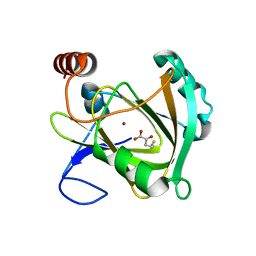 | | Structure of Mouse Acirecutone dioxygenase with to Ni2+ and 2-keto-4-(methylthio)-butyric acid in the active site | | 分子名称: | 1,2-dihydroxy-3-keto-5-methylthiopentene dioxygenase, 4-(METHYLSULFANYL)-2-OXOBUTANOIC ACID, NICKEL (II) ION | | 著者 | Deshpande, A.R, Robinson, H, Wagenpfeil, K, Pochapsky, T.C, Petsko, G.A, Ringe, D. | | 登録日 | 2016-02-19 | | 公開日 | 2016-03-09 | | 最終更新日 | 2023-09-27 | | 実験手法 | X-RAY DIFFRACTION (1.76 Å) | | 主引用文献 | Metal-Dependent Function of a Mammalian Acireductone Dioxygenase.
Biochemistry, 55, 2016
|
|
4QU4
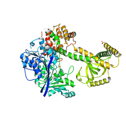 | |
3L2K
 | | Structure of phenazine antibiotic biosynthesis protein with substrate | | 分子名称: | EhpF, phenazine-1,6-dicarboxylic acid | | 著者 | Bera, A.K, Atanasova, V, Parsons, J.F. | | 登録日 | 2009-12-15 | | 公開日 | 2010-05-26 | | 最終更新日 | 2023-09-06 | | 実験手法 | X-RAY DIFFRACTION (2.8 Å) | | 主引用文献 | Structure of the D-alanylgriseoluteic acid biosynthetic protein EhpF, an atypical member of the ANL superfamily of adenylating enzymes.
Acta Crystallogr.,Sect.D, 66, 2010
|
|
3D34
 | |
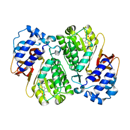
3D3K
 | | Crystal structure of human Edc3p | | 分子名称: | Enhancer of mRNA-decapping protein 3 | | 著者 | Ling, S.H.M. | | 登録日 | 2008-05-12 | | 公開日 | 2008-08-26 | | 最終更新日 | 2024-03-20 | | 実験手法 | X-RAY DIFFRACTION (2.2 Å) | | 主引用文献 | Crystal structure of human Edc3 and its functional implications
Mol.Cell.Biol., 28, 2008
|
|


