7LUH
 
 | |
7LUJ
 | |
1G9S
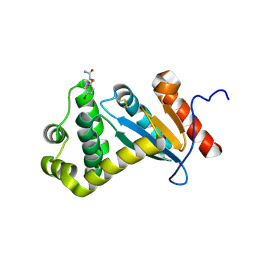
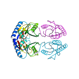
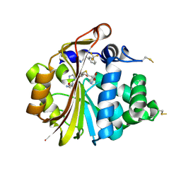
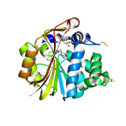
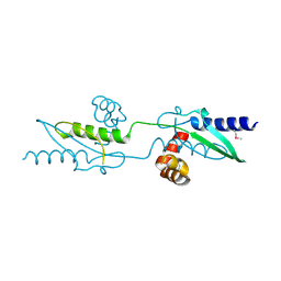
 | | CRYSTAL STRUCTURE OF A COMPLEX BETWEEN E.COLI HPRT AND IMP | | 分子名称: | ANY 5'-MONOPHOSPHATE NUCLEOTIDE, HYPOXANTHINE PHOSPHORIBOSYLTRANSFERASE, INOSINIC ACID | | 著者 | Guddat, L.W, Vos, S, Martin, J.L, Keough, D.T, de Jersey, J. | | 登録日 | 2000-11-27 | | 公開日 | 2002-08-28 | | 最終更新日 | 2024-04-03 | | 実験手法 | X-RAY DIFFRACTION (2.8 Å) | | 主引用文献 | Crystal structures of free, IMP-, and GMP-bound Escherichia coli hypoxanthine phosphoribosyltransferase.
Protein Sci., 11, 2002
|
|
1G9T
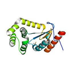
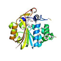
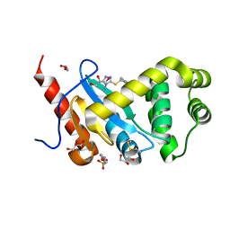
 | | CRYSTAL STRUCTURE OF E.COLI HPRT-GMP COMPLEX | | 分子名称: | ANY 5'-MONOPHOSPHATE NUCLEOTIDE, GUANOSINE-5'-MONOPHOSPHATE, HYPOXANTHINE PHOSPHORIBOSYLTRANSFERASE | | 著者 | Guddat, L.W, Vos, S, Martin, J.L, keough, D.T, de Jersey, J. | | 登録日 | 2000-11-28 | | 公開日 | 2002-08-28 | | 最終更新日 | 2024-04-03 | | 実験手法 | X-RAY DIFFRACTION (2.8 Å) | | 主引用文献 | Crystal structures of free, IMP-, and GMP-bound Escherichia coli hypoxanthine phosphoribosyltransferase.
Protein Sci., 11, 2002
|
|
1GRV
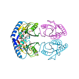
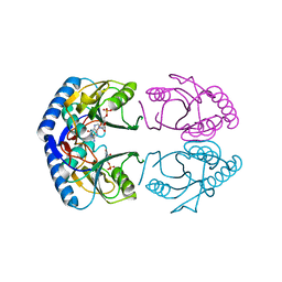
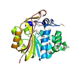
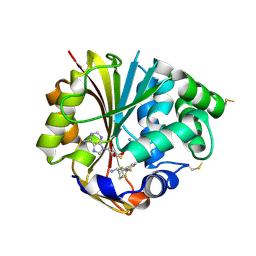
 | | Hypoxanthine Phosphoribosyltransferase from E. coli | | 分子名称: | HYPOXANTHINE PHOSPHORIBOSYLTRANSFERASE, MAGNESIUM ION | | 著者 | Guddat, L.W, Vos, S, Martin, J.L, Keough, D.T, De Jersey, J. | | 登録日 | 2001-12-17 | | 公開日 | 2002-12-13 | | 最終更新日 | 2023-12-13 | | 実験手法 | X-RAY DIFFRACTION (2.9 Å) | | 主引用文献 | Crystal Structures of Free, Imp-, and Gmp- Bound Escherichia Coli Hypoxanthine Phosphoribosyltransferase
Protein Sci., 11, 2002
|
|
2V1O
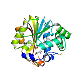
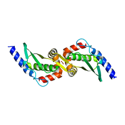
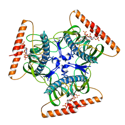 | | Crystal structure of N-terminal domain of acyl-CoA thioesterase 7 | | 分子名称: | COENZYME A, CYTOSOLIC ACYL COENZYME A THIOESTER HYDROLASE | | 著者 | Forwood, J.K, Thakur, A.S, Guncar, G, Marfori, M, Mouradov, D, Meng, W.N, Robinson, J, Huber, T, Kellie, S, Martin, J.L, Hume, D.A, Kobe, B. | | 登録日 | 2007-05-28 | | 公開日 | 2007-06-26 | | 最終更新日 | 2024-05-08 | | 実験手法 | X-RAY DIFFRACTION (1.78 Å) | | 主引用文献 | Structural Basis for Recruitment of Tandem Hotdog Domains in Acyl-Coa Thioesterase 7 and its Role in Inflammation.
Proc.Natl.Acad.Sci.USA, 104, 2007
|
|
3PUK
 | | Re-refinement of the crystal structure of Munc18-3 and Syntaxin4 N-peptide complex | | 分子名称: | Syntaxin-4 N-terminal peptide, Syntaxin-binding protein 3 | | 著者 | Hu, S.-H, Christie, M.P, Saez, N.J, Latham, C.F, Jarrott, R, Lua, L.H.L, Collins, B.M, Martin, J.L. | | 登録日 | 2010-12-05 | | 公開日 | 2011-01-19 | | 最終更新日 | 2023-11-01 | | 実験手法 | X-RAY DIFFRACTION (3.054 Å) | | 主引用文献 | Possible roles for Munc18-1 domain 3a and Syntaxin1 N-peptide and C-terminal anchor in SNARE complex formation
Proc.Natl.Acad.Sci.USA, 108, 2011
|
|
6EEZ
 | |
3HCF
 | |
3HCE
 | |
3HCA
 | |
3HCB
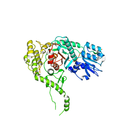
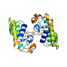
 | | Crystal Structure of hPNMT in Complex With Noradrenochrome and AdoHcy | | 分子名称: | (3S)-3-hydroxy-2,3-dihydro-1H-indole-5,6-dione, Phenylethanolamine N-methyltransferase, S-ADENOSYL-L-HOMOCYSTEINE | | 著者 | Drinkwater, N, Martin, J.L, Gee, C.L. | | 登録日 | 2009-05-06 | | 公開日 | 2009-08-25 | | 最終更新日 | 2023-09-06 | | 実験手法 | X-RAY DIFFRACTION (2.4 Å) | | 主引用文献 | Molecular recognition of physiological substrate noradrenaline by the adrenaline-synthesizing enzyme PNMT and factors influencing its methyltransferase activity.
Biochem.J., 422, 2009
|
|
3HCD
 | |
5DCH
 | | Crystal structure of Pseudomonas aeruginosa DsbA E82I in complex with MIPS-0000851 (3-[(2-METHYLBENZYL)SULFANYL]-4H-1,2,4-TRIAZOL-4-AMINE) | | 分子名称: | 2-(N-MORPHOLINO)-ETHANESULFONIC ACID, 3-[(2-methylbenzyl)sulfanyl]-4H-1,2,4-triazol-4-amine, GLYCEROL, ... | | 著者 | McMahon, R.M, Martin, J.L. | | 登録日 | 2015-08-24 | | 公開日 | 2016-10-05 | | 最終更新日 | 2023-09-27 | | 実験手法 | X-RAY DIFFRACTION (1.447 Å) | | 主引用文献 | Fragment library screening identifies hits that bind to the non-catalytic surface of Pseudomonas aeruginosa DsbA1.
PLoS ONE, 12, 2017
|
|
3HCC
 | | Crystal Structure of hPNMT in Complex With anti-9-amino-5-(trifluromethyl) benzonorbornene and AdoHcy | | 分子名称: | (1S,4R,9S)-5-(trifluoromethyl)-1,2,3,4-tetrahydro-1,4-methanonaphthalen-9-amine, Phenylethanolamine N-methyltransferase, S-ADENOSYL-L-HOMOCYSTEINE | | 著者 | Drinkwater, N, Martin, J.L, Gee, C.L, Puri, M. | | 登録日 | 2009-05-06 | | 公開日 | 2009-08-25 | | 最終更新日 | 2023-09-06 | | 実験手法 | X-RAY DIFFRACTION (2.3 Å) | | 主引用文献 | Molecular recognition of physiological substrate noradrenaline by the adrenaline-synthesizing enzyme PNMT and factors influencing its methyltransferase activity.
Biochem.J., 422, 2009
|
|
3UX3
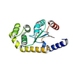
 | | Crystal Structure of Domain-Swapped Fam96a minor dimer | | 分子名称: | ACETATE ION, MIP18 family protein FAM96A, ZINC ION | | 著者 | Chen, K.-E, Kobe, B, Martin, J.L. | | 登録日 | 2011-12-03 | | 公開日 | 2012-05-30 | | 最終更新日 | 2023-11-08 | | 実験手法 | X-RAY DIFFRACTION (1.8 Å) | | 主引用文献 | The mammalian DUF59 protein Fam96a forms two distinct types of domain-swapped dimer.
Acta Crystallogr.,Sect.D, 68, 2012
|
|
3UX2
 | |
1J1A
 | | PANCREATIC SECRETORY PHOSPHOLIPASE A2 (IIa) WITH ANTI-INFLAMMATORY ACTIVITY | | 分子名称: | (S)-5-(4-BENZYLOXY-PHENYL)-4-(7-PHENYL-HEPTANOYLAMINO)-PENTANOIC ACID, CALCIUM ION, Phospholipase A2 | | 著者 | Hansford, K.A, Reid, R.C, Clark, C.I, Tyndall, J.D.A, Whitehouse, M.W, Guthrie, T, McGeary, R.P, Schafer, K, Martin, J.L, Fairlie, D.P. | | 登録日 | 2002-12-03 | | 公開日 | 2003-03-18 | | 最終更新日 | 2023-10-25 | | 実験手法 | X-RAY DIFFRACTION (2.2 Å) | | 主引用文献 | D-Tyrosine as a Chiral Precusor to Potent Inhibitors of Human Nonpancreatic Secretory Phospholipase A2 (IIa) with Antiinflammatory Activity
Chembiochem, 4, 2003
|
|
5U3E
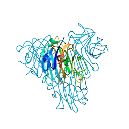 | | Crystal Structure of Native Lectin from Canavalia bonariensis Seeds (CaBo) complexed with alpha-methyl-D-mannoside | | 分子名称: | CALCIUM ION, Canavalia bonariensis seed lectin, MANGANESE (II) ION, ... | | 著者 | Silva, M.T.L, Osterne, V.J.S, Pinto-Junior, V.R, Santiago, M.Q, Araripe, D.A, Neco, A.H.B, Silva-Filho, J.C, Martins, J.L, Rocha, C.R.C, Leal, R.B, Nascimento, K.S, Cavada, B.S. | | 登録日 | 2016-12-02 | | 公開日 | 2017-08-23 | | 最終更新日 | 2023-10-04 | | 実験手法 | X-RAY DIFFRACTION (2.3 Å) | | 主引用文献 | Canavalia bonariensis lectin: Molecular bases of glycoconjugates interaction and antiglioma potential.
Int. J. Biol. Macromol., 106, 2018
|
|
5VYO
 | |
6C29
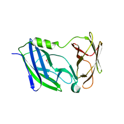 | | Crystal structure of the N-terminal periplasmic domain of ScsB from Proteus mirabilis | | 分子名称: | Putative metal resistance protein | | 著者 | Furlong, E.J, Choudhury, H.G, Kurth, F, Martin, J.L. | | 登録日 | 2018-01-07 | | 公開日 | 2018-03-07 | | 最終更新日 | 2020-01-01 | | 実験手法 | X-RAY DIFFRACTION (1.538 Å) | | 主引用文献 | Disulfide isomerase activity of the dynamic, trimericProteus mirabilisScsC protein is primed by the tandem immunoglobulin-fold domain of ScsB.
J. Biol. Chem., 293, 2018
|
|
1LS6
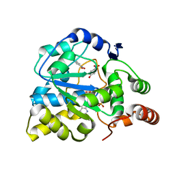 | | Human SULT1A1 complexed with PAP and p-Nitrophenol | | 分子名称: | ADENOSINE-3'-5'-DIPHOSPHATE, P-NITROPHENOL, aryl sulfotransferase | | 著者 | Gamage, N.U, Barnett, A.C, Tresillian, M, Latham, C.F, Liyou, N.E, McManus, M.E, Martin, J.L. | | 登録日 | 2002-05-17 | | 公開日 | 2003-08-05 | | 最終更新日 | 2024-02-14 | | 実験手法 | X-RAY DIFFRACTION (1.9 Å) | | 主引用文献 | Structure of a human carcinogen-converting enzyme, SULT1A1. Structural and kinetic implications of substrate inhibition.
J.Biol.Chem., 278, 2003
|
|
5TLQ
 | | Model structure of the oxidized PaDsbA1 and 3-[(2-methylbenzyl)sulfanyl]-4H-1,2,4-triazol-4-amine complex | | 分子名称: | 3-[(2-methylbenzyl)sulfanyl]-4H-1,2,4-triazol-4-amine, Thiol:disulfide interchange protein DsbA | | 著者 | Mohanty, B, Rimmer, K.A, McMahon, R.M, Headey, S.J, Vazirani, M, Shouldice, S.R, Coincon, M, Tay, S, Morton, C.J, Simpson, J.S, Martin, J.L, Scanlon, M.S. | | 登録日 | 2016-10-11 | | 公開日 | 2017-04-12 | | 最終更新日 | 2023-06-14 | | 実験手法 | SOLUTION NMR | | 主引用文献 | Fragment library screening identifies hits that bind to the non-catalytic surface of Pseudomonas aeruginosa DsbA1.
PLoS ONE, 12, 2017
|
|
1PEN
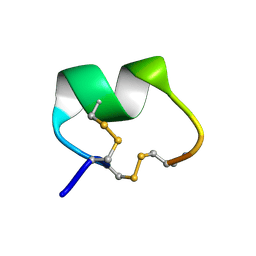 | | ALPHA-CONOTOXIN PNI1 | | 分子名称: | ALPHA-CONOTOXIN PNIA | | 著者 | Hu, S.-H, Gehrmann, J, Guddat, L.W, Alewood, P.F, Craik, D.J, Martin, J.L. | | 登録日 | 1996-01-29 | | 公開日 | 1997-04-21 | | 最終更新日 | 2011-07-13 | | 実験手法 | X-RAY DIFFRACTION (1.1 Å) | | 主引用文献 | The 1.1 A crystal structure of the neuronal acetylcholine receptor antagonist, alpha-conotoxin PnIA from Conus pennaceus.
Structure, 4, 1996
|
|
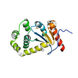
1N7I
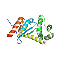
 | | The structure of Phenylethanolamine N-methyltransferase in complex with S-adenosylhomocysteine and the inhibitor LY134046 | | 分子名称: | 8,9-DICHLORO-2,3,4,5-TETRAHYDRO-1H-BENZO[C]AZEPINE, Phenylethanolamine N-methyltransferase, S-ADENOSYL-L-HOMOCYSTEINE | | 著者 | McMillan, F.M, Archbold, J, McLeish, M.J, Caine, J.M, Criscione, K.R, Grunewald, G.L, Martin, J.L. | | 登録日 | 2002-11-15 | | 公開日 | 2003-12-23 | | 最終更新日 | 2024-02-14 | | 実験手法 | X-RAY DIFFRACTION (2.8 Å) | | 主引用文献 | Molecular recognition of sub-micromolar inhibitors by the epinephrine-synthesizing enzyme phenylethanolamine N-methyltransferase.
J.Med.Chem., 47, 2004
|
|


