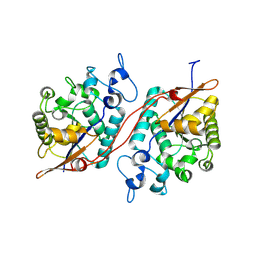
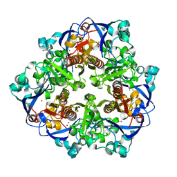
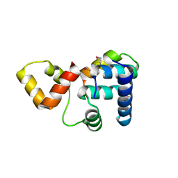
4IK9
 
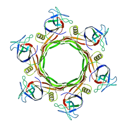
 | | High resolution structure of GCaMP3 dimer form 2 at pH 7.5 | | 分子名称: | CALCIUM ION, DI(HYDROXYETHYL)ETHER, RCaMP, ... | | 著者 | Chen, Y, Song, X, Miao, L, Zhu, Y, Ji, G. | | 登録日 | 2012-12-25 | | 公開日 | 2014-01-29 | | 最終更新日 | 2024-11-13 | | 実験手法 | X-RAY DIFFRACTION (1.8 Å) | | 主引用文献 | Structural insight into enhanced calcium indicator GCaMP3 and GCaMPJ to promote further improvement.
Protein Cell, 4, 2013
|
|
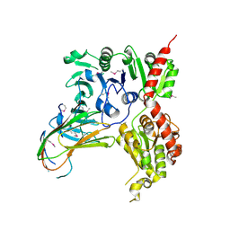
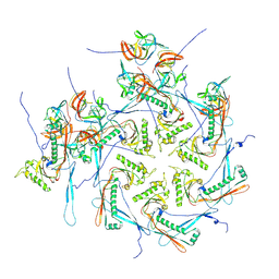
4IK8
 | | High resolution structure of GCaMP3 dimer form 1 at pH 7.5 | | 分子名称: | CALCIUM ION, RCaMP, Green fluorescent protein | | 著者 | Chen, Y, Song, X, Miao, L, Zhu, Y, Ji, G. | | 登録日 | 2012-12-25 | | 公開日 | 2014-02-05 | | 最終更新日 | 2024-11-13 | | 実験手法 | X-RAY DIFFRACTION (1.55 Å) | | 主引用文献 | Structural insight into enhanced calcium indicator GCaMP3 and GCaMPJ to promote further improvement.
Protein Cell, 4, 2013
|
|
9BRF
 | |
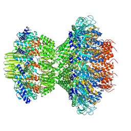
9B1X
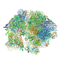 | | HWS19 strain gidB mutant mycobacterial ribosome | | 分子名称: | 16S rRNA, 23S rRNA, 50S ribosomal protein L36, ... | | 著者 | Chen, Y, Young, I.D, Fraser, J.S, Javid, B. | | 登録日 | 2024-03-14 | | 公開日 | 2025-03-26 | | 実験手法 | ELECTRON MICROSCOPY (3.07 Å) | | 主引用文献 | Ribosomal RNA methylation by GidB is a capacitor for discrimination of mischarged tRNA
To be published
|
|
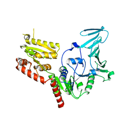
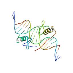
4IK4
 | | High resolution structure of GCaMP3 at pH 5.0 | | 分子名称: | CALCIUM ION, RCaMP, Green fluorescent protein | | 著者 | Chen, Y, Song, X, Miao, L, Zhu, Y, Ji, G. | | 登録日 | 2012-12-25 | | 公開日 | 2014-02-05 | | 最終更新日 | 2024-10-30 | | 実験手法 | X-RAY DIFFRACTION (2.01 Å) | | 主引用文献 | Structural insight into enhanced calcium indicator GCaMP3 and GCaMPJ to promote further improvement.
Protein Cell, 4, 2013
|
|
2MCQ
 | | NMR structure of a BolA-like hypothetical protein RP812 from Rickettsia prowazekii, Seattle structural genomics center for infectious disease (SSGCID) | | 分子名称: | Uncharacterized protein RP812 | | 著者 | Chen, Y, Barnwal, R, Yang, F, Varani, G, Seattle Structural Genomics Center for Infectious Disease (SSGCID) | | 登録日 | 2013-08-22 | | 公開日 | 2013-10-09 | | 最終更新日 | 2024-05-15 | | 実験手法 | SOLUTION NMR | | 主引用文献 | NMR structure of a BolA-like hypothetical protein RP812 from Rickettsia prowazekii, Seattle structural genomics center for infectious disease (SSGCID)
To be Published
|
|
4L3N
 | | Crystal structure of the receptor-binding domain from newly emerged Middle East respiratory syndrome coronavirus | | 分子名称: | 1,2-ETHANEDIOL, 2-acetamido-2-deoxy-beta-D-glucopyranose-(1-4)-2-acetamido-2-deoxy-beta-D-glucopyranose, S protein | | 著者 | Chen, Y, Rajashankar, K.R, Yang, Y, Agnihothram, S.S, Liu, C, Lin, Y.-L, Baric, R.S, Li, F. | | 登録日 | 2013-06-06 | | 公開日 | 2013-07-31 | | 最終更新日 | 2024-10-30 | | 実験手法 | X-RAY DIFFRACTION (2.13 Å) | | 主引用文献 | Crystal structure of the receptor-binding domain from newly emerged middle East respiratory syndrome coronavirus.
J.Virol., 87, 2013
|
|
3C7T
 | | Crystal structure of the ecdysone phosphate phosphatase, EPPase, from Bombix mori in complex with tungstate | | 分子名称: | CHLORIDE ION, Ecdysteroid-phosphate phosphatase, IODIDE ION, ... | | 著者 | Chen, Y, Carpino, N, Nassar, N. | | 登録日 | 2008-02-08 | | 公開日 | 2009-03-24 | | 最終更新日 | 2024-02-21 | | 実験手法 | X-RAY DIFFRACTION (1.76 Å) | | 主引用文献 | Structural and functional characterization of the c-terminal domain of the ecdysteroid phosphate phosphatase from Bombyx mori reveals a new enzymatic activity.
Biochemistry, 47, 2008
|
|
3D4I
 | |
5VAE
 | |
5VAF
 | |
5X8F
 | |
9L0F
 | |
9KZJ
 | |
9L01
 | |
9L0E
 | |
9KHQ
 | | Crystal structure of N-acyl homoserine lactonase AhlX | | 分子名称: | MAGNESIUM ION, N-acylhomoserine lactonase, NICKEL (II) ION | | 著者 | Chen, Y, Chu, X.H. | | 登録日 | 2024-11-11 | | 公開日 | 2025-04-23 | | 実験手法 | X-RAY DIFFRACTION (2.2 Å) | | 主引用文献 | An AHL-lactonase mutant featuring a unique "tri-His" motif exhibits enhanced activity, stability and effectively controls plant soft rot.
Int.J.Biol.Macromol., 308, 2025
|
|
9KHO
 | |
3KMD
 | |
4HCA
 | | DNA binding by GATA transcription factor-complex 1 | | 分子名称: | DNA (5'-D(*AP*AP*TP*GP*TP*CP*CP*AP*TP*CP*TP*GP*AP*TP*AP*AP*GP*AP*CP*G)-3'), DNA (5'-D(*TP*TP*CP*GP*TP*CP*TP*TP*AP*TP*CP*AP*GP*AP*TP*GP*GP*AP*CP*A)-3'), Trans-acting T-cell-specific transcription factor GATA-3, ... | | 著者 | Chen, Y, Bates, D.L, Dey, R, Chen, L. | | 登録日 | 2012-09-28 | | 公開日 | 2012-12-05 | | 最終更新日 | 2024-02-28 | | 実験手法 | X-RAY DIFFRACTION (2.8 Å) | | 主引用文献 | DNA Binding by GATA Transcription Factor Suggests Mechanisms of DNA Looping and Long-Range Gene Regulation.
Cell Rep, 2, 2012
|
|
4HC7
 | | Crystal structure of the full DNA binding domain of GATA3-complex 2 | | 分子名称: | DNA (5'-D(*AP*AP*GP*GP*TP*TP*AP*TP*CP*TP*CP*TP*GP*AP*TP*TP*TP*AP*GP*G)-3'), DNA (5'-D(*TP*TP*CP*CP*TP*AP*AP*AP*TP*CP*AP*GP*AP*GP*AP*TP*AP*AP*CP*C)-3'), Trans-acting T-cell-specific transcription factor GATA-3, ... | | 著者 | Chen, Y, Bates, D.L, Dey, R, Chen, L. | | 登録日 | 2012-09-28 | | 公開日 | 2012-12-05 | | 最終更新日 | 2024-02-28 | | 実験手法 | X-RAY DIFFRACTION (2.65 Å) | | 主引用文献 | DNA Binding by GATA Transcription Factor Suggests Mechanisms of DNA Looping and Long-Range Gene Regulation.
Cell Rep, 2, 2012
|
|
7YJ4
 | | Cryo-EM structure of the INSL5-bound human relaxin family peptidereceptor 4 (RXFP4)-Gi complex | | 分子名称: | Guanine nucleotide-binding protein G(I)/G(S)/G(O) subunit gamma-2, Guanine nucleotide-binding protein G(I)/G(S)/G(T) subunit beta-1, Guanine nucleotide-binding protein G(i) subunit alpha-2, ... | | 著者 | Chen, Y, Zhou, Q.T, Wang, J, Xu, Y.W, Wang, Y, Yan, J.H, Wang, Y.B, Zhu, Q, Zhao, F.H, Li, C.H, Chen, C.W, Cai, X.Q, Bathgate, R.A.D, Shen, C, Liu, H, Xu, H.E, Yang, D.H, Wang, M.W. | | 登録日 | 2022-07-19 | | 公開日 | 2023-03-01 | | 最終更新日 | 2024-10-23 | | 実験手法 | ELECTRON MICROSCOPY (3.19 Å) | | 主引用文献 | Ligand recognition mechanism of the human relaxin family peptide receptor 4 (RXFP4).
Nat Commun, 14, 2023
|
|
7YK7
 | | Cryo-EM structure of the DC591053-bound human relaxin family peptide receptor 4 (RXFP4)-Gi complex | | 分子名称: | Guanine nucleotide-binding protein G(I)/G(S)/G(O) subunit gamma-2, Guanine nucleotide-binding protein G(I)/G(S)/G(T) subunit beta-1, Guanine nucleotide-binding protein G(i) subunit alpha-2, ... | | 著者 | Chen, Y, Zhou, Q.T, Wang, J, Xu, Y.W, Wang, Y, Yan, J.H, Wang, Y.B, Zhu, Q, Zhao, F.H, Li, C.H, Chen, C.W, Cai, X.Q, Bathgate, R.A.D, Shen, C, Xu, H.E, Yang, D.H, Liu, H, Wang, M.W. | | 登録日 | 2022-07-21 | | 公開日 | 2023-03-01 | | 最終更新日 | 2024-11-06 | | 実験手法 | ELECTRON MICROSCOPY (2.75 Å) | | 主引用文献 | Ligand recognition mechanism of the human relaxin family peptide receptor 4 (RXFP4).
Nat Commun, 14, 2023
|
|
7YK6
 | | Cryo-EM structure of the compound 4-bound human relaxin family peptide receptor 4 (RXFP4)-Gi complex | | 分子名称: | 1-[2-(4-chlorophenyl)ethyl]-3-[(7-ethyl-5-oxidanyl-1H-indol-3-yl)methylideneamino]guanidine, Guanine nucleotide-binding protein G(I)/G(S)/G(O) subunit gamma-2, Guanine nucleotide-binding protein G(I)/G(S)/G(T) subunit beta-1, ... | | 著者 | Chen, Y, Zhou, Q.T, Wang, J, Xu, Y.W, Wang, Y, Yan, J.H, Wang, Y.B, Zhu, Q, Zhao, F.H, Li, C.H, Chen, C.W, Cai, X.Q, Bathgate, R.A.D, Shen, C, Liu, H, Xu, H.E, Yang, D.H, Wang, M.W. | | 登録日 | 2022-07-21 | | 公開日 | 2023-03-01 | | 最終更新日 | 2025-06-25 | | 実験手法 | ELECTRON MICROSCOPY (3.03 Å) | | 主引用文献 | Ligand recognition mechanism of the human relaxin family peptide receptor 4 (RXFP4).
Nat Commun, 14, 2023
|
|
3K6G
 | | Crystal structure of Rap1 and TRF2 complex | | 分子名称: | Telomeric repeat-binding factor 2, Telomeric repeat-binding factor 2-interacting protein 1 | | 著者 | Chen, Y, Rai, R, Yang, Y.T, Zheng, H, Chang, S, Lei, M. | | 登録日 | 2009-10-08 | | 公開日 | 2010-10-13 | | 最終更新日 | 2024-11-20 | | 実験手法 | X-RAY DIFFRACTION (1.95 Å) | | 主引用文献 | A conserved motif within RAP1 has diversified roles in telomere protection and regulation in different organisms.
Nat.Struct.Mol.Biol., 18, 2011
|
|


