5TAB
 
 | |
2LVM
 | |
2K2W
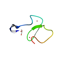
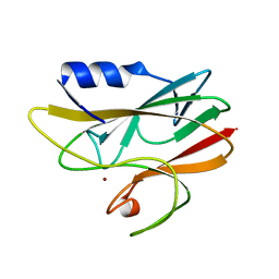
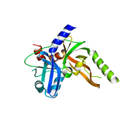
 | | Second BRCT domain of NBS1 | | 分子名称: | Recombination and DNA repair protein | | 著者 | Xu, C, Cui, G, Botuyan, M, Mer, G. | | 登録日 | 2008-04-14 | | 公開日 | 2008-06-17 | | 最終更新日 | 2024-05-29 | | 実験手法 | SOLUTION NMR | | 主引用文献 | Structure of a second BRCT domain identified in the nijmegen breakage syndrome protein Nbs1 and its function in an MDC1-dependent localization of Nbs1 to DNA damage sites.
J.Mol.Biol., 381, 2008
|
|
2LDM
 | |
2KRE
 | |
3L1X
 | |
3L1Z
 | |
3L1Y
 | |
1A3Z
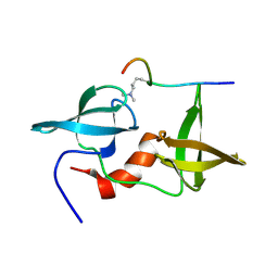
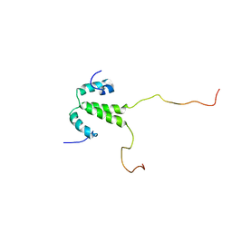
 | | REDUCED RUSTICYANIN AT 1.9 ANGSTROMS | | 分子名称: | COPPER (I) ION, RUSTICYANIN | | 著者 | Zhao, D, Shoham, M. | | 登録日 | 1998-01-27 | | 公開日 | 1998-07-29 | | 最終更新日 | 2024-05-22 | | 実験手法 | X-RAY DIFFRACTION (1.9 Å) | | 主引用文献 | Rusticyanin: Extremes in acid stability and redox potential explained by the crystal structure.
Biophys.J., 74, 1998
|
|
2LH0
 | |
2K3Y
 | |
2K3X
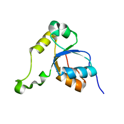
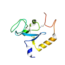
 | | Solution structure of EAF3 chromo barrel domain | | 分子名称: | Chromatin modification-related protein EAF3 | | 著者 | Mer, G, Xu, C. | | 登録日 | 2008-05-19 | | 公開日 | 2008-09-16 | | 最終更新日 | 2024-05-29 | | 実験手法 | SOLUTION NMR | | 主引用文献 | Structural Basis for the Recognition of Methylated Histone H3K36 by the Eaf3 Subunit of Histone Deacetylase Complex Rpd3S.
Structure, 16, 2008
|
|
2L0G
 | |
2L0F
 | |
2KTF
 | |
8S9K
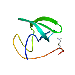
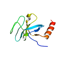
 | | Structure of dimeric FAM111A SPD S541A Mutant | | 分子名称: | GLYCEROL, Serine protease FAM111A | | 著者 | Palani, S, Alvey, J.A, Cong, A.T.Q, Schellenberg, M.J, Machida, Y. | | 登録日 | 2023-03-29 | | 公開日 | 2024-03-20 | | 実験手法 | X-RAY DIFFRACTION (2.72 Å) | | 主引用文献 | Dimerization-dependent serine protease activity of FAM111A prevents replication fork stalling at topoisomerase 1 cleavage complexes.
Nat Commun, 15, 2024
|
|
8S9L
 | | Structure of monomeric FAM111A SPD V347D Mutant | | 分子名称: | SULFATE ION, Serine protease FAM111A | | 著者 | Palani, S, Alvey, J.A, Cong, A.T.Q, Schellenberg, M.J, Machida, Y. | | 登録日 | 2023-03-29 | | 公開日 | 2024-03-20 | | 実験手法 | X-RAY DIFFRACTION (1.85 Å) | | 主引用文献 | Dimerization-dependent serine protease activity of FAM111A prevents replication fork stalling at topoisomerase 1 cleavage complexes.
Nat Commun, 15, 2024
|
|


