7TFS
 
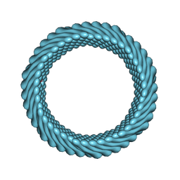
 | | Cryo-EM of the OmcE nanowires from Geobacter sulfurreducens | | 分子名称: | Cytochrome c, HEME C | | 著者 | Wang, F, Mustafa, K, Chan, C.H, Joshi, K, Hochbaum, A.I, Bond, D.R, Egelman, E.H. | | 登録日 | 2022-01-07 | | 公開日 | 2022-05-04 | | 最終更新日 | 2022-11-16 | | 実験手法 | ELECTRON MICROSCOPY (4.3 Å) | | 主引用文献 | Cryo-EM structure of an extracellular Geobacter OmcE cytochrome filament reveals tetrahaem packing.
Nat Microbiol, 7, 2022
|
|
7TGG
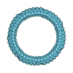
 | | Cryo-EM structure of PilA-N and PilA-C from Geobacter sulfurreducens | | 分子名称: | Geopilin domain 1 protein, Geopilin domain 2 protein | | 著者 | Wang, F, Mustafa, K, Chan, C.H, Joshi, K, Bond, D.R, Hochbaum, A.I, Egelman, E.H. | | 登録日 | 2022-01-07 | | 公開日 | 2022-02-23 | | 最終更新日 | 2024-06-05 | | 実験手法 | ELECTRON MICROSCOPY (4.1 Å) | | 主引用文献 | Cryo-EM structure of an extracellular Geobacter OmcE cytochrome filament reveals tetrahaem packing.
Nat Microbiol, 7, 2022
|
|
5UG6
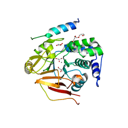
 | | Perforin C2 Domain - T431D | | 分子名称: | IODIDE ION, Perforin-1 | | 著者 | Law, R.H.P, Conroy, P.J, Voskoboinik, I, Whisstock, J.C. | | 登録日 | 2017-01-07 | | 公開日 | 2018-02-07 | | 最終更新日 | 2023-10-04 | | 実験手法 | X-RAY DIFFRACTION (2 Å) | | 主引用文献 | Perforin proteostasis is regulated through its C2 domain: supra-physiological cell death mediated by T431D-perforin.
Cell Death Differ., 25, 2018
|
|
7RO4
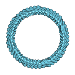
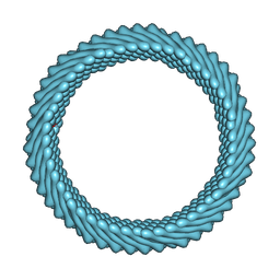
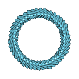
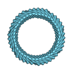 | | Cryo-EM reconstruction of Sulfolobus monocaudavirus SMV1, symmetry 3 | | 分子名称: | major capsid protein | | 著者 | Wang, F, Cvirkaite-Krupovic, V, Krupovic, M, Egelman, E.H. | | 登録日 | 2021-07-30 | | 公開日 | 2022-03-30 | | 最終更新日 | 2024-06-05 | | 実験手法 | ELECTRON MICROSCOPY (4.3 Å) | | 主引用文献 | Spindle-shaped archaeal viruses evolved from rod-shaped ancestors to package a larger genome.
Cell, 185, 2022
|
|
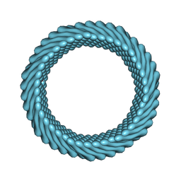
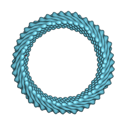
7ROI
 | | Cryo-EM reconstruction of Sulfolobus monocaudavirus SMV1, symmetry 12 | | 分子名称: | major capsid protein | | 著者 | Wang, F, Cvirkaite-Krupovic, V, Krupovic, M, Egelman, E.H. | | 登録日 | 2021-07-30 | | 公開日 | 2022-03-30 | | 最終更新日 | 2024-06-05 | | 実験手法 | ELECTRON MICROSCOPY (4.3 Å) | | 主引用文献 | Spindle-shaped archaeal viruses evolved from rod-shaped ancestors to package a larger genome.
Cell, 185, 2022
|
|
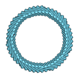
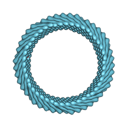
7ROE
 | | Cryo-EM reconstruction of Sulfolobus monocaudavirus SMV1, symmetry 9 | | 分子名称: | major capsid protein | | 著者 | Wang, F, Cvirkaite-Krupovic, V, Krupovic, M, Egelman, E.H. | | 登録日 | 2021-07-30 | | 公開日 | 2022-03-30 | | 最終更新日 | 2024-06-05 | | 実験手法 | ELECTRON MICROSCOPY (3.7 Å) | | 主引用文献 | Spindle-shaped archaeal viruses evolved from rod-shaped ancestors to package a larger genome.
Cell, 185, 2022
|
|
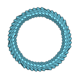
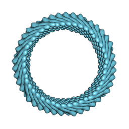
7RO2
 | | Cryo-EM reconstruction of Sulfolobus monocaudavirus SMV1, symmetry 1 | | 分子名称: | major capsid protein | | 著者 | Wang, F, Cvirkaite-Krupovic, V, Krupovic, M, Egelman, E.H. | | 登録日 | 2021-07-30 | | 公開日 | 2022-03-30 | | 最終更新日 | 2024-06-05 | | 実験手法 | ELECTRON MICROSCOPY (5.1 Å) | | 主引用文献 | Spindle-shaped archaeal viruses evolved from rod-shaped ancestors to package a larger genome.
Cell, 185, 2022
|
|
7ROH
 | | Cryo-EM reconstruction of Sulfolobus monocaudavirus SMV1, symmetry 11 | | 分子名称: | major capsid protein | | 著者 | Wang, F, Cvirkaite-Krupovic, V, Krupovic, M, Egelman, E.H. | | 登録日 | 2021-07-30 | | 公開日 | 2022-03-30 | | 最終更新日 | 2024-06-05 | | 実験手法 | ELECTRON MICROSCOPY (4 Å) | | 主引用文献 | Spindle-shaped archaeal viruses evolved from rod-shaped ancestors to package a larger genome.
Cell, 185, 2022
|
|
7ROG
 | | Cryo-EM reconstruction of Sulfolobus monocaudavirus SMV1, symmetry 10 | | 分子名称: | major capsid protein | | 著者 | Wang, F, Cvirkaite-Krupovic, V, Krupovic, M, Egelman, E.H. | | 登録日 | 2021-07-30 | | 公開日 | 2022-03-30 | | 最終更新日 | 2024-06-05 | | 実験手法 | ELECTRON MICROSCOPY (3.8 Å) | | 主引用文献 | Spindle-shaped archaeal viruses evolved from rod-shaped ancestors to package a larger genome.
Cell, 185, 2022
|
|
7ROC
 | | Cryo-EM reconstruction of Sulfolobus monocaudavirus SMV1, symmetry 7 | | 分子名称: | major capsid protein | | 著者 | Wang, F, Cvirkaite-Krupovic, V, Krupovic, M, Egelman, E.H. | | 登録日 | 2021-07-30 | | 公開日 | 2022-03-30 | | 最終更新日 | 2024-06-05 | | 実験手法 | ELECTRON MICROSCOPY (3.7 Å) | | 主引用文献 | Spindle-shaped archaeal viruses evolved from rod-shaped ancestors to package a larger genome.
Cell, 185, 2022
|
|
7ROB
 | | Cryo-EM reconstruction of Sulfolobus monocaudavirus SMV1, symmetry 6 | | 分子名称: | major capsid protein | | 著者 | Wang, F, Cvirkaite-Krupovic, V, Krupovic, M, Egelman, E.H. | | 登録日 | 2021-07-30 | | 公開日 | 2022-03-30 | | 最終更新日 | 2024-06-05 | | 実験手法 | ELECTRON MICROSCOPY (3.9 Å) | | 主引用文献 | Spindle-shaped archaeal viruses evolved from rod-shaped ancestors to package a larger genome.
Cell, 185, 2022
|
|
7RO6
 | | Cryo-EM reconstruction of Sulfolobus monocaudavirus SMV1, symmetry 5 | | 分子名称: | major capsid protein | | 著者 | Wang, F, Cvirkaite-Krupovic, V, Krupovic, M, Egelman, E.H. | | 登録日 | 2021-07-30 | | 公開日 | 2022-03-30 | | 最終更新日 | 2024-06-05 | | 実験手法 | ELECTRON MICROSCOPY (4.1 Å) | | 主引用文献 | Spindle-shaped archaeal viruses evolved from rod-shaped ancestors to package a larger genome.
Cell, 185, 2022
|
|
7RO5
 | | Cryo-EM reconstruction of Sulfolobus monocaudavirus SMV1, symmetry 4 | | 分子名称: | major capsid protein | | 著者 | Wang, F, Cvirkaite-Krupovic, V, Krupovic, M, Egelman, E.H. | | 登録日 | 2021-07-30 | | 公開日 | 2022-03-30 | | 最終更新日 | 2024-06-05 | | 実験手法 | ELECTRON MICROSCOPY (4.1 Å) | | 主引用文献 | Spindle-shaped archaeal viruses evolved from rod-shaped ancestors to package a larger genome.
Cell, 185, 2022
|
|
7RO3
 | | Cryo-EM reconstruction of Sulfolobus monocaudavirus SMV1, symmetry 2 | | 分子名称: | major capsid protein | | 著者 | Wang, F, Cvirkaite-Krupovic, V, Krupovic, M, Egelman, E.H. | | 登録日 | 2021-07-30 | | 公開日 | 2022-03-30 | | 最終更新日 | 2024-06-05 | | 実験手法 | ELECTRON MICROSCOPY (4.8 Å) | | 主引用文献 | Spindle-shaped archaeal viruses evolved from rod-shaped ancestors to package a larger genome.
Cell, 185, 2022
|
|
7ROD
 | | Cryo-EM reconstruction of Sulfolobus monocaudavirus SMV1, symmetry 8 | | 分子名称: | major capsid protein | | 著者 | Wang, F, Cvirkaite-Krupovic, V, Krupovic, M, Egelman, E.H. | | 登録日 | 2021-07-30 | | 公開日 | 2022-03-30 | | 最終更新日 | 2024-06-05 | | 実験手法 | ELECTRON MICROSCOPY (3.8 Å) | | 主引用文献 | Spindle-shaped archaeal viruses evolved from rod-shaped ancestors to package a larger genome.
Cell, 185, 2022
|
|
3V4Y
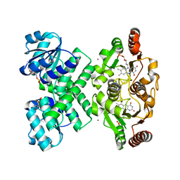
 | | Crystal Structure of the first Nuclear PP1 holoenzyme | | 分子名称: | GLYCEROL, MANGANESE (II) ION, Nuclear inhibitor of protein phosphatase 1, ... | | 著者 | Page, R, Peti, W, O'Connell, N.E, Nichols, S. | | 登録日 | 2011-12-15 | | 公開日 | 2012-10-31 | | 最終更新日 | 2024-02-28 | | 実験手法 | X-RAY DIFFRACTION (2.098 Å) | | 主引用文献 | The Molecular Basis for Substrate Specificity of the Nuclear NIPP1:PP1 Holoenzyme.
Structure, 20, 2012
|
|
3EYW
 | | Crystal structure of the C-terminal domain of E. coli KefC in complex with KefF | | 分子名称: | C-terminal domain of Glutathione-regulated potassium-efflux system protein kefC fused to full length Glutathione-regulated potassium-efflux system ancillary protein kefF, FLAVIN MONONUCLEOTIDE, MAGNESIUM ION, ... | | 著者 | Roosild, T.P. | | 登録日 | 2008-10-22 | | 公開日 | 2009-06-30 | | 最終更新日 | 2023-09-06 | | 実験手法 | X-RAY DIFFRACTION (2.4 Å) | | 主引用文献 | KTN (RCK) Domains Regulate K(+) Channels and Transporters by Controlling the Dimer-Hinge Conformation.
Structure, 17, 2009
|
|
5UG7
 | | Calcium bound Perforin C2 Domain - T431D | | 分子名称: | CALCIUM ION, Perforin-1 | | 著者 | Law, R.H.P, Conroy, P.J, Voskoboinik, I, Whisstock, J.C. | | 登録日 | 2017-01-07 | | 公開日 | 2018-02-07 | | 最終更新日 | 2023-10-04 | | 実験手法 | X-RAY DIFFRACTION (1.8 Å) | | 主引用文献 | Perforin proteostasis is regulated through its C2 domain: supra-physiological cell death mediated by T431D-perforin.
Cell Death Differ., 25, 2018
|
|
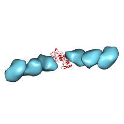
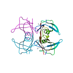
7Z1H
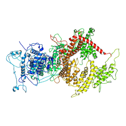 | | VAR2CSA APO | | 分子名称: | VAR2CSA APO | | 著者 | Raghavan, S.S.R, Wang, K.T. | | 登録日 | 2022-02-24 | | 公開日 | 2022-11-02 | | 最終更新日 | 2022-11-30 | | 実験手法 | ELECTRON MICROSCOPY (3.12 Å) | | 主引用文献 | Cryo-EM reveals the conformational epitope of human monoclonal antibody PAM1.4 broadly reacting with polymorphic malarial protein VAR2CSA.
Plos Pathog., 18, 2022
|
|
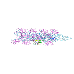
7Z12
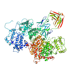 | | VAR2 complex with PAM1.4 | | 分子名称: | PAM1.4, Heavy Chain, light Chain, ... | | 著者 | Raghavan, S.S.R, Wang, K.T. | | 登録日 | 2022-02-24 | | 公開日 | 2022-11-02 | | 最終更新日 | 2022-11-30 | | 実験手法 | ELECTRON MICROSCOPY (3 Å) | | 主引用文献 | Cryo-EM reveals the conformational epitope of human monoclonal antibody PAM1.4 broadly reacting with polymorphic malarial protein VAR2CSA.
Plos Pathog., 18, 2022
|
|
4L1T
 | |
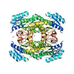
4ZP1
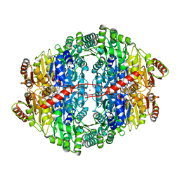 | | Crystal structure of Zymomonas mobilis pyruvate decarboxylase variant Glu473Ala | | 分子名称: | GLYCEROL, MAGNESIUM ION, NICKEL (II) ION, ... | | 著者 | Wechsler, C, Neumann, P, Tittmann, K. | | 登録日 | 2015-05-07 | | 公開日 | 2015-11-04 | | 最終更新日 | 2024-05-08 | | 実験手法 | X-RAY DIFFRACTION (2.205 Å) | | 主引用文献 | Tuning and Switching Enantioselectivity of Asymmetric Carboligation in an Enzyme through Mutational Analysis of a Single Hot Spot.
Chembiochem, 16, 2015
|
|
4L1S
 | |
4E6P
 | |
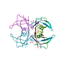
2PBW
 | | Crystal Structure of the Ligand-Binding Core of iGluR5 in Complex with the Partial agonist Domoic Acid at 2.5 A Resolution | | 分子名称: | (2S,3S,4S)-2-CARBOXY-4-[(1Z,3E,5R)-5-CARBOXY-1-METHYL-1,3-HEXADIENYL]-3-PYRROLIDINEACETIC ACID, Glutamate receptor, ionotropic kainate 1 | | 著者 | Hald, H, Naur, P, Gajhede, M, Kastrup, J.S. | | 登録日 | 2007-03-29 | | 公開日 | 2007-07-03 | | 最終更新日 | 2023-08-30 | | 実験手法 | X-RAY DIFFRACTION (2.5 Å) | | 主引用文献 | Partial agonism and antagonism of the ionotropic glutamate receptor iGLuR5: structures of the ligand-binding core in complex with domoic acid and 2-amino-3-[5-tert-butyl-3-(phosphonomethoxy)-4-isoxazolyl]propionic acid.
J.Biol.Chem., 282, 2007
|
|


