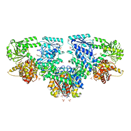
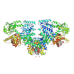
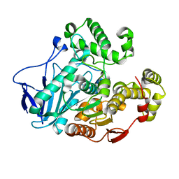
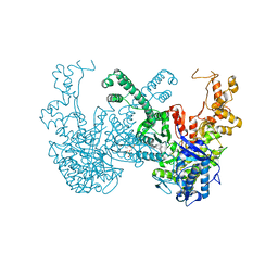
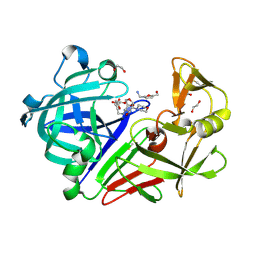
8OQS
 
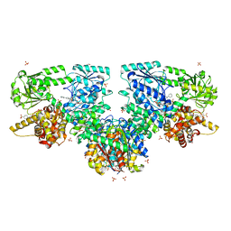 | | Structure of Mycobacterium tuberculosis beta-oxidation trifunctional enzyme in complex with Fragment-M-83 | | 分子名称: | 3-hydroxyacyl-CoA dehydrogenase, 4-phenylbenzenesulfonic acid, GLYCEROL, ... | | 著者 | Dalwani, S, Wierenga, R.K, Venkatesan, R. | | 登録日 | 2023-04-12 | | 公開日 | 2024-01-24 | | 最終更新日 | 2024-08-14 | | 実験手法 | X-RAY DIFFRACTION (2.33 Å) | | 主引用文献 | Crystallographic fragment-binding studies of the Mycobacterium tuberculosis trifunctional enzyme suggest binding pockets for the tails of the acyl-CoA substrates at its active sites and a potential substrate-channeling path between them.
Acta Crystallogr D Struct Biol, 80, 2024
|
|
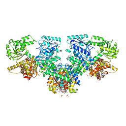
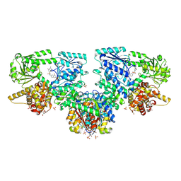
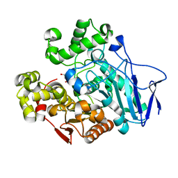
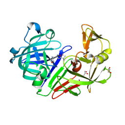
8OQV
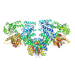 | | Structure of Mycobacterium tuberculosis beta-oxidation trifunctional enzyme in complex with Fragment-M-109 | | 分子名称: | 3-hydroxyacyl-CoA dehydrogenase, 4-nitrobenzenesulfonic acid, GLYCEROL, ... | | 著者 | Dalwani, S, Wierenga, R.K, Venkatesan, R. | | 登録日 | 2023-04-12 | | 公開日 | 2024-01-24 | | 最終更新日 | 2024-08-14 | | 実験手法 | X-RAY DIFFRACTION (2.78 Å) | | 主引用文献 | Crystallographic fragment-binding studies of the Mycobacterium tuberculosis trifunctional enzyme suggest binding pockets for the tails of the acyl-CoA substrates at its active sites and a potential substrate-channeling path between them.
Acta Crystallogr D Struct Biol, 80, 2024
|
|
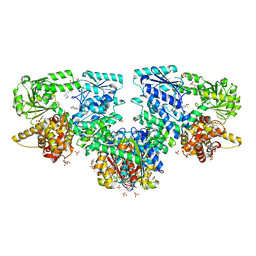
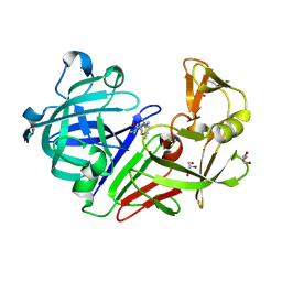
8OQR
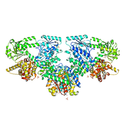 | | Structure of Mycobacterium tuberculosis beta-oxidation trifunctional enzyme in complex with Fragment-M-80 | | 分子名称: | 3-hydroxyacyl-CoA dehydrogenase, 4-cyanobenzenesulfonic acid, GLYCEROL, ... | | 著者 | Dalwani, S, Wierenga, R.K, Venkatesan, R. | | 登録日 | 2023-04-12 | | 公開日 | 2024-01-24 | | 最終更新日 | 2024-08-14 | | 実験手法 | X-RAY DIFFRACTION (2.4 Å) | | 主引用文献 | Crystallographic fragment-binding studies of the Mycobacterium tuberculosis trifunctional enzyme suggest binding pockets for the tails of the acyl-CoA substrates at its active sites and a potential substrate-channeling path between them.
Acta Crystallogr D Struct Biol, 80, 2024
|
|
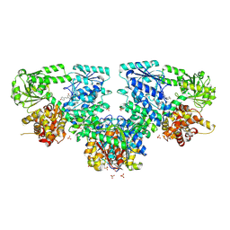
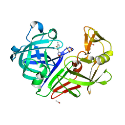
8OQL
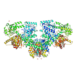 | | Structure of Mycobacterium tuberculosis beta-oxidation trifunctional enzyme in complex with Fragment-M-1 | | 分子名称: | 3-hydroxyacyl-CoA dehydrogenase, GLYCEROL, Hexafluorophosphate anion, ... | | 著者 | Dalwani, S, Wierenga, R.K, Venkatesan, R. | | 登録日 | 2023-04-12 | | 公開日 | 2024-01-24 | | 最終更新日 | 2024-10-16 | | 実験手法 | X-RAY DIFFRACTION (2.7 Å) | | 主引用文献 | Crystallographic fragment-binding studies of the Mycobacterium tuberculosis trifunctional enzyme suggest binding pockets for the tails of the acyl-CoA substrates at its active sites and a potential substrate-channeling path between them.
Acta Crystallogr D Struct Biol, 80, 2024
|
|
8OQQ
 | | Structure of Mycobacterium tuberculosis beta-oxidation trifunctional enzyme in complex with Fragment-M-79 | | 分子名称: | 2-fluoranyl-5-sulfo-benzoic acid, 3-hydroxyacyl-CoA dehydrogenase, GLYCEROL, ... | | 著者 | Dalwani, S, Wierenga, R.K, Venkatesan, R. | | 登録日 | 2023-04-12 | | 公開日 | 2024-01-24 | | 最終更新日 | 2024-08-14 | | 実験手法 | X-RAY DIFFRACTION (2.59 Å) | | 主引用文献 | Crystallographic fragment-binding studies of the Mycobacterium tuberculosis trifunctional enzyme suggest binding pockets for the tails of the acyl-CoA substrates at its active sites and a potential substrate-channeling path between them.
Acta Crystallogr D Struct Biol, 80, 2024
|
|
8OQN
 | | Structure of Mycobacterium tuberculosis beta-oxidation trifunctional enzyme in complex with Fragment-M-53 | | 分子名称: | 1-benzyl-1H-pyrazole-4-carboxylic acid, 3-hydroxyacyl-CoA dehydrogenase, Putative acyltransferase Rv0859, ... | | 著者 | Dalwani, S, Wierenga, R.K, Venkatesan, R. | | 登録日 | 2023-04-12 | | 公開日 | 2024-01-24 | | 最終更新日 | 2024-08-14 | | 実験手法 | X-RAY DIFFRACTION (2.2 Å) | | 主引用文献 | Crystallographic fragment-binding studies of the Mycobacterium tuberculosis trifunctional enzyme suggest binding pockets for the tails of the acyl-CoA substrates at its active sites and a potential substrate-channeling path between them.
Acta Crystallogr D Struct Biol, 80, 2024
|
|
8OQP
 | | Structure of Mycobacterium tuberculosis beta-oxidation trifunctional enzyme in complex with Fragment-M-76 | | 分子名称: | 2-azanyl-5-sulfo-benzoic acid, 3-hydroxyacyl-CoA dehydrogenase, GLYCEROL, ... | | 著者 | Dalwani, S, Wierenga, R.K, Venkatesan, R. | | 登録日 | 2023-04-12 | | 公開日 | 2024-01-24 | | 最終更新日 | 2024-08-14 | | 実験手法 | X-RAY DIFFRACTION (2.18 Å) | | 主引用文献 | Crystallographic fragment-binding studies of the Mycobacterium tuberculosis trifunctional enzyme suggest binding pockets for the tails of the acyl-CoA substrates at its active sites and a potential substrate-channeling path between them.
Acta Crystallogr D Struct Biol, 80, 2024
|
|
8OPV
 | | Structure of Mycobacterium tuberculosis beta-oxidation trifunctional enzyme in complex with Resveratrol (Fragment-B-H11) | | 分子名称: | 3-hydroxyacyl-CoA dehydrogenase, GLYCEROL, Putative acyltransferase Rv0859, ... | | 著者 | Dalwani, S, Wierenga, R.K, Venkatesan, R. | | 登録日 | 2023-04-10 | | 公開日 | 2024-01-24 | | 最終更新日 | 2024-08-14 | | 実験手法 | X-RAY DIFFRACTION (2.8 Å) | | 主引用文献 | Crystallographic fragment-binding studies of the Mycobacterium tuberculosis trifunctional enzyme suggest binding pockets for the tails of the acyl-CoA substrates at its active sites and a potential substrate-channeling path between them.
Acta Crystallogr D Struct Biol, 80, 2024
|
|
8OQU
 | | Structure of Mycobacterium tuberculosis beta-oxidation trifunctional enzyme in complex with Fragment-M-92 | | 分子名称: | 3-hydroxyacyl-CoA dehydrogenase, 4-chloranylbenzenesulfonic acid, GLYCEROL, ... | | 著者 | Dalwani, S, Wierenga, R.K, Venkatesan, R. | | 登録日 | 2023-04-12 | | 公開日 | 2024-01-24 | | 最終更新日 | 2024-08-14 | | 実験手法 | X-RAY DIFFRACTION (2.89 Å) | | 主引用文献 | Crystallographic fragment-binding studies of the Mycobacterium tuberculosis trifunctional enzyme suggest binding pockets for the tails of the acyl-CoA substrates at its active sites and a potential substrate-channeling path between them.
Acta Crystallogr D Struct Biol, 80, 2024
|
|
8OQM
 | | Structure of Mycobacterium tuberculosis beta-oxidation trifunctional enzyme in complex with Fragment-M-10 | | 分子名称: | 3-hydroxyacyl-CoA dehydrogenase, 6-[(6-azanyl-4-oxidanyl-naphthalen-2-yl)sulfonylamino]-4-oxidanyl-naphthalene-2-sulfonic acid, 6-azanyl-4-oxidanyl-naphthalene-2-sulfonic acid, ... | | 著者 | Dalwani, S, Wierenga, R.K, Venkatesan, R. | | 登録日 | 2023-04-12 | | 公開日 | 2024-01-24 | | 最終更新日 | 2024-08-14 | | 実験手法 | X-RAY DIFFRACTION (3.2 Å) | | 主引用文献 | Crystallographic fragment-binding studies of the Mycobacterium tuberculosis trifunctional enzyme suggest binding pockets for the tails of the acyl-CoA substrates at its active sites and a potential substrate-channeling path between them.
Acta Crystallogr D Struct Biol, 80, 2024
|
|
7B83
 | | Structure of SARS-CoV-2 Main Protease bound to pyrithione zinc | | 分子名称: | 3C-like proteinase, 9-oxa-7-thia-1-azonia-8$l^{2}-zincabicyclo[4.3.0]nona-1,3,5-triene, CHLORIDE ION, ... | | 著者 | Guenther, S, Reinke, P, Oberthuer, D, Yefanov, O, Gelisio, L, Ginn, H, Lieske, J, Domaracky, M, Brehm, W, Rahmani Mashour, A, White, T.A, Knoska, J, Pena Esperanza, G, Koua, F, Tolstikova, A, Groessler, M, Fischer, P, Hennicke, V, Fleckenstein, H, Trost, F, Galchenkova, M, Gevorkov, Y, Li, C, Awel, S, Paulraj, L.X, Ullah, N, Falke, S, Alves Franca, B, Schwinzer, M, Brognaro, H, Werner, N, Perbandt, M, Tidow, H, Seychell, B, Beck, T, Meier, S, Doyle, J.J, Giseler, H, Melo, D, Dunkel, I, Lane, T.J, Peck, A, Saouane, S, Hakanpaeae, J, Meyer, J, Noei, H, Gribbon, P, Ellinger, B, Kuzikov, M, Wolf, M, Zhang, L, Ehrt, C, Pletzer-Zelgert, J, Wollenhaupt, J, Feiler, C, Weiss, M, Schulz, E.C, Mehrabi, P, Norton-Baker, B, Schmidt, C, Lorenzen, K, Schubert, R, Han, H, Chari, A, Fernandez Garcia, Y, Turk, D, Hilgenfeld, R, Rarey, M, Zaliani, A, Chapman, H.N, Pearson, A, Betzel, C, Meents, A. | | 登録日 | 2020-12-12 | | 公開日 | 2021-01-13 | | 最終更新日 | 2024-01-31 | | 実験手法 | X-RAY DIFFRACTION (1.8 Å) | | 主引用文献 | X-ray screening identifies active site and allosteric inhibitors of SARS-CoV-2 main protease.
Science, 372, 2021
|
|
7W1I
 | | Crystal structure of carboxylesterase mutant from Thermobifida fusca with C8X and C9C | | 分子名称: | 4-(2-hydroxyethyloxycarbonyl)benzoic acid, Carboxylesterase, bis(2-hydroxyethyl) benzene-1,4-dicarboxylate | | 著者 | Han, X, Gerlis, H, Li, Z, Gao, J, Wei, R, Liu, W. | | 登録日 | 2021-11-19 | | 公開日 | 2022-11-23 | | 最終更新日 | 2023-11-29 | | 実験手法 | X-RAY DIFFRACTION (1.67 Å) | | 主引用文献 | Structural Insights into (Tere)phthalate-Ester Hydrolysis by a Carboxylesterase and Its Role in Promoting PET Depolymerization
Acs Catalysis, 12, 2022
|
|
7W1J
 | | Crystal structure of carboxylesterase from Thermobifida fusca with J1K | | 分子名称: | 4-(2-hydroxyethylcarbamoyl)benzoic acid, Carboxylesterase | | 著者 | Han, X, Gerlis, H, Li, Z, Gao, J, Wei, R, Liu, W. | | 登録日 | 2021-11-19 | | 公開日 | 2022-11-23 | | 最終更新日 | 2023-11-29 | | 実験手法 | X-RAY DIFFRACTION (1.92 Å) | | 主引用文献 | Structural Insights into (Tere)phthalate-Ester Hydrolysis by a Carboxylesterase and Its Role in Promoting PET Depolymerization
Acs Catalysis, 12, 2022
|
|
7W1K
 | | Crystal structure of carboxylesterase from Thermobifida fusca | | 分子名称: | Carboxylesterase | | 著者 | Han, X, Gerlis, H, Li, Z, Gao, J, Wei, R, Liu, W. | | 登録日 | 2021-11-19 | | 公開日 | 2022-11-23 | | 最終更新日 | 2023-11-29 | | 実験手法 | X-RAY DIFFRACTION (1.39 Å) | | 主引用文献 | Structural Insights into (Tere)phthalate-Ester Hydrolysis by a Carboxylesterase and Its Role in Promoting PET Depolymerization
Acs Catalysis, 12, 2022
|
|
7W1L
 | | Crystal structure of carboxylesterase mutant from Thermobifida fusca with C8X | | 分子名称: | Carboxylesterase, bis(2-hydroxyethyl) benzene-1,4-dicarboxylate | | 著者 | Han, X, Gerlis, H, Li, Z, Gao, J, Wei, R, Liu, W. | | 登録日 | 2021-11-19 | | 公開日 | 2022-11-23 | | 最終更新日 | 2023-11-29 | | 実験手法 | X-RAY DIFFRACTION (2.44 Å) | | 主引用文献 | Structural Insights into (Tere)phthalate-Ester Hydrolysis by a Carboxylesterase and Its Role in Promoting PET Depolymerization
Acs Catalysis, 12, 2022
|
|
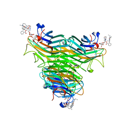
5DIS
 | | Crystal structure of a CRM1-RanGTP-SPN1 export complex bound to a 113 amino acid FG-repeat containing fragment of Nup214 | | 分子名称: | Exportin-1, GTP-binding nuclear protein Ran, GUANOSINE-5'-TRIPHOSPHATE, ... | | 著者 | Monecke, T, Port, S.A, Dickmanns, A, Kehlenbach, R.H, Ficner, R. | | 登録日 | 2015-09-01 | | 公開日 | 2015-11-04 | | 最終更新日 | 2024-01-10 | | 実験手法 | X-RAY DIFFRACTION (2.85 Å) | | 主引用文献 | Structural and Functional Characterization of CRM1-Nup214 Interactions Reveals Multiple FG-Binding Sites Involved in Nuclear Export.
Cell Rep, 13, 2015
|
|
7B9Q
 | | The SERp optimized structure of Ribonucleotide reductase from Rhodobacter sphaeroides | | 分子名称: | 2'-DEOXYADENOSINE 5'-TRIPHOSPHATE, MAGNESIUM ION, Vitamin B12-dependent ribonucleotide reductase | | 著者 | Loderer, C, Feiler, C, Wilk, P, Kabinger, F. | | 登録日 | 2020-12-14 | | 公開日 | 2022-01-12 | | 最終更新日 | 2024-01-31 | | 実験手法 | X-RAY DIFFRACTION (2.78 Å) | | 主引用文献 | HUG Domain Is Responsible for Active Dimer Stabilization in an NrdJd Ribonucleotide Reductase.
Biochemistry, 61, 2022
|
|
7B9P
 | | Structure of Ribonucleotide reductase from Rhodobacter sphaeroides | | 分子名称: | 2'-DEOXYADENOSINE 5'-TRIPHOSPHATE, MAGNESIUM ION, Vitamin B12-dependent ribonucleotide reductase | | 著者 | Wilk, P, Feiler, C, Loderer, C, Kabinger, F. | | 登録日 | 2020-12-14 | | 公開日 | 2022-01-12 | | 最終更新日 | 2024-11-06 | | 実験手法 | X-RAY DIFFRACTION (2.646 Å) | | 主引用文献 | HUG Domain Is Responsible for Active Dimer Stabilization in an NrdJd Ribonucleotide Reductase.
Biochemistry, 61, 2022
|
|
4P9X
 | | Structure of ConA/Rh3Glu complex | | 分子名称: | 2-[2-(2-{4-[(alpha-D-glucopyranosyloxy)methyl]-1H-1,2,3-triazol-1-yl}ethoxy)ethoxy]ethyl 2-[3,6-bis(diethylamino)-9H-xanthen-9-yl]benzoate, CALCIUM ION, Concanavalin-A, ... | | 著者 | Sakai, F, Weiss, M.S, Chen, G. | | 登録日 | 2014-04-06 | | 公開日 | 2014-08-27 | | 最終更新日 | 2023-12-27 | | 実験手法 | X-RAY DIFFRACTION (2.06 Å) | | 主引用文献 | Structure of ConA/Rh3Glu complex
To Be Published
|
|
6TTM
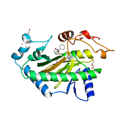 | | Hyoscyamine 6-hydroxylase in complex with N-oxalylglycine and hyoscyamine | | 分子名称: | 1,2-ETHANEDIOL, DI(HYDROXYETHYL)ETHER, Hyoscyamine 6 beta-hydroxylase, ... | | 著者 | Kluza, A, Kurpiewska, K, Mrugala, B, Porebski, P.J, Niedzialkowska, E, Minor, W, Borowski, T. | | 登録日 | 2019-12-29 | | 公開日 | 2020-03-18 | | 最終更新日 | 2024-01-24 | | 実験手法 | X-RAY DIFFRACTION (1.91 Å) | | 主引用文献 | Regioselectivity of hyoscyamine 6 beta-hydroxylase-catalysed hydroxylation as revealed by high-resolution structural information and QM/MM calculations.
Dalton Trans, 49, 2020
|
|
5QBC
 | |
5QBL
 | | Crystal structure of Endothiapepsin-NAT17-347147 complex | | 分子名称: | (1S,2S,3S,4R,5R)-2-({[5-(hydroxymethyl)furan-2-yl]methyl}amino)-4-(morpholin-4-yl)-6,8-dioxabicyclo[3.2.1]octan-3-ol, 1,2-ETHANEDIOL, ACETATE ION, ... | | 著者 | Huschmann, F. | | 登録日 | 2017-08-04 | | 公開日 | 2020-04-22 | | 最終更新日 | 2024-10-30 | | 実験手法 | X-RAY DIFFRACTION (1.619 Å) | | 主引用文献 | Crystal structure of Endothiapepsin
To be published
|
|
5QBB
 | |
5QBJ
 | |
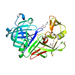
5QBR
 | | Crystal structure of Endothiapepsin | | 分子名称: | (3R,5R)-5-[3-(3-fluoro-4-methylphenyl)-1,2,4-oxadiazol-5-yl]pyrrolidin-3-ol, ACETATE ION, DIMETHYL SULFOXIDE, ... | | 著者 | Huschmann, F. | | 登録日 | 2017-08-04 | | 公開日 | 2020-04-22 | | 最終更新日 | 2024-11-06 | | 実験手法 | X-RAY DIFFRACTION (1.418 Å) | | 主引用文献 | Crystal structure of Endothiapepsin
To be published
|
|


