4KMV
 
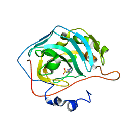
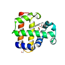 | | Structure of the L100F MUTANT OF DEHALOPEROXIDASE-HEMOGLOBIN A FROM AMPHITRITE ORNATA WITH 2,4,6-TRICHLOROPHENOL | | 分子名称: | 1,2-ETHANEDIOL, 2,4,6-trichlorophenol, Dehaloperoxidase A, ... | | 著者 | Wang, C, Lovelace, L, Lebioda, L. | | 登録日 | 2013-05-08 | | 公開日 | 2013-09-04 | | 最終更新日 | 2024-02-28 | | 実験手法 | X-RAY DIFFRACTION (1.44 Å) | | 主引用文献 | Complexes of dual-function hemoglobin/dehaloperoxidase with substrate 2,4,6-trichlorophenol are inhibitory and indicate binding of halophenol to compound I.
Biochemistry, 52, 2013
|
|
4UBY
 | |
2EUC
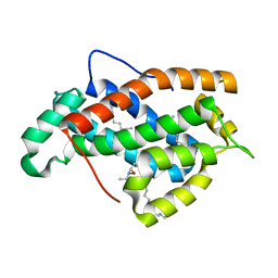
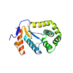
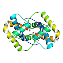 | | Crystal structure of YfmB from Bacillus subtilis. NESG TARGET SR324 | | 分子名称: | Hypothetical protein yfmB | | 著者 | Benach, J, Abashidze, M, Jayaraman, S, Janjua, H, Cooper, B, Rong, X, Acton, T.B, Montelione, G.T, Hunt, J.F, Tong, L, Northeast Structural Genomics Consortium (NESG) | | 登録日 | 2005-10-28 | | 公開日 | 2005-11-08 | | 最終更新日 | 2011-07-13 | | 実験手法 | X-RAY DIFFRACTION (2.5 Å) | | 主引用文献 | Crystal structure of YfmB from bacillus subtilis NESG TARGET SR324.
To be Published
|
|
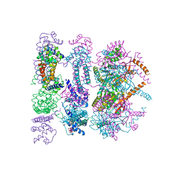
4Z9M
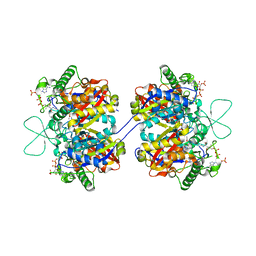
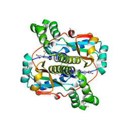
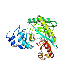 | | Crystal structure of human sarcomeric mitochondrial creatine kinase | | 分子名称: | ADENOSINE-5'-DIPHOSPHATE, Creatine kinase S-type, mitochondrial, ... | | 著者 | Rabeh, W.M, Tempel, W, Nedyalkova, L, Landry, R, Arrowsmith, C.H, Edwards, A.M, Bountra, C, Bochkarev, A, Park, H, Structural Genomics Consortium (SGC) | | 登録日 | 2015-04-10 | | 公開日 | 2015-04-22 | | 最終更新日 | 2023-09-27 | | 実験手法 | X-RAY DIFFRACTION (2.1 Å) | | 主引用文献 | Crystal structure of human sarcomeric mitochondrial creatine kinase
To Be Published
|
|
5L35
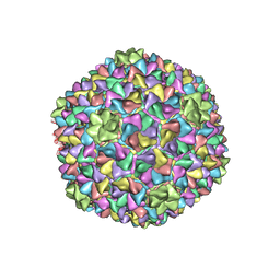 | |
4ZC0
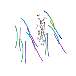
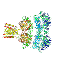
 | | Structure of a dodecameric bacterial helicase | | 分子名称: | HEXATANTALUM DODECABROMIDE, Replicative DNA helicase | | 著者 | Bazin, A, Cherrier, M.V, Gutsche, I, Timmins, J, Terradot, L. | | 登録日 | 2015-04-15 | | 公開日 | 2015-10-21 | | 最終更新日 | 2024-05-08 | | 実験手法 | X-RAY DIFFRACTION (6.7 Å) | | 主引用文献 | Structure and primase-mediated activation of a bacterial dodecameric replicative helicase.
Nucleic Acids Res., 43, 2015
|
|
2F06
 | |
2ERV
 | | Crystal structure of the outer membrane enzyme PagL | | 分子名称: | CALCIUM ION, PENTAETHYLENE GLYCOL MONODECYL ETHER, hypothetical protein Paer03002360 | | 著者 | Rutten, L, Geurtsen, J, Lambert, W, Smolenaers, J.J, Bonvin, A.M, van der Ley, P, Egmond, M.R, Gros, P, Tommassen, J. | | 登録日 | 2005-10-25 | | 公開日 | 2006-04-11 | | 最終更新日 | 2023-08-23 | | 実験手法 | X-RAY DIFFRACTION (2 Å) | | 主引用文献 | Crystal structure and catalytic mechanism of the LPS 3-O-deacylase PagL from Pseudomonas aeruginosa.
Proc.Natl.Acad.Sci.USA, 103, 2006
|
|
2F1W
 | | Crystal structure of the TRAF-like domain of HAUSP/USP7 | | 分子名称: | CALCIUM ION, Ubiquitin carboxyl-terminal hydrolase 7 | | 著者 | Hu, M, Gu, L, Jeffrey, P.D, Shi, Y. | | 登録日 | 2005-11-15 | | 公開日 | 2006-02-07 | | 最終更新日 | 2024-02-14 | | 実験手法 | X-RAY DIFFRACTION (1.65 Å) | | 主引用文献 | Structural Basis of Competitive Recognition of p53 and MDM2 by HAUSP/USP7: Implications for the Regulation of the p53-MDM2 Pathway.
Plos Biol., 4, 2006
|
|
2EU2
 | | Human Carbonic Anhydrase II in complex with novel inhibitors | | 分子名称: | (R)-1-AMINO-1-[5-(DIMETHYLAMINO)-1,3,4-THIADIAZOL-2-YL]METHANESULFONAMIDE, Carbonic anhydrase 2, ZINC ION | | 著者 | Fisher, S.Z, Govindasamy, L, Boyle, N, Agbandje-McKenna, M, Silverman, D.N, Blackburn, G.M, McKenna, R. | | 登録日 | 2005-10-28 | | 公開日 | 2006-07-11 | | 最終更新日 | 2023-08-23 | | 実験手法 | X-RAY DIFFRACTION (1.15 Å) | | 主引用文献 | X-ray crystallographic studies reveal that the incorporation of spacer groups in carbonic anhydrase inhibitors causes alternate binding modes.
Acta Crystallogr.,Sect.F, 62, 2006
|
|
4U2Q
 | | Full-length AMPA subtype ionotropic glutamate receptor GluA2 in complex with partial agonist kainate | | 分子名称: | 2-acetamido-2-deoxy-beta-D-glucopyranose, 3-(CARBOXYMETHYL)-4-ISOPROPENYLPROLINE, Glutamate receptor 2 | | 著者 | Duerr, K.L, Chen, L, Gouaux, E. | | 登録日 | 2014-07-17 | | 公開日 | 2014-08-20 | | 最終更新日 | 2023-12-27 | | 実験手法 | X-RAY DIFFRACTION (3.5247 Å) | | 主引用文献 | Structure and Dynamics of AMPA Receptor GluA2 in Resting, Pre-Open, and Desensitized States.
Cell, 158, 2014
|
|
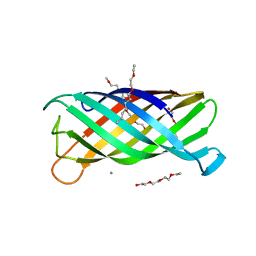
2EUM
 | | Crystal structure of human Glycolipid Transfer Protein complexed with 8:0 Lactosylceramide | | 分子名称: | DECANE, Glycolipid transfer protein, N-OCTANE, ... | | 著者 | Malinina, L, Malakhova, M.L, Kanack, A.T, Abagyan, R, Brown, R.E, Patel, D.J. | | 登録日 | 2005-10-28 | | 公開日 | 2006-11-14 | | 最終更新日 | 2023-08-23 | | 実験手法 | X-RAY DIFFRACTION (2.3 Å) | | 主引用文献 | The liganding of glycolipid transfer protein is controlled by glycolipid acyl structure.
Plos Biol., 4, 2006
|
|
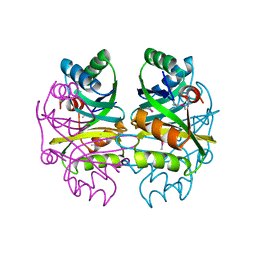
2F2S
 | | Human mitochondrial acetoacetyl-CoA thiolase | | 分子名称: | Acetyl-CoA acetyltransferase, mitochondrial, CHLORIDE ION, ... | | 著者 | Min, J.R, Dombrovski, L, Antoshenko, T, Wu, H, Loppnau, P, Weigelt, J, Sundstrom, M, Arrowsmith, C.H, Edwards, A.M, Bochkarev, A, Plotnikov, A.N, Structural Genomics Consortium (SGC) | | 登録日 | 2005-11-17 | | 公開日 | 2005-11-29 | | 最終更新日 | 2011-07-13 | | 実験手法 | X-RAY DIFFRACTION (2 Å) | | 主引用文献 | The Crystal Structure of Human Mitochondrial Acetoacetyl-Coa Thiolase Acat1.
To be Published
|
|
1DCM
 | | STRUCTURE OF UNPHOSPHORYLATED FIXJ-N WITH AN ATYPICAL CONFORMER (MONOMER A) | | 分子名称: | MAGNESIUM ION, TRANSCRIPTIONAL REGULATORY PROTEIN FIXJ | | 著者 | Gouet, P, Fabry, B, Guillet, V, Birck, C, Mourey, L, Kahn, D, Samama, J.P. | | 登録日 | 1999-11-05 | | 公開日 | 2000-11-08 | | 最終更新日 | 2023-08-09 | | 実験手法 | X-RAY DIFFRACTION (3 Å) | | 主引用文献 | Structural transitions in the FixJ receiver domain.
Structure Fold.Des., 7, 1999
|
|
2BBE
 | |
4U49
 | |
4U4B
 | |
2B29
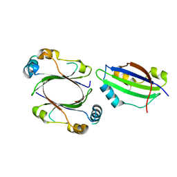
 | | N-terminal domain of the RPA70 subunit of human replication protein A. | | 分子名称: | Replication protein A 70 kDa DNA-binding subunit | | 著者 | Bochkareva, E, Kaustov, L, Ayed, A, Okorokov, A, Milner, J, Arrowsmith, C.H, Bochkarev, A. | | 登録日 | 2005-09-18 | | 公開日 | 2005-10-04 | | 最終更新日 | 2024-02-14 | | 実験手法 | X-RAY DIFFRACTION (1.6 Å) | | 主引用文献 | Single-stranded DNA mimicry in the p53 transactivation domain interaction with replication protein A.
Proc.Natl.Acad.Sci.Usa, 102, 2005
|
|
2B2E
 | | RNA stemloop from bacteriophage MS2 complexed with an N87S,E89K mutant MS2 capsid | | 分子名称: | 5'-R(*AP*CP*AP*UP*GP*AP*GP*GP*AP*UP*UP*AP*CP*CP*CP*AP*UP*GP*U)-3', Coat protein | | 著者 | Horn, W.T, Tars, K, Grahn, E, Helgstrand, C, Baron, A.J, Lago, H, Adams, C.J, Peabody, D.S, Phillips, S.E.V, Stonehouse, N.J, Liljas, L, Stockley, P.G. | | 登録日 | 2005-09-19 | | 公開日 | 2006-05-09 | | 最終更新日 | 2023-10-25 | | 実験手法 | X-RAY DIFFRACTION (3.15 Å) | | 主引用文献 | Structural basis of RNA binding discrimination between bacteriophages Qbeta and MS2
Structure, 14, 2006
|
|
4U2R
 | | Crystal structure of the GLUR2 ligand binding core (S1S2J, flip variant) in the apo state | | 分子名称: | Glutamate receptor 2, SULFATE ION | | 著者 | Duerr, K.L, Chen, L, Gouaux, E. | | 登録日 | 2014-07-17 | | 公開日 | 2014-08-20 | | 最終更新日 | 2023-12-27 | | 実験手法 | X-RAY DIFFRACTION (1.4114 Å) | | 主引用文献 | Structure and Dynamics of AMPA Receptor GluA2 in Resting, Pre-Open, and Desensitized States.
Cell, 158, 2014
|
|
2B0O
 | | Crystal structure of UPLC1 GAP domain | | 分子名称: | UPLC1, ZINC ION | | 著者 | Ismail, S, Shen, L, Arrowsmith, C, Edwards, A, Sundstrom, M, Weigelt, J, Bochkarev, A, Park, H, Structural Genomics Consortium (SGC) | | 登録日 | 2005-09-14 | | 公開日 | 2005-09-20 | | 最終更新日 | 2023-08-23 | | 実験手法 | X-RAY DIFFRACTION (2.06 Å) | | 主引用文献 | Structural analysis of GAP and ankyrin domains of UPLC1
To be Published
|
|
4ZA1
 | | Crystal Structure of NosA Involved in Nosiheptide Biosynthesis | | 分子名称: | 2,3-DIHYDROXY-1,4-DITHIOBUTANE, NosA | | 著者 | Liu, S, Guo, H, Zhang, T, Han, L, Yao, P, Zhang, Y, Rong, N, Yu, Y, Lan, W, Wang, C, Ding, J, Wang, R, Liu, W, Cao, C. | | 登録日 | 2015-04-13 | | 公開日 | 2015-08-19 | | 最終更新日 | 2024-03-20 | | 実験手法 | X-RAY DIFFRACTION (2.5 Å) | | 主引用文献 | Structure-based Mechanistic Insights into Terminal Amide Synthase in Nosiheptide-Represented Thiopeptides Biosynthesis
Sci Rep, 5, 2015
|
|
2B67
 | | Crystal structure of the Nitroreductase Family Protein from Streptococcus pneumoniae TIGR4 | | 分子名称: | ACETIC ACID, COG0778: Nitroreductase, FLAVIN MONONUCLEOTIDE | | 著者 | Kim, Y, Volkart, L, Abdullah, J, Collart, F, Joachimiak, A, Midwest Center for Structural Genomics (MCSG) | | 登録日 | 2005-09-30 | | 公開日 | 2005-11-15 | | 最終更新日 | 2017-10-11 | | 実験手法 | X-RAY DIFFRACTION (2.05 Å) | | 主引用文献 | Crystal Structure of the Nitroreductase Family Protein from Streptococcus pneumoniae TIGR4
To be Published
|
|
2B3S
 | |
2B9C
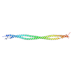 | | Structure of tropomyosin's mid-region: bending and binding sites for actin | | 分子名称: | striated-muscle alpha tropomyosin | | 著者 | Brown, J.H, Zhou, Z, Reshetnikova, L, Robinson, H, Yammani, R.D, Tobacman, L.S, Cohen, C. | | 登録日 | 2005-10-11 | | 公開日 | 2006-01-03 | | 最終更新日 | 2022-12-21 | | 実験手法 | X-RAY DIFFRACTION (2.3 Å) | | 主引用文献 | Structure of the mid-region of tropomyosin: Bending and binding sites for actin.
Proc.Natl.Acad.Sci.Usa, 102, 2005
|
|


