3KKQ
 
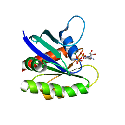
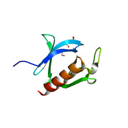
 | | Crystal structure of M-Ras P40D in complex with GDP | | 分子名称: | GUANOSINE-5'-DIPHOSPHATE, MAGNESIUM ION, Ras-related protein M-Ras | | 著者 | Muraoka, S, Shima, F, Liao, J, Ijiri, Y, Matsumoto, K, Ye, M, Inoue, T, Kataoka, T. | | 登録日 | 2009-11-06 | | 公開日 | 2010-06-16 | | 最終更新日 | 2023-11-01 | | 実験手法 | X-RAY DIFFRACTION (1.2 Å) | | 主引用文献 | Structural basis for conformational dynamics of GTP-bound Ras protein
J.Biol.Chem., 285, 2010
|
|
3KKO
 | | Crystal structure of M-Ras P40D/D41E/L51R in complex with GppNHp | | 分子名称: | MAGNESIUM ION, PHOSPHOAMINOPHOSPHONIC ACID-GUANYLATE ESTER, Ras-related protein M-Ras, ... | | 著者 | Muraoka, S, Shima, F, Liao, J, Ijiri, Y, Matsumoto, K, Ye, M, Inoue, T, Kataoka, T. | | 登録日 | 2009-11-06 | | 公開日 | 2010-06-16 | | 最終更新日 | 2023-11-01 | | 実験手法 | X-RAY DIFFRACTION (1.9 Å) | | 主引用文献 | Structural basis for conformational dynamics of GTP-bound Ras protein
J.Biol.Chem., 285, 2010
|
|
3KKM
 | | Crystal structure of H-Ras T35S in complex with GppNHp | | 分子名称: | GTPase HRas, MAGNESIUM ION, PHOSPHOAMINOPHOSPHONIC ACID-GUANYLATE ESTER, ... | | 著者 | Muraoka, S, Shima, F, Liao, J, Ijiri, Y, Matsumoto, K, Ye, M, Inoue, T, Kataoka, T. | | 登録日 | 2009-11-06 | | 公開日 | 2010-06-16 | | 最終更新日 | 2023-11-01 | | 実験手法 | X-RAY DIFFRACTION (1.7 Å) | | 主引用文献 | Structural basis for conformational dynamics of GTP-bound Ras protein
J.Biol.Chem., 285, 2010
|
|
3KKP
 | | Crystal structure of M-Ras P40D in complex with GppNHp | | 分子名称: | MAGNESIUM ION, PHOSPHOAMINOPHOSPHONIC ACID-GUANYLATE ESTER, Ras-related protein M-Ras | | 著者 | Muraoka, S, Shima, F, Liao, J, Ijiri, Y, Matsumoto, K, Ye, M, Inoue, T, Kataoka, T. | | 登録日 | 2009-11-06 | | 公開日 | 2010-06-16 | | 最終更新日 | 2023-11-01 | | 実験手法 | X-RAY DIFFRACTION (1.35 Å) | | 主引用文献 | Structural basis for conformational dynamics of GTP-bound Ras protein
J.Biol.Chem., 285, 2010
|
|
3KKN
 | | Crystal structure of H-Ras T35S in complex with GppNHp | | 分子名称: | GTPase HRas, MAGNESIUM ION, PHOSPHOAMINOPHOSPHONIC ACID-GUANYLATE ESTER | | 著者 | Muraoka, S, Shima, F, Liao, J, Ijiri, Y, Matsumoto, K, Ye, M, Inoue, T, Kataoka, T. | | 登録日 | 2009-11-06 | | 公開日 | 2010-06-16 | | 最終更新日 | 2023-11-01 | | 実験手法 | X-RAY DIFFRACTION (2.09 Å) | | 主引用文献 | Structural basis for conformational dynamics of GTP-bound Ras protein
J.Biol.Chem., 285, 2010
|
|
8PS0
 | |
3WVM
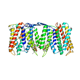
 | | The 0.88 angstrom X-ray structure of the human heart fatty acid-binding protein complexed with stearic acid | | 分子名称: | Fatty acid-binding protein, heart, HEXAETHYLENE GLYCOL, ... | | 著者 | Sugiyama, S, Matsuoka, S, Mizohata, E, Matsuoka, D, Ishida, H, Hirose, M, Kakinouchi, K, Hara, T, Matsumura, H, Murakami, S, Inoue, T, Murata, M. | | 登録日 | 2014-05-25 | | 公開日 | 2015-01-28 | | 最終更新日 | 2024-05-29 | | 実験手法 | X-RAY DIFFRACTION (0.88 Å) | | 主引用文献 | Water-mediated recognition of simple alkyl chains by heart-type fatty-acid-binding protein.
Angew.Chem.Int.Ed.Engl., 54, 2015
|
|
6PX1
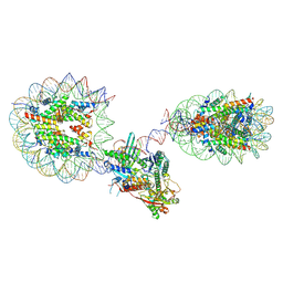
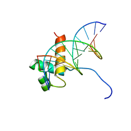
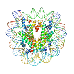 | | Set2 bound to nucleosome | | 分子名称: | DNA (149-MER), Histone H2B 1.1, Histone H3, ... | | 著者 | Halic, M, Bilokapic, S. | | 登録日 | 2019-07-24 | | 公開日 | 2019-08-28 | | 最終更新日 | 2024-03-20 | | 実験手法 | ELECTRON MICROSCOPY (3.3 Å) | | 主引用文献 | Nucleosome and ubiquitin position Set2 to methylate H3K36.
Nat Commun, 10, 2019
|
|
6X0L
 | |
6X0M
 | |
6PX3
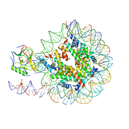
 | | Set2 bound to nucleosome | | 分子名称: | DNA (145-MER), Histone H2B 1.1, Histone H3, ... | | 著者 | Halic, M, Bilokapic, S. | | 登録日 | 2019-07-24 | | 公開日 | 2019-08-28 | | 最終更新日 | 2019-09-04 | | 実験手法 | ELECTRON MICROSCOPY (4.1 Å) | | 主引用文献 | Nucleosome and ubiquitin position Set2 to methylate H3K36.
Nat Commun, 10, 2019
|
|
6WZ5
 | |
6WZ9
 | |
6X0N
 | |
6N88
 | |
6NZO
 | | Set2 bound to nucleosome | | 分子名称: | DNA (149-MER), Histone H2B 1.1, Histone H3, ... | | 著者 | Halic, M, Bilokapic, S. | | 登録日 | 2019-02-14 | | 公開日 | 2019-08-28 | | 最終更新日 | 2019-09-04 | | 実験手法 | ELECTRON MICROSCOPY (3.8 Å) | | 主引用文献 | Nucleosome and ubiquitin position Set2 to methylate H3K36.
Nat Commun, 10, 2019
|
|
6N89
 | |
3AJ4
 | | Crystal structure of the PH domain of Evectin-2 from human complexed with O-phospho-L-serine | | 分子名称: | 1,2-ETHANEDIOL, PHOSPHOSERINE, Pleckstrin homology domain-containing family B member 2 | | 著者 | Okazaki, S, Kato, R, Wakatsuki, S. | | 登録日 | 2010-05-21 | | 公開日 | 2011-05-25 | | 最終更新日 | 2023-11-01 | | 実験手法 | X-RAY DIFFRACTION (1 Å) | | 主引用文献 | Intracellular phosphatidylserine is essential for retrograde membrane traffic through endosomes
Proc.Natl.Acad.Sci.USA, 108, 2011
|
|
1VFC
 | |
1VF9
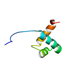 | | Solution Structure Of Human Trf2 | | 分子名称: | Telomeric repeat binding factor 2 | | 著者 | Nishimura, Y, Hanaoka, S. | | 登録日 | 2004-04-12 | | 公開日 | 2005-05-17 | | 最終更新日 | 2023-12-27 | | 実験手法 | SOLUTION NMR | | 主引用文献 | Comparison between TRF2 and TRF1 of their telomeric DNA-bound structures and DNA-binding activities
Protein Sci., 14, 2005
|
|
7EGO
 | | X-ray structure of the human heart fatty acid-binding protein complexed with the fluorescent probe HA527 | | 分子名称: | 3-[methyl-(4-nitro-2,1,3-benzoxadiazol-7-yl)amino]propanoic acid, Fatty acid-binding protein, heart, ... | | 著者 | Takabayashi, M, Yokota, J, Matsuoka, S, Tsuchikawa, H, Sonoyama, M, Inoue, Y, Hayashi, F, Sugiyama, S. | | 登録日 | 2021-03-24 | | 公開日 | 2022-03-30 | | 最終更新日 | 2023-11-29 | | 実験手法 | X-RAY DIFFRACTION (1.21 Å) | | 主引用文献 | X-ray structure of the human heart fatty acid-binding protein complexed with the fluorescent probe HA527
To Be Published
|
|
3WQH
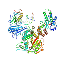
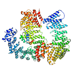
 | | Crystal Structure of human DPP-IV in complex with Anagliptin | | 分子名称: | 2-acetamido-2-deoxy-beta-D-glucopyranose, Dipeptidyl peptidase 4, N-[2-({2-[(2S)-2-cyanopyrrolidin-1-yl]-2-oxoethyl}amino)-2-methylpropyl]-2-methylpyrazolo[1,5-a]pyrimidine-6-carboxamide | | 著者 | Watanabe, Y.S, Okada, S, Motoyama, T, Takahashi, R, Adachi, H, Oka, M. | | 登録日 | 2014-01-27 | | 公開日 | 2015-07-15 | | 最終更新日 | 2023-11-08 | | 実験手法 | X-RAY DIFFRACTION (2.85 Å) | | 主引用文献 | Anagliptin, a potent dipeptidyl peptidase IV inhibitor: its single-crystal structure and enzyme interactions.
J Enzyme Inhib Med Chem, 30, 2015
|
|
1ISE
 | | Crystal structure of a mutant of ribosome recycling factor from Escherichia coli, Arg132Gly | | 分子名称: | Ribosome Recycling Factor | | 著者 | Nakano, H, Yoshida, T, Oka, S, Uchiyama, S, Nishina, K, Ohkubo, T, Kato, H, Yamagata, Y, Kobayashi, Y. | | 登録日 | 2001-11-30 | | 公開日 | 2003-10-07 | | 最終更新日 | 2023-12-27 | | 実験手法 | X-RAY DIFFRACTION (2.2 Å) | | 主引用文献 | Crystal structure of a mutant of ribosome recycling factor from Escherichia coli, Arg132Gly
To be Published
|
|
3W39
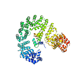
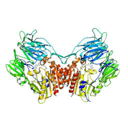
 | | Crystal structure of HLA-B*5201 in complexed with HIV immunodominant epitope (TAFTIPSI) | | 分子名称: | Beta-2-microglobulin, HLA class I histocompatibility antigen, B-52 alpha chain, ... | | 著者 | Yagita, Y, Kuse, N, Kuroki, K, Gatanaga, H, Carlson, J.M, Chikata, T, Brumme, Z.L, Murakoshi, H, Akahoshi, T, Pfeifer, N, Mallal, S, John, M, Ose, T, Matsubara, H, Kanda, R, Fukunaga, Y, Honda, K, Kawashima, Y, Ariumi, Y, Oka, S, Maenaka, K, Takiguchi, M. | | 登録日 | 2012-12-13 | | 公開日 | 2013-02-13 | | 最終更新日 | 2023-11-08 | | 実験手法 | X-RAY DIFFRACTION (3.1 Å) | | 主引用文献 | Distinct HIV-1 Escape Patterns Selected by Cytotoxic T Cells with Identical Epitope Specificity
J.Virol., 87, 2013
|
|
2D0J
 | | Crystal Structure of Human GlcAT-S Apo Form | | 分子名称: | Galactosylgalactosylxylosylprotein 3-beta-glucuronosyltransferase 2 | | 著者 | Shiba, T, Kakuda, S, Ishiguro, M, Oka, S, Kawasaki, T, Wakatsuki, S, Kato, R. | | 登録日 | 2005-08-03 | | 公開日 | 2006-07-18 | | 最終更新日 | 2023-10-25 | | 実験手法 | X-RAY DIFFRACTION (2 Å) | | 主引用文献 | Crystal structure of GlcAT-S, a human glucuronyltransferase, involved in the biosynthesis of the HNK-1 carbohydrate epitope
To be published
|
|


