2Y46
 
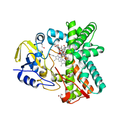
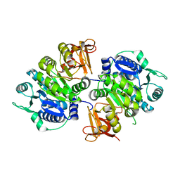
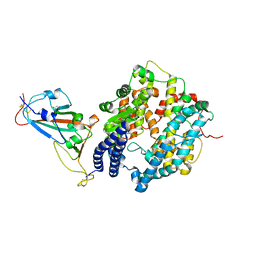
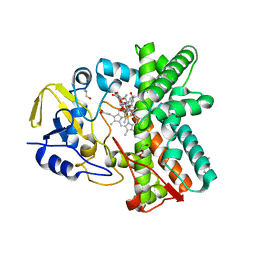 | | Structure of the mixed-function P450 MycG in complex with mycinamicin IV in C 2 2 21 space group | | 分子名称: | BENZAMIDINE, GLYCEROL, MYCINAMICIN IV, ... | | 著者 | Li, S, Kells, P.M, Sherman, D.H, Podust, L.M. | | 登録日 | 2011-01-05 | | 公開日 | 2012-01-25 | | 最終更新日 | 2023-12-20 | | 実験手法 | X-RAY DIFFRACTION (1.83 Å) | | 主引用文献 | Substrate Recognition by the Multifunctional Cytochrome P450 Mycg in Mycinamicin Hydroxylation and Epoxidation Reactions.
J.Biol.Chem., 287, 2012
|
|
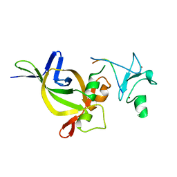
4I16
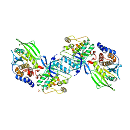
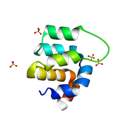 | | Crystal structure of CARMA1 CARD | | 分子名称: | Caspase recruitment domain-containing protein 11, SULFATE ION | | 著者 | Li, S, Yang, X, Shen, Y. | | 登録日 | 2012-11-20 | | 公開日 | 2012-12-26 | | 最終更新日 | 2024-03-20 | | 実験手法 | X-RAY DIFFRACTION (1.751 Å) | | 主引用文献 | Structural insights into the assembly of CARMA1 and BCL10
Plos One, 7, 2012
|
|
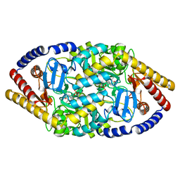
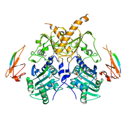
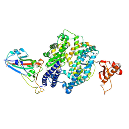
1EGU
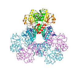
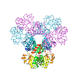
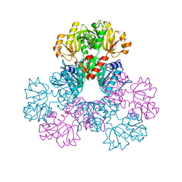
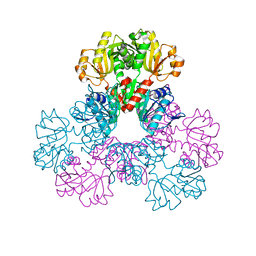
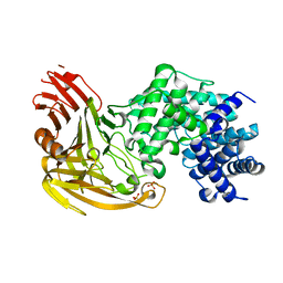 | | CRYSTAL STRUCTURE OF STREPTOCOCCUS PNEUMONIAE HYALURONATE LYASE AT 1.56 A RESOLUTION | | 分子名称: | HYALURONATE LYASE, SULFATE ION | | 著者 | Li, S, Kelly, S.J, Lamani, E, Ferraroni, M, Jedrzejas, M.J. | | 登録日 | 2000-02-16 | | 公開日 | 2001-02-16 | | 最終更新日 | 2024-02-07 | | 実験手法 | X-RAY DIFFRACTION (1.56 Å) | | 主引用文献 | Structural basis of hyaluronan degradation by Streptococcus pneumoniae hyaluronate lyase.
EMBO J., 19, 2000
|
|
1F1S
 | |
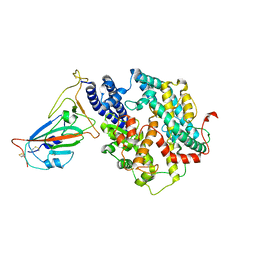
3ZSN
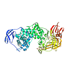
 | | Structure of the mixed-function P450 MycG F286A mutant in complex with mycinamicin IV | | 分子名称: | BENZAMIDINE, GLYCEROL, MYCINAMICIN IV, ... | | 著者 | Li, S, Kells, P.M, Rutaganira, F.U, Anzai, Y, Kato, F, Sherman, D.H, Podust, L.M. | | 登録日 | 2011-06-29 | | 公開日 | 2012-05-09 | | 最終更新日 | 2023-12-20 | | 実験手法 | X-RAY DIFFRACTION (1.9 Å) | | 主引用文献 | Substrate Recognition by the Multifunctional Cytochrome P450 Mycg in Mycinamicin Hydroxylation and Epoxidation Reactions.
J.Biol.Chem., 287, 2012
|
|
5YKR
 | |
5YKT
 | |
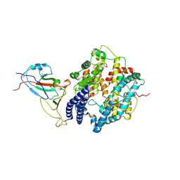
4AW3
 | | Structure of the mixed-function P450 MycG F286V mutant in complex with mycinamicin V in P1 space group | | 分子名称: | GLYCEROL, MYCINAMICIN V, P-450-LIKE PROTEIN, ... | | 著者 | Li, S, Tietz, D.R, Rutaganira, F.U, Kells, P.M, Anzai, Y, Kato, F, Pochapsky, T.C, Sherman, D.H, Podust, L.M. | | 登録日 | 2012-05-30 | | 公開日 | 2012-09-05 | | 最終更新日 | 2023-12-20 | | 実験手法 | X-RAY DIFFRACTION (2.05 Å) | | 主引用文献 | Substrate Recognition by the Multifunctional Cytochrome P450 Mycg in Mycinamicin Hydroxylation and Epoxidation Reactions.
J.Biol.Chem., 287, 2012
|
|
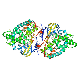
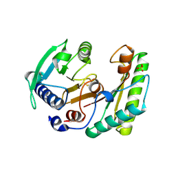
2HCR
 | | crystal structure of human phosphoribosyl pyrophosphate synthetase 1 in complex with AMP(ATP), cadmium and sulfate ion | | 分子名称: | ADENOSINE MONOPHOSPHATE, CADMIUM ION, Ribose-phosphate pyrophosphokinase I, ... | | 著者 | Li, S, Peng, B, Ding, J. | | 登録日 | 2006-06-18 | | 公開日 | 2006-10-24 | | 最終更新日 | 2023-10-25 | | 実験手法 | X-RAY DIFFRACTION (2.2 Å) | | 主引用文献 | Crystal structure of human phosphoribosylpyrophosphate synthetase 1 reveals a novel allosteric site
Biochem.J., 401, 2007
|
|
2GV5
 | | crystal structure of Sfi1p/Cdc31p complex | | 分子名称: | Cell division control protein 31, Sfi1p | | 著者 | Li, S, Sandercock, A.M, Conduit, P.T, Robinson, C.V, Williams, R.L, Kilmartin, J.V. | | 登録日 | 2006-05-02 | | 公開日 | 2006-06-27 | | 最終更新日 | 2017-10-18 | | 実験手法 | X-RAY DIFFRACTION (3 Å) | | 主引用文献 | Structural role of Sfi1p-centrin filaments in budding yeast spindle pole body duplication.
J.Cell Biol., 173, 2006
|
|
2H06
 | |
2H08
 | |
2H07
 | |
5FYN
 | | Sub-tomogram averaging of Tula virus glycoprotein spike | | 分子名称: | 2-acetamido-2-deoxy-beta-D-glucopyranose-(1-4)-2-acetamido-2-deoxy-beta-D-glucopyranose, PUUMALA VIRUS GN GLYCOPROTEIN, alpha-D-mannopyranose-(1-3)-beta-D-mannopyranose-(1-4)-2-acetamido-2-deoxy-beta-D-glucopyranose-(1-4)-2-acetamido-2-deoxy-beta-D-glucopyranose, ... | | 著者 | Li, S, Rissanen, I, Zeltina, A, Hepojoki, J, Raghwani, J, Harlos, K, Pybus, O.G, Huiskonen, J.T, Bowden, T.A. | | 登録日 | 2016-03-08 | | 公開日 | 2016-06-08 | | 最終更新日 | 2020-07-29 | | 実験手法 | ELECTRON MICROSCOPY (15.6 Å) | | 主引用文献 | A Molecular-Level Account of the Antigenic Hantaviral Surface.
Cell Rep., 15, 2016
|
|
5HH7
 | |
5IX2
 | | Crystal structure of mouse Morc3 ATPase-CW cassette in complex with AMPPNP and unmodified H3 peptide | | 分子名称: | MAGNESIUM ION, MORC family CW-type zinc finger protein 3, PHOSPHOAMINOPHOSPHONIC ACID-ADENYLATE ESTER, ... | | 著者 | Li, S, Du, J, Patel, D.J. | | 登録日 | 2016-03-23 | | 公開日 | 2016-08-17 | | 最終更新日 | 2023-11-08 | | 実験手法 | X-RAY DIFFRACTION (2.9 Å) | | 主引用文献 | Mouse MORC3 is a GHKL ATPase that localizes to H3K4me3 marked chromatin
Proc.Natl.Acad.Sci.USA, 113, 2016
|
|
6IKJ
 | | Crystal structure of YfiB(F48S) | | 分子名称: | GLYCEROL, SULFATE ION, YfiB | | 著者 | Li, S, Zhang, Q, Bartlam, M. | | 登録日 | 2018-10-16 | | 公開日 | 2019-03-13 | | 最終更新日 | 2023-11-22 | | 実験手法 | X-RAY DIFFRACTION (1.76 Å) | | 主引用文献 | Structural analysis of activating mutants of YfiB from Pseudomonas aeruginosa PAO1.
Biochem. Biophys. Res. Commun., 506, 2018
|
|
6IKK
 | |
6IKI
 | | Crystal structure of YfiB(W55L) | | 分子名称: | GLYCEROL, SULFATE ION, YfiB | | 著者 | Li, S, Zhang, Q, Bartlam, M. | | 登録日 | 2018-10-16 | | 公開日 | 2019-03-13 | | 最終更新日 | 2023-11-22 | | 実験手法 | X-RAY DIFFRACTION (2.204 Å) | | 主引用文献 | Structural analysis of activating mutants of YfiB from Pseudomonas aeruginosa PAO1.
Biochem. Biophys. Res. Commun., 506, 2018
|
|
8IVI
 | |
7WSF
 | |
7WSE
 | |
7WSG
 | |
7WSH
 | | Cryo-EM structure of SARS-CoV-2 spike receptor-binding domain in complex with sea lion ACE2 | | 分子名称: | 2-acetamido-2-deoxy-beta-D-glucopyranose, Angiotensin-converting enzyme, Spike protein S1, ... | | 著者 | Li, S, Han, P, Qi, J. | | 登録日 | 2022-01-29 | | 公開日 | 2022-11-09 | | 実験手法 | ELECTRON MICROSCOPY (2.89 Å) | | 主引用文献 | Cross-species recognition and molecular basis of SARS-CoV-2 and SARS-CoV binding to ACE2s of marine animals.
Natl Sci Rev, 9, 2022
|
|
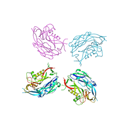
2B4S
 | | Crystal structure of a complex between PTP1B and the insulin receptor tyrosine kinase | | 分子名称: | Insulin receptor, SULFATE ION, Tyrosine-protein phosphatase, ... | | 著者 | Li, S, Depetris, R.S, Barford, D, Chernoff, J, Hubbard, S.R. | | 登録日 | 2005-09-26 | | 公開日 | 2005-11-15 | | 最終更新日 | 2023-11-15 | | 実験手法 | X-RAY DIFFRACTION (2.3 Å) | | 主引用文献 | Crystal Structure of a Complex between Protein Tyrosine Phosphatase 1B and the Insulin Receptor Tyrosine Kinase.
Structure, 13, 2005
|
|


