5V8Z
 
 | |
5V90
 | |
5VRQ
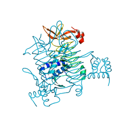
 | | Crystal structure of Legionella pneumophila effector AnkC | | 分子名称: | Ankyrin repeat-containing protein | | 著者 | Kozlov, G, Wong, K, Wang, W, Skubak, P, Munoz-Escobar, J, Liu, Y, Pannu, N.S, Gehring, K, Montreal-Kingston Bacterial Structural Genomics Initiative (BSGI) | | 登録日 | 2017-05-11 | | 公開日 | 2017-11-29 | | 最終更新日 | 2024-03-13 | | 実験手法 | X-RAY DIFFRACTION (3.205 Å) | | 主引用文献 | Ankyrin repeats as a dimerization module.
Biochem. Biophys. Res. Commun., 495, 2018
|
|
5WD8
 | |
5WD9
 | |
6DFD
 | |
4F26
 | |
4F25
 | |
8F6D
 | |
3UVT
 | |
8SMO
 | |
1GH9
 | |
5BTY
 | | Structure of the middle domain of lpg1496 from Legionella pneumophila in P21 space group | | 分子名称: | lpg1496 | | 著者 | Kozlov, G, Zhang, Y, Gehring, K, Montreal-Kingston Bacterial Structural Genomics Initiative (BSGI) | | 登録日 | 2015-06-03 | | 公開日 | 2015-08-26 | | 最終更新日 | 2020-01-08 | | 実験手法 | X-RAY DIFFRACTION (1.15 Å) | | 主引用文献 | Structure of the Legionella Effector, lpg1496, Suggests a Role in Nucleotide Metabolism.
J.Biol.Chem., 290, 2015
|
|
3BCY
 | | Crystal structure of YER067W | | 分子名称: | Protein YER067W | | 著者 | Kozlov, G, Gehring, K. | | 登録日 | 2007-11-13 | | 公開日 | 2008-11-18 | | 最終更新日 | 2011-07-13 | | 実験手法 | X-RAY DIFFRACTION (1.7 Å) | | 主引用文献 | Structural and functional study of YER067W, a new protein involved in yeast metabolism control and drug resistance.
Plos One, 5, 2010
|
|
3BXY
 | | Crystal structure of tetrahydrodipicolinate N-succinyltransferase from E. coli | | 分子名称: | 2,3,4,5-tetrahydropyridine-2,6-dicarboxylate N-succinyltransferase | | 著者 | Kozlov, G, Gehring, K, Montreal-Kingston Bacterial Structural Genomics Initiative (BSGI) | | 登録日 | 2008-01-15 | | 公開日 | 2008-01-29 | | 最終更新日 | 2023-08-30 | | 実験手法 | X-RAY DIFFRACTION (2 Å) | | 主引用文献 | Structure of Escherichia coli tetrahydrodipicolinate N-succinyltransferase reveals the role of a conserved C-terminal helix in cooperative substrate binding.
Febs Lett., 582, 2008
|
|
4I6X
 | |
4IOT
 | | High-resolution Structure of Triosephosphate isomerase from E. coli | | 分子名称: | SULFATE ION, Triosephosphate isomerase | | 著者 | Vinaik, R, Kozlov, G, Gehring, K, Montreal-Kingston Bacterial Structural Genomics Initiative (BSGI) | | 登録日 | 2013-01-08 | | 公開日 | 2013-01-23 | | 最終更新日 | 2023-09-20 | | 実験手法 | X-RAY DIFFRACTION (1.85 Å) | | 主引用文献 | Triosephosphate isomerase is a common crystallization contaminant of soluble His-tagged proteins produced in Escherichia coli.
Acta Crystallogr.,Sect.F, 69, 2013
|
|
6B3Y
 | | Crystal structure of the PH-like domain from DENND3 | | 分子名称: | DENN domain-containing protein 3 | | 著者 | Kozlov, G, Xu, J, Menade, M, Beaugrand, M, Pan, T, McPherson, P.S, Gehring, K. | | 登録日 | 2017-09-25 | | 公開日 | 2018-01-24 | | 最終更新日 | 2024-03-13 | | 実験手法 | X-RAY DIFFRACTION (1.852 Å) | | 主引用文献 | A PH-like domain of the Rab12 guanine nucleotide exchange factor DENND3 binds actin and is required for autophagy.
J. Biol. Chem., 293, 2018
|
|
3RG0
 | | Structural and functional relationships between the lectin and arm domains of calreticulin | | 分子名称: | CALCIUM ION, Calreticulin | | 著者 | Kozlov, G, Pocanschi, C.L, Brockmeier, U, Williams, D.B, Gehring, K. | | 登録日 | 2011-04-07 | | 公開日 | 2011-06-01 | | 最終更新日 | 2023-09-13 | | 実験手法 | X-RAY DIFFRACTION (2.57 Å) | | 主引用文献 | Structural and Functional Relationships between the Lectin and Arm Domains of Calreticulin.
J.Biol.Chem., 286, 2011
|
|
5K24
 | |
5K22
 | |
5KDG
 | | Crystal Structure of Salmonella Typhimurium Effector GtgE | | 分子名称: | GLYCEROL, Gifsy-2 prophage protein, SULFATE ION | | 著者 | Kozlov, G, Xu, C, Wong, K, Gehring, K, Cygler, M, Montreal-Kingston Bacterial Structural Genomics Initiative (BSGI) | | 登録日 | 2016-06-08 | | 公開日 | 2016-11-16 | | 最終更新日 | 2023-09-27 | | 実験手法 | X-RAY DIFFRACTION (1.73 Å) | | 主引用文献 | Crystal Structure of the Salmonella Typhimurium Effector GtgE.
PLoS ONE, 11, 2016
|
|
2ILX
 | |
2I3E
 | |
3NTW
 | |


