5ZWR
 
 | | Structural Basis for the Enantioselectivity of Est-Y29 toward (S)-ketoprofen | | 分子名称: | (2S)-2-[3-(benzenecarbonyl)phenyl]propanoic acid, Est-Y29, GLYCEROL | | 著者 | Ngo, D.T, Oh, C, Park, K, Nguyen, L, Byun, H.M, Kim, S, Yoon, S, Ryu, Y, Ryu, B.H, Kim, T.D, Kim, K.K. | | 登録日 | 2018-05-16 | | 公開日 | 2019-03-13 | | 最終更新日 | 2023-11-22 | | 実験手法 | X-RAY DIFFRACTION (1.69 Å) | | 主引用文献 | Structural Basis for the Enantioselectivity of Esterase Est-Y29 toward (S)-Ketoprofen
Acs Catalysis, 9, 2019
|
|
5ZWQ
 | | Structural Basis for the Enantioselectivity of Est-Y29 toward (S)-ketoprofen | | 分子名称: | Est-Y29, GLYCEROL, ethyl (2S)-2-[3-(benzenecarbonyl)phenyl]propanoate | | 著者 | Ngo, D.T, Oh, C, Park, K, Nguyen, L, Byun, H.M, Kim, S, Yoon, S, Ryu, Y, Ryu, B.H, Kim, T.D, Yang, J.W, Kim, K.K. | | 登録日 | 2018-05-16 | | 公開日 | 2019-03-13 | | 最終更新日 | 2024-03-27 | | 実験手法 | X-RAY DIFFRACTION (1.797 Å) | | 主引用文献 | Structural Basis for the Enantioselectivity of Esterase Est-Y29 toward (S)-Ketoprofen
Acs Catalysis, 9, 2019
|
|
4NJQ
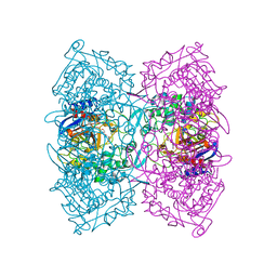
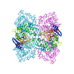
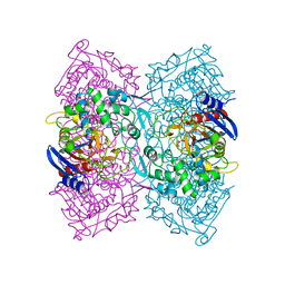
 | | Structural and kinetic bases for the metal preference of the M18 aminopeptidase from Pseudomonas aeruginosa | | 分子名称: | 2-[N-CYCLOHEXYLAMINO]ETHANE SULFONIC ACID, CARBONATE ION, COBALT (II) ION, ... | | 著者 | Nguyen, D.D, Pandian, R, Kim, D.Y, Ha, S.C, Yun, K.H, Kim, K.S, Kim, J.H, Kim, K.K. | | 登録日 | 2013-11-11 | | 公開日 | 2014-04-02 | | 最終更新日 | 2024-03-20 | | 実験手法 | X-RAY DIFFRACTION (2.702 Å) | | 主引用文献 | Structural and kinetic bases for the metal preference of the M18 aminopeptidase from Pseudomonas aeruginosa
Biochem.Biophys.Res.Commun., 447, 2014
|
|
2L4M
 | |
4KMF
 | |
1BCC
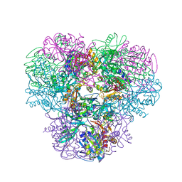
 | | CYTOCHROME BC1 COMPLEX FROM CHICKEN | | 分子名称: | 1,2-Dioleoyl-sn-glycero-3-phosphoethanolamine, FE2/S2 (INORGANIC) CLUSTER, PROTOPORPHYRIN IX CONTAINING FE, ... | | 著者 | Zhang, Z, Huang, L, Shulmeister, V.M, Chi, Y.-I, Kim, K.K, Hung, L.-W, Crofts, A.R, Berry, E.A, Kim, S.-H. | | 登録日 | 1998-03-23 | | 公開日 | 1998-08-19 | | 最終更新日 | 2020-07-29 | | 実験手法 | X-RAY DIFFRACTION (3.16 Å) | | 主引用文献 | Electron transfer by domain movement in cytochrome bc1.
Nature, 392, 1998
|
|
3EYI
 | |
4YGQ
 | | Crystal structure of HAD phosphatase from Thermococcus onnurineus | | 分子名称: | Hydrolase, TERTIARY-BUTYL ALCOHOL | | 著者 | Ngo, T.D, Le, B.V, Subramani, V.K, Nguyen, C.M.T, Lee, H.S, Cho, Y, Kim, K.K, Hwang, H.Y. | | 登録日 | 2015-02-26 | | 公開日 | 2015-04-22 | | 最終更新日 | 2024-03-20 | | 実験手法 | X-RAY DIFFRACTION (2 Å) | | 主引用文献 | Structural basis for the substrate selectivity of a HAD phosphatase from Thermococcus onnurineus NA1
Biochem.Biophys.Res.Commun., 461, 2015
|
|
4YGS
 | | Crystal structure of HAD phosphatase from Thermococcus onnurineus | | 分子名称: | CITRIC ACID, Hydrolase, MAGNESIUM ION | | 著者 | Ngo, T.D, Le, B.V, Subramani, V.K, Nguyen, C.M.T, Lee, H.S, Cho, Y, Kim, K.K, Hwang, H.Y. | | 登録日 | 2015-02-26 | | 公開日 | 2015-04-22 | | 最終更新日 | 2024-03-20 | | 実験手法 | X-RAY DIFFRACTION (1.7 Å) | | 主引用文献 | Structural basis for the substrate selectivity of a HAD phosphatase from Thermococcus onnurineus NA1
Biochem.Biophys.Res.Commun., 461, 2015
|
|
2P52
 | |
4YGR
 | | Crystal structure of HAD phosphatase from Thermococcus onnurineus | | 分子名称: | 2-[N-CYCLOHEXYLAMINO]ETHANE SULFONIC ACID, Hydrolase, MAGNESIUM ION | | 著者 | Ngo, T.D, Le, B.V, Subramani, V.K, Nguyen, C.M.T, Lee, H.S, Cho, Y, Kim, K.K, Hwang, H.Y. | | 登録日 | 2015-02-26 | | 公開日 | 2015-04-22 | | 最終更新日 | 2024-03-20 | | 実験手法 | X-RAY DIFFRACTION (1.703 Å) | | 主引用文献 | Structural basis for the substrate selectivity of a HAD phosphatase from Thermococcus onnurineus NA1
Biochem.Biophys.Res.Commun., 461, 2015
|
|
2P4B
 | | Crystal structure of E.coli RseB | | 分子名称: | Sigma-E factor regulatory protein rseB, octyl beta-D-glucopyranoside | | 著者 | Kim, D.Y, Kim, K.K. | | 登録日 | 2007-03-12 | | 公開日 | 2007-05-22 | | 最終更新日 | 2024-03-13 | | 実験手法 | X-RAY DIFFRACTION (2.4 Å) | | 主引用文献 | Crystal structure of RseB and a model of its binding mode to RseA
Proc.Natl.Acad.Sci.Usa, 104, 2007
|
|
3SH5
 | |
3SH4
 | |
3M4W
 | | Structural basis for the negative regulation of bacterial stress response by RseB | | 分子名称: | Sigma-E factor negative regulatory protein, Sigma-E factor regulatory protein rseB, ZINC ION | | 著者 | Kim, D.Y, Kwon, E, Choi, J.K, Hwang, H.-Y, Kim, K.K. | | 登録日 | 2010-03-12 | | 公開日 | 2010-05-05 | | 最終更新日 | 2023-11-01 | | 実験手法 | X-RAY DIFFRACTION (2.3 Å) | | 主引用文献 | Structural basis for the negative regulation of bacterial stress response by RseB
Protein Sci., 19, 2010
|
|
4P85
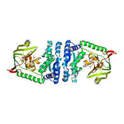
 | | Crystal structure of Est-Y29, a novel penicillin-binding protein/beta-lactamase homolog from a metagenomic library | | 分子名称: | DIETHYL PHOSPHONATE, Est-Y29 | | 著者 | Ngo, T.D, Ryu, B.H, Ju, H.S, Jang, E.J, Kim, K.K, Kim, D.H. | | 登録日 | 2014-03-30 | | 公開日 | 2014-09-10 | | 最終更新日 | 2023-12-27 | | 実験手法 | X-RAY DIFFRACTION (2 Å) | | 主引用文献 | Crystallographic analysis and biochemical applications of a novel penicillin-binding protein/ beta-lactamase homologue from a metagenomic library.
Acta Crystallogr.,Sect.D, 70, 2014
|
|
4P6B
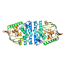
 | | Crystal structure of Est-Y29,a novel penicillin-binding protein/beta-lactamase homolog from a metagenomic library | | 分子名称: | Est-Y29 | | 著者 | Ngo, T.D, Ryu, B.H, Ju, H.S, Jang, E.J, Kim, K.K, Kim, D.H. | | 登録日 | 2014-03-24 | | 公開日 | 2014-09-10 | | 最終更新日 | 2023-12-27 | | 実験手法 | X-RAY DIFFRACTION (1.7 Å) | | 主引用文献 | Crystallographic analysis and biochemical applications of a novel penicillin-binding protein/ beta-lactamase homologue from a metagenomic library.
Acta Crystallogr.,Sect.D, 70, 2014
|
|
4ICS
 | | Crystal structure of PepS from Streptococcus pneumoniae in complex with a substrate | | 分子名称: | Aminopeptidase PepS, GLYCINE, TRYPTOPHAN, ... | | 著者 | Lee, S, Kim, K.K, Ta, M.H. | | 登録日 | 2012-12-11 | | 公開日 | 2013-10-23 | | 最終更新日 | 2024-02-28 | | 実験手法 | X-RAY DIFFRACTION (1.97 Å) | | 主引用文献 | Structure-based elucidation of the regulatory mechanism for aminopeptidase activity.
Acta Crystallogr.,Sect.D, 69, 2013
|
|
4P87
 | | Crystal structure of Est-Y29, a novel penicillin-binding protein/beta-lactamase homolog from a metagenomic library | | 分子名称: | 4-NITROPHENYL PHOSPHATE, Est-Y29 | | 著者 | Ngo, T.D, Ryu, B.H, Ju, H.S, Jang, E.J, Kim, K.K, Kim, D.H. | | 登録日 | 2014-03-30 | | 公開日 | 2014-09-10 | | 最終更新日 | 2023-12-27 | | 実験手法 | X-RAY DIFFRACTION (1.999 Å) | | 主引用文献 | Crystallographic analysis and biochemical applications of a novel penicillin-binding protein/ beta-lactamase homologue from a metagenomic library.
Acta Crystallogr.,Sect.D, 70, 2014
|
|
3OEO
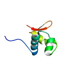
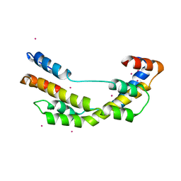 | | The crystal structure E. coli Spy | | 分子名称: | CADMIUM ION, Spheroplast protein Y | | 著者 | Kwon, E, Kim, D.Y, Gross, C.A, Gross, J.D, Kim, K.K. | | 登録日 | 2010-08-13 | | 公開日 | 2010-09-22 | | 最終更新日 | 2024-02-21 | | 実験手法 | X-RAY DIFFRACTION (2.7 Å) | | 主引用文献 | The crystal structure Escherichia coli Spy.
Protein Sci., 19, 2010
|
|
4ICR
 | | Structural basis for substrate recognition and reaction mechanism of bacterial aminopeptidase peps | | 分子名称: | Aminopeptidase PepS, CACODYLATE ION, ZINC ION | | 著者 | Lee, S, Kim, K.K, Ta, M.H. | | 登録日 | 2012-12-11 | | 公開日 | 2013-10-23 | | 最終更新日 | 2024-02-28 | | 実験手法 | X-RAY DIFFRACTION (2.17 Å) | | 主引用文献 | Structure-based elucidation of the regulatory mechanism for aminopeptidase activity.
Acta Crystallogr.,Sect.D, 69, 2013
|
|
4ICQ
 | |
1PCV
 | | Crystal structure of osmotin, a plant antifungal protein | | 分子名称: | osmotin | | 著者 | Min, K, Ha, S.C, Yun, D.-J, Bressan, R.A, Kim, K.K. | | 登録日 | 2003-05-16 | | 公開日 | 2004-02-17 | | 最終更新日 | 2023-10-25 | | 実験手法 | X-RAY DIFFRACTION (2.3 Å) | | 主引用文献 | Crystal structure of osmotin, a plant antifungal protein
PROTEINS: STRUCT.,FUNCT.,GENET., 54, 2004
|
|
4LFU
 | | Crystal structure of Escherichia coli SdiA in the space group C2 | | 分子名称: | CHLORIDE ION, Regulatory protein SdiA, TETRAETHYLENE GLYCOL | | 著者 | Kim, T, Duong, T, Wu, C.A, Choi, J, Lan, N, Kang, S.W, Lokanath, N.K, Shin, D, Hwang, H.Y, Kim, K.K. | | 登録日 | 2013-06-27 | | 公開日 | 2014-03-19 | | 最終更新日 | 2024-03-20 | | 実験手法 | X-RAY DIFFRACTION (2.26 Å) | | 主引用文献 | Structural insights into the molecular mechanism of Escherichia coli SdiA, a quorum-sensing receptor
Acta Crystallogr.,Sect.D, 70, 2014
|
|
4LGW
 | | Crystal structure of Escherichia coli SdiA in the space group P6522 | | 分子名称: | GLYCEROL, Regulatory protein SdiA | | 著者 | Kim, T, Duong, T, Wu, C.A, Choi, J, Lan, N, Kang, S.W, Lokanath, N.K, Shin, D, Hwang, H.Y, Kim, K.K. | | 登録日 | 2013-06-28 | | 公開日 | 2014-03-19 | | 最終更新日 | 2024-03-20 | | 実験手法 | X-RAY DIFFRACTION (2.7 Å) | | 主引用文献 | Structural insights into the molecular mechanism of Escherichia coli SdiA, a quorum-sensing receptor
Acta Crystallogr.,Sect.D, 70, 2014
|
|


