2M1B
 
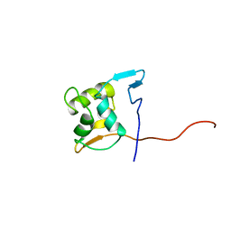 | |
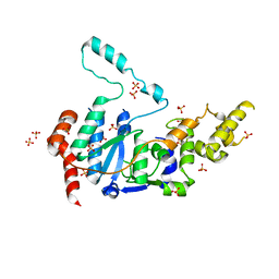
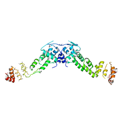
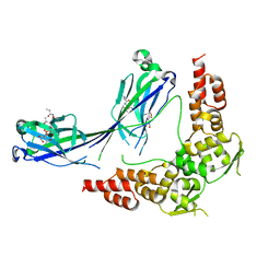
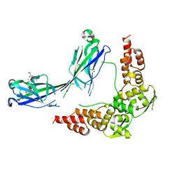
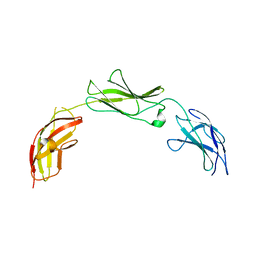
4KC3
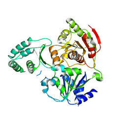
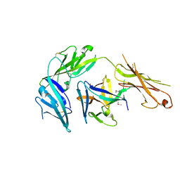 | | Cytokine/receptor binary complex | | 分子名称: | 2-acetamido-2-deoxy-beta-D-glucopyranose, Interleukin-1 receptor-like 1, Interleukin-33 | | 著者 | Liu, X, Wang, X.Q. | | 登録日 | 2013-04-24 | | 公開日 | 2013-08-28 | | 最終更新日 | 2023-12-06 | | 実験手法 | X-RAY DIFFRACTION (3.2702 Å) | | 主引用文献 | Structural insights into the interaction of IL-33 with its receptors.
Proc.Natl.Acad.Sci.USA, 110, 2013
|
|
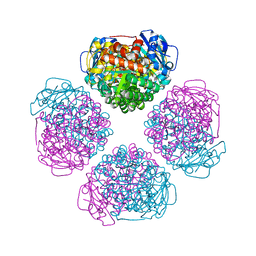
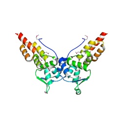
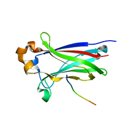
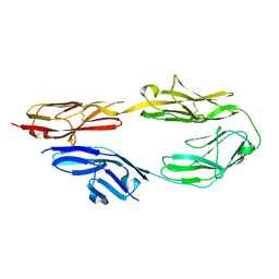
4LMY
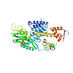
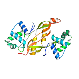 | | Structure of GAS PerR-Zn-Zn | | 分子名称: | Peroxide stress regulator PerR, FUR family, ZINC ION | | 著者 | Lin, C.S, Chao, S.Y, Nix, J.C, Tseng, H.L, Tsou, C.C, Fei, C.H, Ciou, H.S, Jeng, U.S, Lin, Y.S, Chuang, W.J, Wu, J.J, Wang, S. | | 登録日 | 2013-07-11 | | 公開日 | 2014-04-02 | | 最終更新日 | 2024-03-20 | | 実験手法 | X-RAY DIFFRACTION (1.6 Å) | | 主引用文献 | Distinct structural features of the peroxide response regulator from group a streptococcus drive DNA binding
Plos One, 9, 2014
|
|
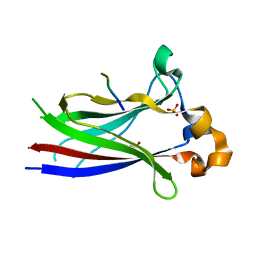
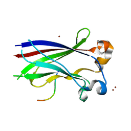
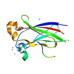
3CRV
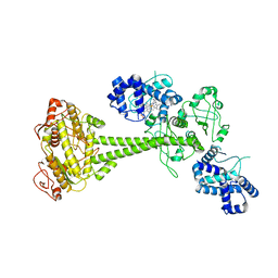
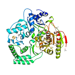 | | XPD_Helicase | | 分子名称: | CITRATE ANION, GLYCEROL, IRON/SULFUR CLUSTER, ... | | 著者 | Fan, L, Arvai, A.S, Tainer, J.A. | | 登録日 | 2008-04-07 | | 公開日 | 2008-06-10 | | 最終更新日 | 2024-02-21 | | 実験手法 | X-RAY DIFFRACTION (2 Å) | | 主引用文献 | XPD helicase structures and activities: insights into the cancer and aging phenotypes from XPD mutations.
Cell(Cambridge,Mass.), 133, 2008
|
|
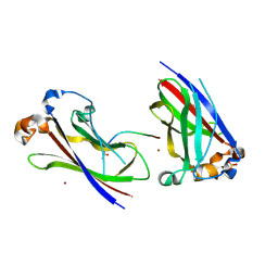
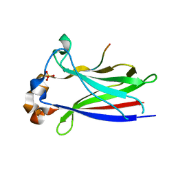
3CRW
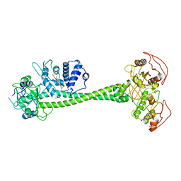
 | | XPD_APO | | 分子名称: | HEXACYANOFERRATE(3-), XPD/Rad3 related DNA helicase | | 著者 | Fan, L, Arvai, A.S, Tainer, J.A. | | 登録日 | 2008-04-07 | | 公開日 | 2008-06-10 | | 最終更新日 | 2023-08-30 | | 実験手法 | X-RAY DIFFRACTION (4 Å) | | 主引用文献 | XPD helicase structures and activities: insights into the cancer and aging phenotypes from XPD mutations.
Cell(Cambridge,Mass.), 133, 2008
|
|
3DPL
 | |
6PAS
 | | Inactive State of Manduca sexta soluble guanylate cyclase | | 分子名称: | PROTOPORPHYRIN IX CONTAINING FE, Soluble guanylyl cyclase alpha-1 subunit, Soluble guanylyl cyclase beta-1 subunit | | 著者 | Yokom, A.L, Horst, B.G, Marletta, M.A, Hurley, J.H. | | 登録日 | 2019-06-11 | | 公開日 | 2019-10-23 | | 最終更新日 | 2024-03-20 | | 実験手法 | ELECTRON MICROSCOPY (5.1 Å) | | 主引用文献 | Allosteric activation of the nitric oxide receptor soluble guanylate cyclase mapped by cryo-electron microscopy.
Elife, 8, 2019
|
|
6PAT
 | | Active State of Manduca sexta soluble Guanylate Cyclase | | 分子名称: | PROTOPORPHYRIN IX CONTAINING FE, Soluble guanylyl cyclase alpha-1 subunit, Soluble guanylyl cyclase beta-1 subunit | | 著者 | Yokom, A.L, Horst, B.G, Marletta, M.A, Hurley, J.H. | | 登録日 | 2019-06-11 | | 公開日 | 2019-10-23 | | 最終更新日 | 2024-03-20 | | 実験手法 | ELECTRON MICROSCOPY (5.8 Å) | | 主引用文献 | Allosteric activation of the nitric oxide receptor soluble guanylate cyclase mapped by cryo-electron microscopy.
Elife, 8, 2019
|
|
6VBH
 | | Human XPG endonuclease catalytic domain | | 分子名称: | DNA repair protein complementing XP-G cells,Flap endonuclease 1, SULFATE ION | | 著者 | Tsutakawa, S.E, Arvai, A.S, Tainer, J.A. | | 登録日 | 2019-12-18 | | 公開日 | 2020-06-17 | | 最終更新日 | 2023-10-11 | | 実験手法 | X-RAY DIFFRACTION (1.995 Å) | | 主引用文献 | Human XPG nuclease structure, assembly, and activities with insights for neurodegeneration and cancer from pathogenic mutations.
Proc.Natl.Acad.Sci.USA, 117, 2020
|
|
6URA
 | | Crystal structure of RUBISCO from Promineofilum breve | | 分子名称: | 2-CARBOXYARABINITOL-1,5-DIPHOSPHATE, MAGNESIUM ION, Ribulose bisphosphate carboxylase large chain | | 著者 | Pereira, J.H, Banda, D.M, Liu, A.K, Shih, P.M, Adams, P.D. | | 登録日 | 2019-10-23 | | 公開日 | 2020-08-19 | | 最終更新日 | 2023-11-15 | | 実験手法 | X-RAY DIFFRACTION (2.17 Å) | | 主引用文献 | Novel bacterial clade reveals origin of form I Rubisco.
Nat.Plants, 6, 2020
|
|
3HQM
 | |
3HSV
 | | Structures of SPOP-Substrate Complexes: Insights into Molecular Architectures of BTB-Cul3 Ubiquitin Ligases: SPOPMATHx-MacroH2ASBCpep2 | | 分子名称: | Core histone macro-H2A.1, SULFATE ION, Speckle-type POZ protein, ... | | 著者 | Zhuang, M, Schulman, B.A, Miller, D. | | 登録日 | 2009-06-10 | | 公開日 | 2009-10-20 | | 最終更新日 | 2023-09-06 | | 実験手法 | X-RAY DIFFRACTION (1.43 Å) | | 主引用文献 | Structures of SPOP-substrate complexes: insights into molecular architectures of BTB-Cul3 ubiquitin ligases.
Mol.Cell, 36, 2009
|
|
3HVE
 | |
3HTM
 | |
3HQH
 | |
3HQL
 | |
3HQI
 | |
3HU6
 | |
3IVQ
 | | Structures of SPOP-Substrate Complexes: Insights into Molecular Architectures of BTB-Cul3 Ubiquitin Ligases: SPOPMATH-CiSBC2 | | 分子名称: | CiSBC2, Speckle-type POZ protein | | 著者 | Schulman, B.A, Miller, D.J, Calabrese, M.F, Seyedin, S. | | 登録日 | 2009-09-01 | | 公開日 | 2009-10-20 | | 最終更新日 | 2024-02-21 | | 実験手法 | X-RAY DIFFRACTION (2.1 Å) | | 主引用文献 | Structures of SPOP-Substrate Complexes: Insights into Molecular Architectures of BTB-Cul3 Ubiquitin Ligases.
Mol.Cell, 36, 2009
|
|
3IVV
 | | Structures of SPOP-Substrate Complexes: Insights into Molecular Architectures of BTB-Cul3 Ubiquitin Ligases: SPOPMATH-PucSBC1_pep1 | | 分子名称: | PucSBC1, Speckle-type POZ protein | | 著者 | Schulman, B.A, Miller, D.J, Calabrese, M.F, Seyedin, S. | | 登録日 | 2009-09-01 | | 公開日 | 2009-10-20 | | 最終更新日 | 2024-02-21 | | 実験手法 | X-RAY DIFFRACTION (1.25 Å) | | 主引用文献 | Structures of SPOP-Substrate Complexes: Insights into Molecular Architectures of BTB-Cul3 Ubiquitin Ligases.
Mol.Cell, 36, 2009
|
|
3IVB
 | | Structures of SPOP-Substrate Complexes: Insights into Architectures of BTB-Cul3 Ubiquitin Ligases: SPOPMATH-MacroH2ASBCpep1 | | 分子名称: | CHLORIDE ION, Core histone macro-H2A.1, Speckle-type POZ protein, ... | | 著者 | Schulman, B.A, Miller, D.J, Calabrese, M.F, Seyedin, S. | | 登録日 | 2009-08-31 | | 公開日 | 2009-10-20 | | 最終更新日 | 2024-02-21 | | 実験手法 | X-RAY DIFFRACTION (1.75 Å) | | 主引用文献 | Structure 9
To be Published
|
|
5E4Q
 | |
5E4S
 | |
5E4I
 | |
5E52
 | |


