Loading
PDBj
メニューPDBj@FacebookPDBj@TwitterPDBj@YouTubewwPDB FoundationwwPDB
RCSB PDBPDBeBMRBAdv. SearchSearch help
Search by PDB author
3H41
DownloadVisualize
BU of 3h41 by Molmil
CRYSTAL STRUCTURE OF A NLPC/P60 FAMILY PROTEIN (BCE_2878) FROM BACILLUS CEREUS ATCC 10987 AT 1.79 A RESOLUTION
分子名称: ALANINE, D-GLUTAMIC ACID, NLP/P60 family protein, ...
著者Joint Center for Structural Genomics (JCSG)
登録日2009-04-17
公開日2009-05-05
最終更新日2023-02-01
実験手法X-RAY DIFFRACTION (1.79 Å)
主引用文献Structure of the gamma-D-glutamyl-L-diamino acid endopeptidase YkfC from Bacillus cereus in complex with L-Ala-[gamma]-D-Glu: insights into substrate recognition by NlpC/P60 cysteine peptidases
Acta Crystallogr.,Sect.F, 66, 2010
3HBZ
DownloadVisualize
BU of 3hbz by Molmil
Crystal structure of a putative glycoside hydrolase (bt_2081) from bacteroides thetaiotaomicron vpi-5482 at 2.05 A resolution
分子名称: ACETATE ION, CACODYLATE ION, CALCIUM ION, ...
著者Joint Center for Structural Genomics (JCSG)
登録日2009-05-05
公開日2009-05-26
最終更新日2023-02-01
実験手法X-RAY DIFFRACTION (2.05 Å)
主引用文献Structure of Bacteroides thetaiotaomicron BT2081 at 2.05 A resolution: the first structural representative of a new protein family that may play a role in carbohydrate metabolism.
Acta Crystallogr.,Sect.F, 66, 2010
3IRB
DownloadVisualize
BU of 3irb by Molmil
Crystal structure of protein with unknown function from DUF35 family (13815350) from SULFOLOBUS SOLFATARICUS at 1.80 A resolution
分子名称: ACETIC ACID, SULFATE ION, ZINC ION, ...
著者Joint Center for Structural Genomics (JCSG)
登録日2009-08-21
公開日2009-09-01
最終更新日2023-02-01
実験手法X-RAY DIFFRACTION (1.8 Å)
主引用文献The structure of SSO2064, the first representative of Pfam family PF01796, reveals a novel two-domain zinc-ribbon OB-fold architecture with a potential acyl-CoA-binding role.
Acta Crystallogr.,Sect.F, 66, 2010
3BYQ
DownloadVisualize
BU of 3byq by Molmil
Crystal structure of a duf1185 family protein (bb2672) from bordetella bronchiseptica rb50 at 1.70 A resolution
分子名称: 1,2-ETHANEDIOL, CHLORIDE ION, TETRAETHYLENE GLYCOL, ...
著者Joint Center for Structural Genomics (JCSG)
登録日2008-01-16
公開日2008-01-29
最終更新日2023-02-01
実験手法X-RAY DIFFRACTION (1.7 Å)
主引用文献Structures of the first representatives of Pfam family PF06684 (DUF1185) reveal a novel variant of the Bacillus chorismate mutase fold and suggest a role in amino-acid metabolism.
Acta Crystallogr.,Sect.F, 66, 2010
3BOS
DownloadVisualize
BU of 3bos by Molmil
Crystal structure of a putative dna replication regulator HDA (SAMA_1916) from Shewanella amazonensis sb2b at 1.75 A resolution
分子名称: 1,2-ETHANEDIOL, CHLORIDE ION, CYTIDINE-5'-DIPHOSPHATE, ...
著者Joint Center for Structural Genomics (JCSG)
登録日2007-12-17
公開日2008-01-15
最終更新日2023-01-25
実験手法X-RAY DIFFRACTION (1.75 Å)
主引用文献A structural basis for the regulatory inactivation of DnaA.
J.Mol.Biol., 385, 2009
3BY7
DownloadVisualize
BU of 3by7 by Molmil
CRYSTAL STRUCTURE OF A PROTEIN STRUCTURALLY SIMILAR TO SM/LSM-LIKE RNA-BINDING PROTEINS (JCVI_PEP_1096686650277) FROM UNCULTURED MARINE ORGANISM AT 2.60 A RESOLUTION
分子名称: uncharacterized protein
著者Joint Center for Structural Genomics (JCSG)
登録日2008-01-15
公開日2008-01-29
最終更新日2023-02-01
実験手法X-RAY DIFFRACTION (2.6 Å)
主引用文献Crystal structure of a novel Sm-like protein of putative cyanophage origin at 2.60 A resolution.
Proteins, 75, 2009
3CGH
DownloadVisualize
BU of 3cgh by Molmil
Crystal structure of a susd homolog (bt_3984) from bacteroides thetaiotaomicron vpi-5482 at 1.70 A resolution
分子名称: ACETATE ION, SusD homolog, ZINC ION
著者Joint Center for Structural Genomics (JCSG)
登録日2008-03-05
公開日2008-03-18
最終更新日2023-02-01
実験手法X-RAY DIFFRACTION (1.7 Å)
主引用文献Structure of BT_3984, a member of the SusD/RagB family of nutrient-binding molecules.
Acta Crystallogr.,Sect.F, 66, 2010
3DEE
DownloadVisualize
BU of 3dee by Molmil
CRYSTAL STRUCTURE OF A PUTATIVE REGULATORY PROTEIN INVOLVED IN TRANSCRIPTION (NGO1945) FROM NEISSERIA GONORRHOEAE FA 1090 AT 2.25 A RESOLUTION
分子名称: CHLORIDE ION, IMIDAZOLE, Putative regulatory protein
著者Joint Center for Structural Genomics (JCSG)
登録日2008-06-09
公開日2008-08-05
最終更新日2023-02-01
実験手法X-RAY DIFFRACTION (2.1 Å)
主引用文献The structure of the first representative of Pfam family PF09836 reveals a two-domain organization and suggests involvement in transcriptional regulation.
Acta Crystallogr.,Sect.F, 66, 2010
3D00
DownloadVisualize
BU of 3d00 by Molmil
Crystal structure of a tungsten formylmethanofuran dehydrogenase subunit e (fmde)-like protein (syn_00638) from syntrophus aciditrophicus at 1.90 A resolution
分子名称: CHLORIDE ION, Tungsten formylmethanofuran dehydrogenase subunit E, ZINC ION
著者Joint Center for Structural Genomics (JCSG)
登録日2008-04-30
公開日2008-05-20
最終更新日2023-02-01
実験手法X-RAY DIFFRACTION (1.9 Å)
主引用文献Structures of three members of Pfam PF02663 (FmdE) implicated in microbial methanogenesis reveal a conserved alpha+beta core domain and an auxiliary C-terminal treble-clef zinc finger.
Acta Crystallogr.,Sect.F, 66, 2010
3B77
DownloadVisualize
BU of 3b77 by Molmil
Crystal structure of a ph domain containing bacterial protein (exig_2160) from exiguobacterium sibiricum 255-15 at 2.42 A resolution
分子名称: Uncharacterized protein
著者Joint Center for Structural Genomics (JCSG)
登録日2007-10-30
公開日2007-11-13
最終更新日2023-01-25
実験手法X-RAY DIFFRACTION (2.42 Å)
主引用文献Bacterial pleckstrin homology domains: a prokaryotic origin for the PH domain.
J.Mol.Biol., 396, 2010
3CM1
DownloadVisualize
BU of 3cm1 by Molmil
Crystal structure of SsgA-like sporulation-specific cell division protein (YP_290167.1) from Thermobifida fusca YX-ER1 at 2.60 A resolution
分子名称: SsgA-like sporulation-specific cell division protein
著者Joint Center for Structural Genomics (JCSG), Chruszcz, M, Minor, W, Wang, S.
登録日2008-03-20
公開日2008-04-01
最終更新日2023-02-01
実験手法X-RAY DIFFRACTION (2.6 Å)
主引用文献Structural and functional characterizations of SsgB, a conserved activator of developmental cell division in morphologically complex actinomycetes.
J.Biol.Chem., 284, 2009
3DCX
DownloadVisualize
BU of 3dcx by Molmil
Crystal structure of a duf1696 family protein with a pleckstrin-homology domain (shew_0819) from shewanella loihica pv-4 at 2.00 A resolution
分子名称: (4S)-2-METHYL-2,4-PENTANEDIOL, CHLORIDE ION, Protein of Unknown Function (DUF1696) with Pleckstrin-homology Domains
著者Joint Center for Structural Genomics (JCSG)
登録日2008-06-04
公開日2008-07-29
最終更新日2023-02-01
実験手法X-RAY DIFFRACTION (2 Å)
主引用文献Bacterial pleckstrin homology domains: a prokaryotic origin for the PH domain.
J.Mol.Biol., 396, 2010
3E0F
DownloadVisualize
BU of 3e0f by Molmil
Crystal structure of a putative metal-dependent phosphoesterase (bad_1165) from bifidobacterium adolescentis atcc 15703 at 2.40 A resolution
分子名称: ACETATE ION, FE (III) ION, PHOSPHATE ION, ...
著者Joint Center for Structural Genomics (JCSG)
登録日2008-07-31
公開日2008-09-02
最終更新日2023-02-01
実験手法X-RAY DIFFRACTION (2.4 Å)
主引用文献Crystal structure of a metal-dependent phosphoesterase (YP_910028.1) from Bifidobacterium adolescentis: Computational prediction and experimental validation of phosphoesterase activity.
Proteins, 79, 2011
3DUE
DownloadVisualize
BU of 3due by Molmil
CRYSTAL STRUCTURE OF A PUTATIVE PERIPLASMIC PROTEIN FROM DUF2874 FAMILY (BVU_2987) FROM BACTEROIDES VULGATUS ATCC 8482 AT 1.85 A RESOLUTION
分子名称: CACODYLATE ION, Putative periplasmic protein
著者Joint Center for Structural Genomics (JCSG)
登録日2008-07-17
公開日2008-08-05
最終更新日2023-02-01
実験手法X-RAY DIFFRACTION (1.85 Å)
主引用文献The structure of BVU2987 from Bacteroides vulgatus reveals a superfamily of bacterial periplasmic proteins with possible inhibitory function.
Acta Crystallogr.,Sect.F, 66, 2010
5T5B
DownloadVisualize
BU of 5t5b by Molmil
CRYSTAL STRUCTURE OF THE COMPLEX OF 10E8 FAB LIGHT CHAIN MUTANT5 AND T117V2 HIV-1 MPER SCAFFOLD
分子名称: 10E8 EPITOPE SCAFFOLD T117V2, Antibody 10E8 FAB HEAVY CHAIN, Antibody 10E8 FAB LIGHT CHAIN MUTATED, ...
著者Irimia, A, Wilson, I.A.
登録日2016-08-30
公開日2017-03-08
最終更新日2023-10-04
実験手法X-RAY DIFFRACTION (2.07 Å)
主引用文献Lipid interactions and angle of approach to the HIV-1 viral membrane of broadly neutralizing antibody 10E8: Insights for vaccine and therapeutic design.
PLoS Pathog., 13, 2017
5T6L
DownloadVisualize
BU of 5t6l by Molmil
Crystal structure of 10E8 Fab in complex with the MPER epitope scaffold T117v2
分子名称: 10E8 EPITOPE SCAFFOLD T117V2, 4-(2-HYDROXYETHYL)-1-PIPERAZINE ETHANESULFONIC ACID, Antibody 10E8 FAB HEAVY CHAIN, ...
著者Irimia, A, Wilson, I.A.
登録日2016-09-01
公開日2017-03-08
最終更新日2023-10-04
実験手法X-RAY DIFFRACTION (2.1 Å)
主引用文献Lipid interactions and angle of approach to the HIV-1 viral membrane of broadly neutralizing antibody 10E8: Insights for vaccine and therapeutic design.
PLoS Pathog., 13, 2017
5TFW
DownloadVisualize
BU of 5tfw by Molmil
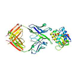
Crystal structure of 10E8 Fab light chain mutant2 against the MPER region of the HIV-1 Env, in complex with T117v2 epitope scaffold
分子名称: 1,2-ETHANEDIOL, 10E8 EPITOPE SCAFFOLD T117V2, Antibody 10E8 FAB HEAVY CHAIN, ...
著者Irimia, A, Wilson, I.A.
登録日2016-09-26
公開日2017-03-08
最終更新日2023-10-04
実験手法X-RAY DIFFRACTION (2.168 Å)
主引用文献Lipid interactions and angle of approach to the HIV-1 viral membrane of broadly neutralizing antibody 10E8: Insights for vaccine and therapeutic design.
PLoS Pathog., 13, 2017
5T29
DownloadVisualize
BU of 5t29 by Molmil
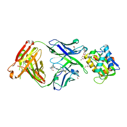
Crystal structure of 10E8 Fab light chain mutant3, against the MPER region of the HIV-1 Env, in complex with the MPER epitope scaffold T117v2
分子名称: 10E8 EPITOPE SCAFFOLD T117V2, Antibody 10E8 FAB HEAVY CHAIN, Antibody 10E8 FAB LIGHT CHAIN, ...
著者Irimia, A, Wilson, I.A.
登録日2016-08-23
公開日2017-03-08
最終更新日2023-10-04
実験手法X-RAY DIFFRACTION (2.03 Å)
主引用文献Lipid interactions and angle of approach to the HIV-1 viral membrane of broadly neutralizing antibody 10E8: Insights for vaccine and therapeutic design.
PLoS Pathog., 13, 2017
5SY8
DownloadVisualize
BU of 5sy8 by Molmil
Crystal structure of the complex of 10E8 Fab light chain mutant1 and T117v2 HIV-1 MPER scaffold
分子名称: 10E8 EPITOPE SCAFFOLD T117v2, 10E8 FAB HEAVY CHAIN, 10E8 FAB LIGHT CHAIN, ...
著者Irimia, A, Wilson, I.A.
登録日2016-08-10
公開日2017-03-08
最終更新日2023-10-04
実験手法X-RAY DIFFRACTION (1.62 Å)
主引用文献Lipid interactions and angle of approach to the HIV-1 viral membrane of broadly neutralizing antibody 10E8: Insights for vaccine and therapeutic design.
PLoS Pathog., 13, 2017
5T80
DownloadVisualize
BU of 5t80 by Molmil
Crystal structure of 10E8 Fab in complex with the MPER epitope scaffold T117v2 and phosphatidic acid (06:0 PA)
分子名称: (2R)-3-(phosphonooxy)propane-1,2-diyl dihexanoate, 10E8 EPITOPE SCAFFOLD T117V2, Antibody 10E8 FAB HEAVY CHAIN, ...
著者Irimia, A, Wilson, I.A.
登録日2016-09-06
公開日2017-03-08
最終更新日2023-10-04
実験手法X-RAY DIFFRACTION (2.62 Å)
主引用文献Lipid interactions and angle of approach to the HIV-1 viral membrane of broadly neutralizing antibody 10E8: Insights for vaccine and therapeutic design.
PLoS Pathog., 13, 2017
5T85
DownloadVisualize
BU of 5t85 by Molmil
Crystal structure of 10E8 Fab in complex with the MPER epitope scaffold T117v2 and phosphatidylglycerol (06:0 PG)
分子名称: (2S)-3-{[(R)-{[(2R)-2,3-dihydroxypropyl]oxy}(hydroxy)phosphoryl]oxy}-2-(hexanoyloxy)propyl hexanoate, 10E8 EPITOPE SCAFFOLD T117V2, Antibody 10E8 FAB HEAVY CHAIN, ...
著者Irimia, A, Wilson, I.A.
登録日2016-09-06
公開日2017-03-08
最終更新日2023-10-04
実験手法X-RAY DIFFRACTION (2.373 Å)
主引用文献Lipid interactions and angle of approach to the HIV-1 viral membrane of broadly neutralizing antibody 10E8: Insights for vaccine and therapeutic design.
PLoS Pathog., 13, 2017
<123


222926

件を2024-07-24に公開中

PDB statisticsPDBj update infoContact PDBjnumon