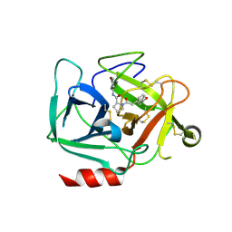
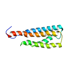
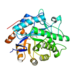
6YWQ
 
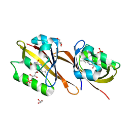 | | Structure of Chloroflexus aggregans flavin based fluorescent protein (CagFbFP) Q148H variant | | 分子名称: | FLAVIN MONONUCLEOTIDE, GLYCEROL, Multi-sensor hybrid histidine kinase | | 著者 | Remeeva, A, Nazarenko, V, Kovalev, K, Gushchin, I. | | 登録日 | 2020-04-30 | | 公開日 | 2021-04-21 | | 最終更新日 | 2024-01-24 | | 実験手法 | X-RAY DIFFRACTION (1.27 Å) | | 主引用文献 | Insights into the mechanisms of light-oxygen-voltage domain color tuning from a set of high-resolution X-ray structures.
Proteins, 2021
|
|
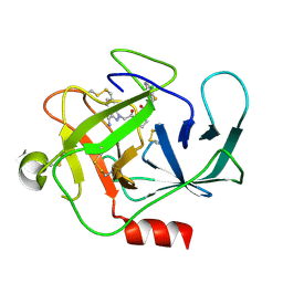
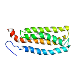
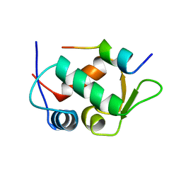
6YWR
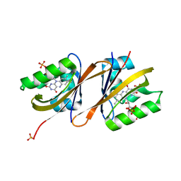 | | Structure of Chloroflexus aggregans flavin based fluorescent protein (CagFbFP) Q148H variant (space group C2) | | 分子名称: | FLAVIN MONONUCLEOTIDE, Multi-sensor hybrid histidine kinase, SULFATE ION | | 著者 | Remeeva, A, Nazarenko, V, Kovalev, K, Gushchin, I. | | 登録日 | 2020-04-30 | | 公開日 | 2021-04-21 | | 最終更新日 | 2024-01-24 | | 実験手法 | X-RAY DIFFRACTION (1.5 Å) | | 主引用文献 | Insights into the mechanisms of light-oxygen-voltage domain color tuning from a set of high-resolution X-ray structures.
Proteins, 2021
|
|
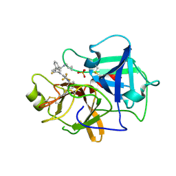
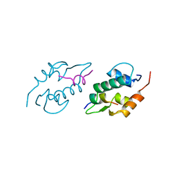
6YWG
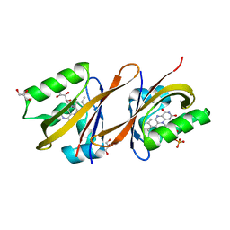 | | Structure of Chloroflexus aggregans flavin based fluorescent protein (CagFbFP) Q148N variant | | 分子名称: | FLAVIN MONONUCLEOTIDE, GLYCEROL, Multi-sensor hybrid histidine kinase | | 著者 | Remeeva, A, Nazarenko, V, Kovalev, K, Gushchin, I. | | 登録日 | 2020-04-29 | | 公開日 | 2021-04-21 | | 最終更新日 | 2024-01-24 | | 実験手法 | X-RAY DIFFRACTION (1.45 Å) | | 主引用文献 | Insights into the mechanisms of light-oxygen-voltage domain color tuning from a set of high-resolution X-ray structures.
Proteins, 2021
|
|
8B7D
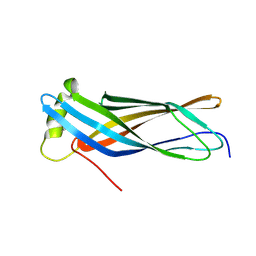 | | Luminal domain of TMEM106B | | 分子名称: | 2-acetamido-2-deoxy-beta-D-glucopyranose, Transmembrane protein 106B | | 著者 | Pye, V.E, Roustan, C, Cherepanov, P. | | 登録日 | 2022-09-29 | | 公開日 | 2023-07-19 | | 最終更新日 | 2024-05-01 | | 実験手法 | X-RAY DIFFRACTION (2.59 Å) | | 主引用文献 | TMEM106B is a receptor mediating ACE2-independent SARS-CoV-2 cell entry.
Cell, 186, 2023
|
|
8DEA
 | |
8DG6
 | |
8D95
 | |
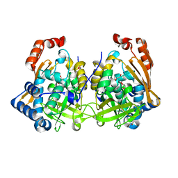
7N7X
 | | Crystal structure of BCX7353(ORLADEYO) in complex with human plasma kallikrein serine protease domain at 2.1 angstrom resolution | | 分子名称: | Orladeyo, PHOSPHATE ION, Plasma kallikrein light chain | | 著者 | Krishnan, R, Yarlagadda, B.S, Kotian, P, Polach, K.J, Zhang, W. | | 登録日 | 2021-06-11 | | 公開日 | 2021-09-15 | | 最終更新日 | 2023-10-18 | | 実験手法 | X-RAY DIFFRACTION (2.097 Å) | | 主引用文献 | Berotralstat (BCX7353): Structure-Guided Design of a Potent, Selective, and Oral Plasma Kallikrein Inhibitor to Prevent Attacks of Hereditary Angioedema (HAE).
J.Med.Chem., 64, 2021
|
|
7QI3
 | | Structure of Fusarium verticillioides NAT1 (FDB2) N-malonyltransferase | | 分子名称: | 1,2-ETHANEDIOL, Arylamine N-acetyltransferase, DI(HYDROXYETHYL)ETHER, ... | | 著者 | Lowe, E.D, Kotomina, E, Karagianni, E, Boukouvala, S. | | 登録日 | 2021-12-14 | | 公開日 | 2022-11-23 | | 最終更新日 | 2024-02-07 | | 実験手法 | X-RAY DIFFRACTION (1.8 Å) | | 主引用文献 | Fusarium verticillioides NAT1 (FDB2) N-malonyltransferase is structurally, functionally and phylogenetically distinct from its N-acetyltransferase (NAT) homologues.
Febs J., 290, 2023
|
|
2LKH
 | | WSA minor conformation | | 分子名称: | Acetylcholine receptor | | 著者 | Xu, Y, Mowrey, D, Cui, T, Perez-Aguilar, J, Saven, J.G, Eckenhoff, R, Tang, P. | | 登録日 | 2011-10-11 | | 公開日 | 2012-01-11 | | 最終更新日 | 2024-05-15 | | 実験手法 | SOLUTION NMR | | 主引用文献 | NMR structure and dynamics of a designed water-soluble transmembrane domain of nicotinic acetylcholine receptor.
Biochim.Biophys.Acta, 1818, 2011
|
|
2LKG
 | | WSA major conformation | | 分子名称: | Acetylcholine receptor | | 著者 | Xu, Y, Mowrey, D, Cui, T, Perez-Aguilar, J.M, Saven, J.G, Eckenhoff, R, Tang, P. | | 登録日 | 2011-10-11 | | 公開日 | 2012-01-11 | | 最終更新日 | 2024-05-15 | | 実験手法 | SOLUTION NMR | | 主引用文献 | NMR structure and dynamics of a designed water-soluble transmembrane domain of nicotinic acetylcholine receptor.
Biochim.Biophys.Acta, 1818, 2011
|
|
7ZBU
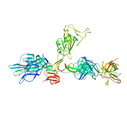 | | CryoEM structure of SARS-CoV-2 spike monomer in complex with neutralising antibody P008_60 | | 分子名称: | 2-acetamido-2-deoxy-beta-D-glucopyranose, 3-[5-[(4-ethyl-3-methyl-5-oxidanylidene-pyrrol-2-yl)methyl]-2-[[5-[(3-ethyl-4-methyl-5-oxidanylidene-pyrrol-2-yl)methyl]-3-(3-hydroxy-3-oxopropyl)-4-methyl-1H-pyrrol-2-yl]methyl]-4-methyl-1H-pyrrol-3-yl]propanoic acid, P008_60 antibody, ... | | 著者 | Rosa, A, Pye, V.E, Cronin, N, Cherepanov, P. | | 登録日 | 2022-03-24 | | 公開日 | 2022-08-17 | | 最終更新日 | 2022-08-31 | | 実験手法 | ELECTRON MICROSCOPY (4.31 Å) | | 主引用文献 | A neutralizing epitope on the SD1 domain of SARS-CoV-2 spike targeted following infection and vaccination.
Cell Rep, 40, 2022
|
|
2M62
 | |
2JRB
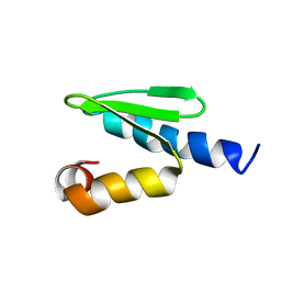 | | C-terminal domain of ORF1p from mouse LINE-1 | | 分子名称: | ORF 1 protein | | 著者 | Januszyk, K, Clubb, R. | | 登録日 | 2007-06-21 | | 公開日 | 2007-07-17 | | 最終更新日 | 2023-12-20 | | 実験手法 | SOLUTION NMR | | 主引用文献 | Identification and solution structure of a highly conserved C-terminal domain within ORF1p required for retrotransposition of long interspersed nuclear element-1.
J.Biol.Chem., 282, 2007
|
|
6W2S
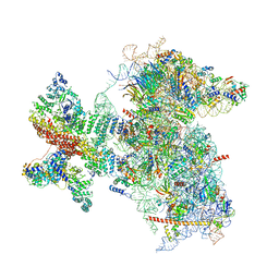 | | Structure of the Cricket Paralysis Virus 5-UTR IRES (CrPV 5-UTR-IRES) bound to the small ribosomal subunit in the open state (Class 1) | | 分子名称: | 18S rRNA, CrPV 5'-UTR IRES, Eukaryotic translation initiation factor 3 subunit A, ... | | 著者 | Neupane, R, Pisareva, V, Rodriguez, C.F, Pisarev, A, Fernandez, I.S. | | 登録日 | 2020-03-08 | | 公開日 | 2020-04-22 | | 最終更新日 | 2024-03-06 | | 実験手法 | ELECTRON MICROSCOPY (3.47 Å) | | 主引用文献 | A complex IRES at the 5'-UTR of a viral mRNA assembles a functional 48S complex via an uAUG intermediate.
Elife, 9, 2020
|
|
6FGE
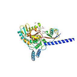 | | Crystal structure of human ZUFSP/ZUP1 in complex with ubiquitin | | 分子名称: | ACETATE ION, AMMONIUM ION, DI(HYDROXYETHYL)ETHER, ... | | 著者 | Kwasna, D, Abdul Rehman, S.A, Kulathu, Y. | | 登録日 | 2018-01-10 | | 公開日 | 2018-04-04 | | 最終更新日 | 2018-04-18 | | 実験手法 | X-RAY DIFFRACTION (1.74 Å) | | 主引用文献 | Discovery and Characterization of ZUFSP/ZUP1, a Distinct Deubiquitinase Class Important for Genome Stability.
Mol. Cell, 70, 2018
|
|
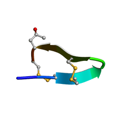
5K9P
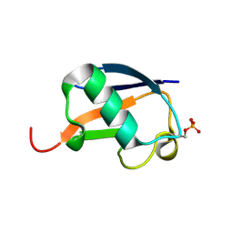 | | Ser20 phosphorylated ubiquitin | | 分子名称: | Polyubiquitin-B | | 著者 | Huguenin-Dezot, N, Chin, J.W. | | 登録日 | 2016-06-01 | | 公開日 | 2016-07-20 | | 最終更新日 | 2024-01-10 | | 実験手法 | X-RAY DIFFRACTION (1.55 Å) | | 主引用文献 | Synthesis of Isomeric Phosphoubiquitin Chains Reveals that Phosphorylation Controls Deubiquitinase Activity and Specificity.
Cell Rep, 16, 2016
|
|
3F03
 | |
1YCR
 | |
1YCQ
 | |


