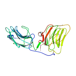
6EJE
 
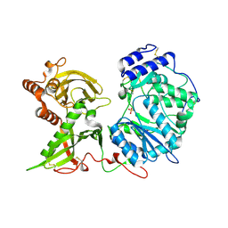 | |
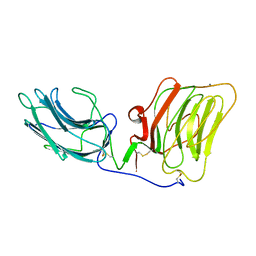
6EJD
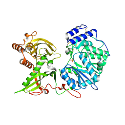 | |
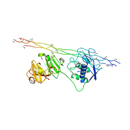
1GR3
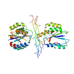
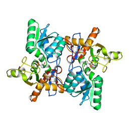
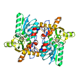
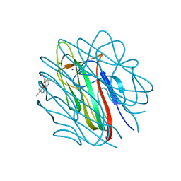 | | Structure of the human collagen X NC1 trimer | | 分子名称: | 3-[(3-CHOLAMIDOPROPYL)DIMETHYLAMMONIO]-1-PROPANESULFONATE, CALCIUM ION, COLLAGEN X, ... | | 著者 | Bogin, O, Kvansakul, M, Rom, E, Singer, J, Yayon, A, Hohenester, E. | | 登録日 | 2001-12-12 | | 公開日 | 2002-02-14 | | 最終更新日 | 2023-12-13 | | 実験手法 | X-RAY DIFFRACTION (2 Å) | | 主引用文献 | Insight Into Schmid Metaphyseal Chondrodysplasia from the Crystal Structure of the Collagen X Nc1 Domain Trimer.
Structure, 10, 2002
|
|
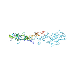
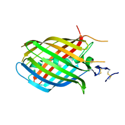
1BNL
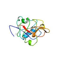 | | ZINC DEPENDENT DIMERS OBSERVED IN CRYSTALS OF HUMAN ENDOSTATIN | | 分子名称: | COLLAGEN XVIII, ZINC ION | | 著者 | Ding, Y.-H, Javaherian, K, Lo, K.-M, Chopra, R, Boehm, T, Lanciotti, J, Harris, B.A, Li, Y, Shapiro, R, Hohenester, E, Timpl, R, Folkman, J, Wiley, D.C. | | 登録日 | 1998-07-30 | | 公開日 | 1998-10-14 | | 最終更新日 | 2024-10-16 | | 実験手法 | X-RAY DIFFRACTION (2.9 Å) | | 主引用文献 | Zinc-dependent dimers observed in crystals of human endostatin.
Proc.Natl.Acad.Sci.USA, 95, 1998
|
|
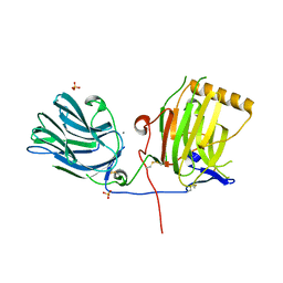
1DYK
 | | Laminin alpha 2 chain LG4-5 domain pair | | 分子名称: | CALCIUM ION, LAMININ ALPHA 2 CHAIN | | 著者 | Tisi, D, Talts, J.F, Timple, R, Hohenester, E. | | 登録日 | 2000-02-01 | | 公開日 | 2001-02-04 | | 最終更新日 | 2024-10-09 | | 実験手法 | X-RAY DIFFRACTION (2 Å) | | 主引用文献 | Structure of the C-Terminal Laminin G-Like Domain Pair of the Laminin Alpha 2 Chain Harbouring Binding Sites for Alpha-Dystroglycan and Heparin
Embo J., 19, 2000
|
|
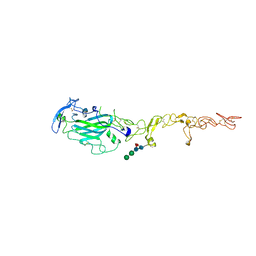
1OKQ
 | | LAMININ ALPHA 2 CHAIN LG4-5 DOMAIN PAIR, CA1 SITE MUTANT | | 分子名称: | CALCIUM ION, LAMININ ALPHA 2 CHAIN | | 著者 | Wizemann, H, Garbe, J.H.O, Friedrich, M.V.K, Timpl, R, Sasaki, T, Hohenester, E. | | 登録日 | 2003-07-28 | | 公開日 | 2003-09-11 | | 最終更新日 | 2024-11-13 | | 実験手法 | X-RAY DIFFRACTION (2.8 Å) | | 主引用文献 | Distinct Requirements for Heparin and Alpha-Dystroglycan Binding Revealed by Structure-Based Mutagenesis of the Laminin Alpha2 Lg4-Lg5 Domain Pair
J.Mol.Biol., 332, 2003
|
|
4AUO
 | | Crystal structure of MMP-1(E200A) in complex with a triple-helical collagen peptide | | 分子名称: | CALCIUM ION, INTERSTITIAL COLLAGENASE, TRIPLE-HELICAL COLLAGEN PEPTIDE, ... | | 著者 | Manka, S.W, Carafoli, F, Visse, R, Bihan, D, Raynal, N, Farndale, R.W, Murphy, G, Enghild, J.J, Hohenester, E, Nagase, H. | | 登録日 | 2012-05-18 | | 公開日 | 2012-07-11 | | 最終更新日 | 2023-12-20 | | 実験手法 | X-RAY DIFFRACTION (3 Å) | | 主引用文献 | Structural Insights Into Triple-Helical Collagen Cleavage by Matrix Metalloproteinase 1
Proc.Natl.Acad.Sci.USA, 109, 2012
|
|
4AQT
 | | Laminin gamma1 LN-LE1-2 structure | | 分子名称: | 2-acetamido-2-deoxy-beta-D-glucopyranose, CALCIUM ION, LAMININ SUBUNIT GAMMA-1, ... | | 著者 | Carafoli, F, Hussain, S, Hohenester, E. | | 登録日 | 2012-04-19 | | 公開日 | 2012-08-15 | | 最終更新日 | 2024-11-20 | | 実験手法 | X-RAY DIFFRACTION (3.2 Å) | | 主引用文献 | Crystal Structures of the Network-Forming Short-Arm Tips of the Laminin Beta1 and Gamma1 Chains.
Plos One, 7, 2012
|
|
4BJ3
 | | Integrin alpha2 I domain E318W-collagen complex | | 分子名称: | 2-[BIS-(2-HYDROXY-ETHYL)-AMINO]-2-HYDROXYMETHYL-PROPANE-1,3-DIOL, CHLORIDE ION, GFOGER PEPTIDE, ... | | 著者 | Carafoli, F, Hamaia, S.W, Bihan, D, Hohenester, E, Farndale, R.W. | | 登録日 | 2013-04-16 | | 公開日 | 2013-11-20 | | 最終更新日 | 2023-12-20 | | 実験手法 | X-RAY DIFFRACTION (3.042 Å) | | 主引用文献 | An Activating Mutation Reveals a Second Binding Mode of the Integrin Alpha2 I Domain to the Gfoger Motif in Collagens.
Plos One, 8, 2013
|
|
1H4U
 | | Domain G2 of mouse nidogen-1 | | 分子名称: | NIDOGEN-1 | | 著者 | Hopf, M, Gohring, W, Ries, A, Timpl, R, Hohenester, E. | | 登録日 | 2001-05-14 | | 公開日 | 2001-06-28 | | 最終更新日 | 2024-11-06 | | 実験手法 | X-RAY DIFFRACTION (2.2 Å) | | 主引用文献 | Crystal Structure and Mutational Analysis of a Perlecan-Binding Fragment of Nidogen-1
Nat.Struct.Biol., 8, 2001
|
|
1H30
 | | C-terminal LG domain pair of human Gas6 | | 分子名称: | CALCIUM ION, GROWTH-ARREST-SPECIFIC PROTEIN, SULFATE ION | | 著者 | Sasaki, T, Knyazev, P.G, Cheburkin, Y, Gohring, W, Tisi, D, Ullrich, A, Timpl, R, Hohenester, E. | | 登録日 | 2002-08-21 | | 公開日 | 2003-01-30 | | 最終更新日 | 2024-11-13 | | 実験手法 | X-RAY DIFFRACTION (2.2 Å) | | 主引用文献 | Crystal Structure of a Carboxy-Terminal Fragment of Growth-Arrest-Specific Protein Gas6: Receptor Tyrosine Kinase Activation by Laminin G-Like Domains
J.Biol.Chem., 277, 2002
|
|
4AQS
 | | Laminin beta1 LN-LE1-4 structure | | 分子名称: | 2-acetamido-2-deoxy-beta-D-glucopyranose, LAMININ SUBUNIT BETA-1, alpha-D-mannopyranose-(1-3)-beta-D-mannopyranose-(1-4)-2-acetamido-2-deoxy-beta-D-glucopyranose-(1-4)-[beta-L-fucopyranose-(1-6)]2-acetamido-2-deoxy-beta-D-glucopyranose | | 著者 | Carafoli, F, Hussain, S, Hohenester, E. | | 登録日 | 2012-04-19 | | 公開日 | 2012-08-15 | | 最終更新日 | 2024-10-23 | | 実験手法 | X-RAY DIFFRACTION (3.109 Å) | | 主引用文献 | Crystal Structures of the Network-Forming Short-Arm Tips of the Laminin Beta1 and Gamma1 Chains.
Plos One, 7, 2012
|
|
4AG4
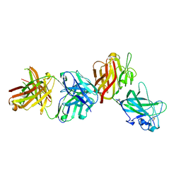 | | Crystal structure of a DDR1-Fab complex | | 分子名称: | 2-acetamido-2-deoxy-beta-D-glucopyranose-(1-4)-2-acetamido-2-deoxy-beta-D-glucopyranose, CALCIUM ION, EPITHELIAL DISCOIDIN DOMAIN-CONTAINING RECEPTOR 1, ... | | 著者 | Carafoli, F, Mayer, M.C, Shiraishi, K, Pecheva, M.A, Chan, L.Y, Nan, R, Leitinger, B, Hohenester, E. | | 登録日 | 2012-01-24 | | 公開日 | 2012-04-18 | | 最終更新日 | 2024-11-20 | | 実験手法 | X-RAY DIFFRACTION (2.8 Å) | | 主引用文献 | Structure of the Discoidin Domain Receptor 1 Extracellular Region Bound to an Inhibitory Fab Fragment Reveals Features Important for Signaling.
Structure, 20, 2012
|
|
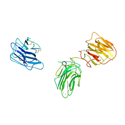
2VKX
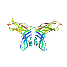 | | Human NCAM, FN3 domains 1 and 2, M610R mutant | | 分子名称: | NEURAL CELL ADHESION MOLECULE, SULFATE ION | | 著者 | Carafoli, F, Saffell, J.L, Hohenester, E. | | 登録日 | 2008-01-04 | | 公開日 | 2008-02-26 | | 最終更新日 | 2024-05-01 | | 実験手法 | X-RAY DIFFRACTION (2.7 Å) | | 主引用文献 | Structure of the Tandem Fibronectin Type 3 Domains of Neural Cell Adhesion Molecule
J.Mol.Biol., 377, 2008
|
|
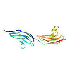
2VKW
 | | Human NCAM, FN3 domains 1 and 2 | | 分子名称: | NEURAL CELL ADHESION MOLECULE 1,140 KDA ISOFORM, SULFATE ION | | 著者 | Carafoli, F, Saffell, J.L, Hohenester, E. | | 登録日 | 2008-01-04 | | 公開日 | 2008-02-26 | | 最終更新日 | 2024-05-08 | | 実験手法 | X-RAY DIFFRACTION (2.3 Å) | | 主引用文献 | Structure of the Tandem Fibronectin Type 3 Domains of Neural Cell Adhesion Molecule
J.Mol.Biol., 377, 2008
|
|
2Y38
 | | LAMININ ALPHA5 CHAIN N-TERMINAL FRAGMENT | | 分子名称: | 2-acetamido-2-deoxy-beta-D-glucopyranose, 2-acetamido-2-deoxy-beta-D-glucopyranose-(1-4)-2-acetamido-2-deoxy-beta-D-glucopyranose, LAMININ SUBUNIT ALPHA-5, ... | | 著者 | Hussain, S.A, Carafoli, F, Hohenester, E. | | 登録日 | 2010-12-19 | | 公開日 | 2011-02-23 | | 最終更新日 | 2024-11-20 | | 実験手法 | X-RAY DIFFRACTION (2.9 Å) | | 主引用文献 | Determinants of Laminin Polymerisation Revealed by the Crystal Structure of the Alpha5 Chain Amino-Terminal Region
Embo Rep., 12, 2011
|
|
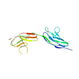
2WJS
 | |
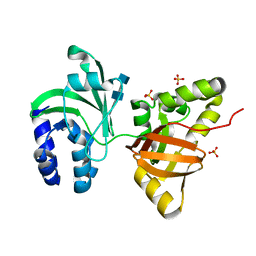
2VRA
 | | Drosophila Robo IG1-2 (monoclinic form) | | 分子名称: | 2-O-sulfo-alpha-L-idopyranuronic acid-(1-4)-2-deoxy-6-O-sulfo-2-(sulfoamino)-alpha-D-glucopyranose-(1-4)-2-O-sulfo-alpha-L-idopyranuronic acid-(1-4)-2-deoxy-6-O-sulfo-2-(sulfoamino)-alpha-D-glucopyranose, ROUNDABOUT 1, SULFATE ION | | 著者 | Fukuhara, N, Howitt, J.A, Hussain, S, Hohenester, E. | | 登録日 | 2008-03-28 | | 公開日 | 2008-04-08 | | 最終更新日 | 2024-10-16 | | 実験手法 | X-RAY DIFFRACTION (3.2 Å) | | 主引用文献 | Structural and Functional Analysis of Slit and Heparin Binding to Immunoglobulin-Like Domains 1 and 2 of Drosophila Robo
J.Biol.Chem., 283, 2008
|
|
2VR9
 | |
1O70
 | |
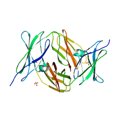
1OAS
 | | O-ACETYLSERINE SULFHYDRYLASE FROM SALMONELLA TYPHIMURIUM | | 分子名称: | O-ACETYLSERINE SULFHYDRYLASE, PYRIDOXAL-5'-PHOSPHATE | | 著者 | Burkhard, P, Rao, G.S.J, Hohenester, E, Schnackerz, K.D, Cook, P.F, Jansonius, J.N. | | 登録日 | 1999-01-29 | | 公開日 | 2000-01-28 | | 最終更新日 | 2023-12-27 | | 実験手法 | X-RAY DIFFRACTION (2.2 Å) | | 主引用文献 | Three-dimensional structure of O-acetylserine sulfhydrylase from Salmonella typhimurium.
J.Mol.Biol., 283, 1998
|
|
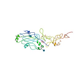
1DKA
 | | DIALKYLGLYCINE DECARBOXYLASE STRUCTURE: BIFUNCTIONAL ACTIVE SITE AND ALKALI METAL BINDING SITES | | 分子名称: | 2,2-DIALKYLGLYCINE DECARBOXYLASE (PYRUVATE), 2-(N-MORPHOLINO)-ETHANESULFONIC ACID, POTASSIUM ION, ... | | 著者 | Toney, M.D, Hohenester, E, Jansonius, J.N. | | 登録日 | 1993-06-18 | | 公開日 | 1994-10-15 | | 最終更新日 | 2025-03-26 | | 実験手法 | X-RAY DIFFRACTION (2.6 Å) | | 主引用文献 | Dialkylglycine decarboxylase structure: bifunctional active site and alkali metal sites.
Science, 261, 1993
|
|
2OAT
 | | ORNITHINE AMINOTRANSFERASE COMPLEXED WITH 5-FLUOROMETHYLORNITHINE | | 分子名称: | 1-AMINO-7-(2-METHYL-3-OXIDO-5-((PHOSPHONOXY)METHYL)-4-PYRIDOXAL-5-OXO-6-HEPTENATE, ORNITHINE AMINOTRANSFERASE | | 著者 | Storici, P, Schirmer, T. | | 登録日 | 1998-05-07 | | 公開日 | 1998-12-09 | | 最終更新日 | 2024-05-22 | | 実験手法 | X-RAY DIFFRACTION (1.95 Å) | | 主引用文献 | Crystal structure of human ornithine aminotransferase complexed with the highly specific and potent inhibitor 5-fluoromethylornithine.
J.Mol.Biol., 285, 1999
|
|
1OAT
 | |
1D6S
 | | CRYSTAL STRUCTURE OF THE K41A MUTANT OF O-ACETYLSERINE SULFHYDRYLASE COMPLEXED IN EXTERNAL ALDIMINE LINKAGE WITH METHIONINE | | 分子名称: | METHIONINE, O-ACETYLSERINE SULFHYDRYLASE, PYRIDOXAL-5'-PHOSPHATE | | 著者 | Burkhard, P, Tai, C.H, Ristroph, C.M, Cook, P.F, Jansonius, J.N. | | 登録日 | 1999-10-15 | | 公開日 | 2000-04-15 | | 最終更新日 | 2024-02-07 | | 実験手法 | X-RAY DIFFRACTION (2.3 Å) | | 主引用文献 | Ligand binding induces a large conformational change in O-acetylserine sulfhydrylase from Salmonella typhimurium.
J.Mol.Biol., 291, 1999
|
|


