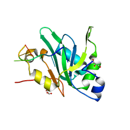
7EIO
 
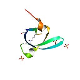
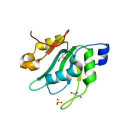 | | Crystal Structure of Mei2 RRM3 | | 分子名称: | GLYCEROL, Meiosis protein mei2, SULFATE ION | | 著者 | Shen, S.Y, Li, F.D. | | 登録日 | 2021-03-31 | | 公開日 | 2022-04-06 | | 最終更新日 | 2024-05-29 | | 実験手法 | X-RAY DIFFRACTION (1.895 Å) | | 主引用文献 | Structural insights reveal the specific recognition of meiRNA by the Mei2 protein.
J Mol Cell Biol, 14, 2022
|
|
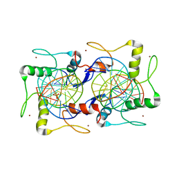
7FIA
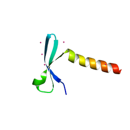
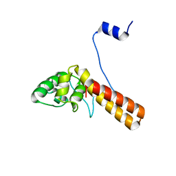 | | Structure of AcrIF23 | | 分子名称: | AcrIF23 | | 著者 | Ren, J, Yue, F. | | 登録日 | 2021-07-30 | | 公開日 | 2022-07-27 | | 最終更新日 | 2024-05-29 | | 実験手法 | X-RAY DIFFRACTION (2.13 Å) | | 主引用文献 | Structural and mechanistic insights into the inhibition of type I-F CRISPR-Cas system by anti-CRISPR protein AcrIF23.
J.Biol.Chem., 298, 2022
|
|
7UMI
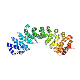 | | Importin a1 bound to Cp183-CTD | | 分子名称: | HBV-NLS, Importin subunit alpha-1 | | 著者 | Yang, R, Cingolani, G. | | 登録日 | 2022-04-07 | | 公開日 | 2023-10-25 | | 最終更新日 | 2024-02-28 | | 実験手法 | X-RAY DIFFRACTION (1.99 Å) | | 主引用文献 | Structural basis for nuclear import of hepatitis B virus (HBV) nucleocapsid core.
Sci Adv, 10, 2024
|
|
7V69
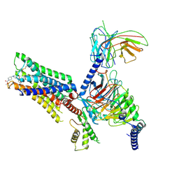
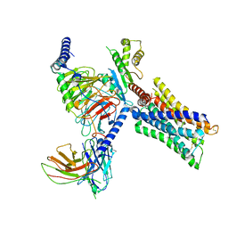 | | Cryo-EM structure of a class A GPCR-G protein complex | | 分子名称: | Guanine nucleotide-binding protein G(I)/G(S)/G(O) subunit gamma-2, Guanine nucleotide-binding protein G(I)/G(S)/G(T) subunit beta-1, Guanine nucleotide-binding protein G(i) subunit alpha-1, ... | | 著者 | Wang, J.J, Wu, M, Wu, L.J, Hua, T, Liu, Z.J, Wang, T. | | 登録日 | 2021-08-20 | | 公開日 | 2022-05-11 | | 最終更新日 | 2022-11-23 | | 実験手法 | ELECTRON MICROSCOPY (3.4 Å) | | 主引用文献 | The unconventional activation of the muscarinic acetylcholine receptor M4R by diverse ligands.
Nat Commun, 13, 2022
|
|
7V6A
 | | Cry-EM structure of M4-c110-G protein complex | | 分子名称: | Guanine nucleotide-binding protein G(I)/G(S)/G(O) subunit gamma-2, Guanine nucleotide-binding protein G(I)/G(S)/G(T) subunit beta-1, Guanine nucleotide-binding protein G(i) subunit alpha-1, ... | | 著者 | Wang, J.J, Wu, M, Wu, L.J, Hua, T, Liu, Z.J, Wang, T. | | 登録日 | 2021-08-20 | | 公開日 | 2022-05-11 | | 最終更新日 | 2022-11-23 | | 実験手法 | ELECTRON MICROSCOPY (3.6 Å) | | 主引用文献 | The unconventional activation of the muscarinic acetylcholine receptor M4R by diverse ligands.
Nat Commun, 13, 2022
|
|
7V68
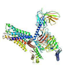 | | An Agonist and PAM-bound Class A GPCR with Gi protein complex structure | | 分子名称: | 3-amino-5-chloro-N-cyclopropyl-4-methyl-6-[2-(4-methylpiperazin-1-yl)-2-oxoethoxy]thieno[2,3-b]pyridine-2-carboxamide, 4-(4,5-dihydro-1,2-oxazol-3-yloxy)-N,N,N-trimethylbut-2-yn-1-aminium, Guanine nucleotide-binding protein G(I)/G(S)/G(O) subunit gamma-2, ... | | 著者 | Wang, J.J, Wu, L.J, Wu, M, Hua, T, Liu, Z.J, Wang, T. | | 登録日 | 2021-08-20 | | 公開日 | 2022-05-11 | | 最終更新日 | 2022-11-23 | | 実験手法 | ELECTRON MICROSCOPY (3.4 Å) | | 主引用文献 | The unconventional activation of the muscarinic acetylcholine receptor M4R by diverse ligands.
Nat Commun, 13, 2022
|
|
7VYW
 | |
7VZ2
 | | Crystal structure of chromodomain of Arabidopsis LHP1 | | 分子名称: | Chromo domain-containing protein LHP1, UNKNOWN ATOM OR ION | | 著者 | Liu, Y, Min, J. | | 登録日 | 2021-11-15 | | 公開日 | 2022-02-02 | | 最終更新日 | 2023-11-29 | | 実験手法 | X-RAY DIFFRACTION (1.7 Å) | | 主引用文献 | Structural basis for the recognition of methylated histone H3 by the Arabidopsis LHP1 chromodomain.
J.Biol.Chem., 298, 2022
|
|
7EIU
 | |
5X0W
 | | Molecular mechanism for the binding between Sharpin and HOIP | | 分子名称: | E3 ubiquitin-protein ligase RNF31, Sharpin | | 著者 | Liu, J, Li, F, Cheng, X, Pan, L. | | 登録日 | 2017-01-23 | | 公開日 | 2017-10-18 | | 実験手法 | X-RAY DIFFRACTION (3 Å) | | 主引用文献 | Structural Insights into SHARPIN-Mediated Activation of HOIP for the Linear Ubiquitin Chain Assembly
Cell Rep, 21, 2017
|
|
7EYI
 | |


